What Causes Pain Behind the Knee? Key Causes Explained
Pain behind the knee, called posterior knee pain, has multiple distinct causes, ranging from a fluid-filled cyst to a torn ligament to a blood clot that can become life-threatening without prompt treatment. Understanding what causes pain behind the knee requires knowing which specific anatomical structure is involved, because the correct treatment, urgency level, and provider type all depend on the diagnosis.
The posterior knee is one of the most diagnostically complex areas in musculoskeletal medicine. According to the National Institute of Arthritis and Musculoskeletal and Skin Diseases (NIAMS), knee problems affect millions of Americans annually, and posterior knee pain represents a clinically distinct subset that is frequently misidentified as general knee pain, leading to delays in appropriate care. Deep vein thrombosis alone, one of the causes that can produce posterior knee and calf pain, is responsible for an estimated 60,000 to 100,000 deaths per year in the United States, according to the Centers for Disease Control and Prevention.
This article covers the nine most clinically important causes of posterior knee pain with the specific anatomical and physiological mechanism behind each, the populations at highest risk for each cause, how each cause is identified clinically, and the specific signs that require same-day or emergency evaluation rather than watchful waiting.
What Causes Pain Behind the Knee: An Overview
The most common causes of pain behind the knee include Baker’s cyst, posterior cruciate ligament (PCL) injury, posterior horn meniscus tears, hamstring tendinopathy, deep vein thrombosis (DVT), knee osteoarthritis, and referred pain from lumbar spine pathology, with the specific cause determining whether the pain is structural, vascular, inflammatory, or neurological in origin.
No single presentation pattern reliably identifies one cause over another without clinical evaluation. Pain that worsens with knee flexion suggests different anatomy than pain that appears at rest at night. Swelling with warmth suggests a different pathway than pain without swelling.
| Cause | Pain Character | Key Associated Sign | Urgency Level |
|---|---|---|---|
| Baker’s cyst | Dull ache, pressure | Posterior fullness or lump | Non-urgent if stable |
| PCL injury | Aching, worse with activity | Instability, posterior tibial sag | Orthopedic referral within days |
| Meniscus tear | Sharp with twisting, locking | Clicking, joint line tenderness | Orthopedic evaluation within 1 to 2 weeks |
| Hamstring tendinopathy | Aching at tendon insertion | Pain worse at speed or incline | Non-urgent, sports medicine referral |
| DVT | Constant ache, calf tightness | Unilateral swelling, warmth, redness | Emergency evaluation same day |
| Osteoarthritis | Chronic ache, stiffness | Bilateral or gradual onset | Primary care referral |
| Lumbar spine referred pain | Vague, deep, often with back pain | No local swelling or tenderness | Primary care or spine specialist |
The one cause that overrides all others in terms of clinical urgency is DVT. Any sudden onset of posterior knee pain with unilateral calf swelling and skin warmth must be evaluated the same day, not scheduled for a routine appointment.
The Anatomy of the Popliteal Fossa and Why It Is Vulnerable
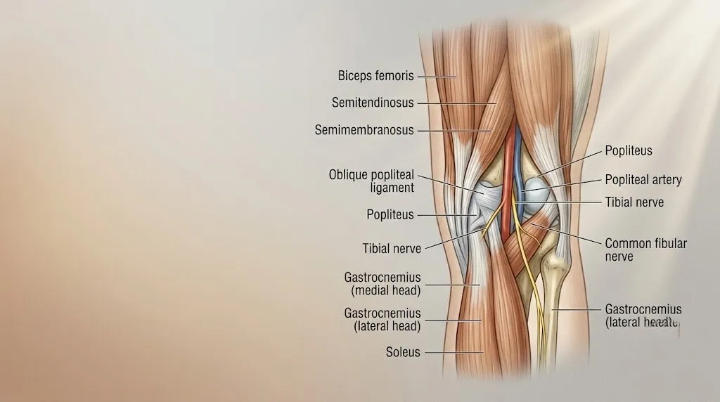
The popliteal fossa is the diamond-shaped anatomical space behind the knee joint, and its concentration of critical vascular, neural, and structural tissues is exactly why so many different conditions produce pain in this specific location.
The popliteal fossa is bounded on the upper medial side by the semimembranosus and semitendinosus muscles, on the upper lateral side by the biceps femoris muscle, and on the lower sides by the medial and lateral heads of the gastrocnemius muscle. Inside this space run the popliteal artery, the popliteal vein, the tibial nerve, and the common fibular nerve (also called the common peroneal nerve). The posterior capsule of the knee joint forms the floor of the fossa.
Think of the popliteal fossa like a crowded utility corridor behind a wall. Every pipe and cable in the building passes through it: arterial blood supply to the lower leg, venous return to the heart, and nerve signals to the foot. When any structure in that corridor becomes inflamed, compressed, or obstructed, the pain registers in the same location: directly behind the knee.
The posterior joint capsule is particularly vulnerable because it is the weakest region of the knee’s fibrous envelope. Elevated intra-articular pressure from any cause, whether a meniscus tear, osteoarthritis, or ligament injury, can force synovial fluid through a natural defect in the posteromedial capsule, producing the fluid-filled sac called a Baker’s cyst. This mechanism explains why Baker’s cysts so often coexist with other knee pathology rather than appearing in isolation.
For adolescents and young adults, the anatomy is the same but the most common causes of posterior knee pain differ. Hamstring strains and PCL injuries from sports contact dominate this age group, according to the American Academy of Orthopaedic Surgeons (AAOS).
Baker’s Cyst: The Most Common Cause of Pain Behind the Knee
A Baker’s cyst, also called a popliteal cyst, causes pain behind the knee when synovial fluid herniates through a defect in the posterior joint capsule and accumulates in the popliteal fossa, creating a fluid-filled sac that produces pressure, aching, and in some cases, a visible lump.
The mechanism is pressure-driven. When the knee joint produces excess synovial fluid, as it does in response to meniscus tears, osteoarthritis, rheumatoid arthritis, or any condition that causes joint inflammation, the intra-articular pressure rises. The path of least resistance is a natural communication between the posteromedial joint capsule and the semimembranosus-gastrocnemius bursa. Fluid flows through this channel and accumulates, forming the cyst.
Baker’s cysts are not independently occurring lesions in most adults. According to the Mayo Clinic, the majority of Baker’s cysts in adults form secondary to an underlying knee condition, most commonly osteoarthritis or a meniscus tear. Treating only the cyst without addressing the underlying cause of joint effusion reliably results in recurrence.
Quick Tip:
- A Baker’s cyst that ruptures sends fluid down between the muscle layers of the calf, mimicking deep vein thrombosis with calf swelling and warmth. This is called a ruptured popliteal cyst or “pseudothrombophlebitis.”
- A ruptured Baker’s cyst typically causes a crescent-shaped bruise below the medial malleolus (the inner ankle bone), which can help distinguish it from DVT clinically.
- Individuals with rheumatoid arthritis have significantly higher Baker’s cyst formation rates than the general population because of their chronic elevated intra-articular inflammation.
Children with Baker’s cysts present differently than adults. Pediatric popliteal cysts typically arise without underlying joint pathology, are more often located in the lateral popliteal fossa, and have a substantially higher rate of spontaneous resolution, according to clinical data published in the Journal of Bone and Joint Surgery.
Posterior Cruciate Ligament Injury and Posterior Knee Pain
Posterior cruciate ligament (PCL) injury causes pain behind the knee by disrupting the primary ligamentous restraint against posterior tibial translation, triggering nociceptor activation in the ligament’s collagen fibers and in the densely innervated posterior joint capsule.
The PCL originates on the posterior surface of the proximal tibia and inserts on the medial femoral condyle inside the joint. Its primary mechanical role is resisting backward displacement of the tibia relative to the femur. PCL tears typically result from a direct blow to the front of the proximal tibia with the knee flexed, the classic “dashboard injury” in motor vehicle collisions, or from hyperflexion of the knee in athletic contact.
The AAOS reports that PCL injuries are less common than ACL injuries overall but account for a substantial proportion of sports-related knee injuries in contact athletes. PCL tears produce a characteristic clinical finding called the posterior tibial sag sign: when the knee is flexed to 90 degrees and observed from the side, the tibia visibly sags backward relative to the femur due to loss of the PCL’s restraining force.
Posterior cruciate ligament injuries range from partial ligament sprains (Grade I and II) that often heal without surgery to complete ruptures (Grade III) that may require surgical reconstruction, particularly when combined with posterolateral corner injuries. Grade I and II injuries in non-athletes typically follow a conservative management path with physical therapy focusing on quadriceps strengthening to compensate for PCL function.
Any acute posterior knee pain following a direct knee trauma, particularly with knee instability, posterior swelling, or the inability to fully bear weight, warrants evaluation by a board-certified orthopedic surgeon or sports medicine physician within 24 to 72 hours. Delaying evaluation after a suspected PCL tear risks compounding posterior capsule damage and increasing the complexity of any subsequent surgical intervention.
Key Takeaway: Baker’s cyst is the single most common cause of pain and a palpable lump behind the knee in adults, but it almost always signals an underlying joint problem (meniscus tear or osteoarthritis) that needs to be diagnosed and treated separately from the cyst itself.
Meniscus Tear Causing Pain Behind the Knee
Posterior horn meniscus tears cause pain specifically at the back of the knee because the posterior horns of both the medial and lateral menisci sit within the posterior compartment of the knee joint, directly adjacent to the popliteal fossa and posterior capsule.
The medial meniscus and lateral meniscus are C-shaped fibrocartilage structures that distribute load across the tibiofemoral joint, absorb shock, and provide secondary stability. The posterior horn of the medial meniscus is the most commonly torn segment of the entire meniscus, accounting for the majority of meniscus tears seen in clinical practice. This anatomical vulnerability exists because the posterior horn of the medial meniscus is tightly anchored to the posteromedial joint capsule, making it less mobile and more susceptible to shear forces during twisting movements.
Research published in the American Journal of Sports Medicine has found that meniscus tears, particularly degenerative horizontal cleavage tears of the posterior horn, occur with rising frequency in adults over 40 and are closely associated with early knee osteoarthritis. In this age group, tears can develop without a specific traumatic event, produced instead by repetitive load accumulation on already-compromised cartilage.
A posterior horn tear classically produces pain with deep knee flexion, squatting, or rotating the knee while the foot is planted. Mechanical symptoms, including clicking, catching, or a sensation of the knee “giving way,” suggest that a torn flap of meniscus is intermittently displacing between the joint surfaces.
People with obesity carry disproportionately elevated meniscal load per unit of activity, and research consistently shows higher meniscus tear rates in individuals with a body mass index above 30. A primary care physician, after initial assessment, will typically refer a suspected meniscus tear to a board-certified orthopedic surgeon for MRI confirmation and management planning.
Hamstring Tendinopathy and Pain at the Back of the Knee
Hamstring tendinopathy causes a deep, aching pain at the back of the knee by producing degenerative changes at the tendon insertion points of the semimembranosus, semitendinosus, and biceps femoris muscles on the posterior tibia and fibular head, with tendon fiber disorganization replacing normal collagen architecture.
Unlike acute hamstring muscle strains in the mid-belly of the thigh, hamstring tendinopathy affects the tendon’s attachment point at the bone, a zone called the enthesis. Tendinopathy at the enthesis is not a primarily inflammatory process in most cases. The underlying mechanism is tendon degeneration: disorganized collagen type III replacing the strong parallel collagen type I fibers, increased vascularity and nerve ingrowth into the pathological tendon tissue, and a loss of the tendon’s mechanical load-bearing capacity. This process is described in the tendinopathy literature as failed healing response rather than classic inflammation.
The British Journal of Sports Medicine has published extensive research establishing that hamstring tendinopathy at the distal (knee) insertion is distinct from proximal hamstring tendinopathy at the ischial tuberosity. Distal hamstring tendinopathy is more common in distance runners, cyclists, and middle-aged recreational athletes who have increased their training load rapidly. Pain is characteristically worst at the start of activity, improves with warm-up, and worsens again after cooling down.
Quick Tip:
- Resisted knee flexion against manual resistance (the “resisted hamstring curl test”) that reproduces posterior knee pain is a reliable clinical indicator of distal hamstring tendinopathy.
- Compression of the tendon against a firm surface, such as a bicycle seat, often aggravates symptoms specifically in biceps femoris tendinopathy.
- Female runners show a slightly higher incidence of distal hamstring tendinopathy than male runners at equivalent training volumes, possibly due to differences in pelvic width and hamstring angle of pull.
Deep Vein Thrombosis Causing Pain Behind the Knee
Deep vein thrombosis (DVT) causes pain behind the knee when a blood clot forms in the popliteal vein or the femoral vein, obstructing venous return from the lower leg and causing venous distension, local inflammatory mediator release, and the characteristic aching pain and swelling in the posterior knee and calf region.
DVT operates through Virchow’s triad: venous stasis (slow or stopped blood flow), endothelial injury (damage to the vein wall), and hypercoagulability (an abnormally activated clotting system). Any of these three conditions can independently trigger the coagulation cascade, converting fibrinogen to fibrin and trapping red blood cells to form a growing thrombus. The inflammatory response around the clot, mediated by prostaglandins and cytokines, produces the warmth, redness, and pain that alert a clinician to DVT’s presence.
According to the CDC, DVT and pulmonary embolism together affect an estimated 900,000 Americans per year and cause between 60,000 and 100,000 deaths annually. The lethality comes from clot propagation: a DVT in the popliteal vein can fragment and travel through the venous system to the pulmonary arteries, causing pulmonary embolism, which restricts blood flow through the lungs and can cause sudden cardiovascular collapse.
Posterior knee and calf pain from DVT is characteristically different from musculoskeletal causes. It tends to come on acutely over hours rather than days, is present at rest and does not improve with position change, is accompanied by unilateral calf or leg swelling, and often occurs in the setting of a known risk factor (recent surgery, prolonged bed rest, long-haul air travel, oral contraceptive use, active cancer, or a personal or family history of blood clots).
Women using combined oral contraceptives have approximately three to four times the baseline DVT risk of non-users, according to research published in the New England Journal of Medicine. This risk is compounded further during pregnancy and in the postpartum period, making posterior knee pain with swelling in these populations particularly urgent.
Key Takeaway: DVT causing posterior knee pain is the single most dangerous cause on this list, and its presentation can closely mimic a Baker’s cyst rupture or a muscle strain, making any combination of acute-onset posterior knee pain with unilateral swelling and warmth a same-day emergency evaluation, not a scheduled appointment.
Osteoarthritis as a Cause of Posterior Knee Pain
Knee osteoarthritis causes pain in the posterior compartment when progressive loss of articular cartilage in the medial or lateral tibiofemoral joint compartment is accompanied by synovitis, joint effusion, osteophyte formation along the posterior femoral condyles, and posterior capsular thickening.
Osteoarthritis is not simply “wear and tear.” The underlying mechanism involves a pathological imbalance between chondrocyte-mediated cartilage synthesis and degradation. Pro-inflammatory cytokines including interleukin-1 beta (IL-1 beta) and tumor necrosis factor alpha (TNF-alpha) stimulate chondrocytes to produce matrix metalloproteinases (MMPs), enzymes that break down the collagen and proteoglycan framework of articular cartilage faster than it can be rebuilt. Over years, this process exposes subchondral bone, triggers bone remodeling, osteophyte formation, and progressive joint deformity.
Posterior osteophytes at the femoral condyles are a particularly common finding on knee radiographs in patients with advanced osteoarthritis and directly contribute to pain during full knee extension, because the osteophytes compress posterior capsular tissue and soft structures in the popliteal fossa.
According to the NIAMS, osteoarthritis affects approximately 32.5 million adults in the United States and is the leading cause of disability among adults over 65. Females develop knee osteoarthritis at higher rates than males after age 55, a difference partly explained by the decline in estrogen during menopause. Estrogen has a protective effect on articular cartilage by modulating the inflammatory response in synovial tissue, and its postmenopausal decline accelerates chondrocyte vulnerability to inflammatory cytokines.
A primary care physician can order weight-bearing knee radiographs as the initial diagnostic step for suspected osteoarthritis, and referral to a board-certified orthopedic surgeon is appropriate when conservative management has not reduced symptoms after 8 to 12 weeks.
Referred Pain to the Back of the Knee from the Lumbar Spine
Referred pain to the back of the knee from the lumbar spine occurs when disc herniation at L4-L5 or L5-S1 compresses the tibial nerve root (primarily the S1 or S2 root), producing pain that travels along the sciatic nerve pathway through the posterior thigh and into the posterior knee region without any local knee pathology.
This cause is absent from every major competitor article on this topic, yet it is a clinically important differential diagnosis that changes the entire management approach. Referred pain from the lumbar spine does not produce local knee swelling. It does not cause isolated joint-line tenderness. The knee examination is typically normal. The pain is vague, deep, and often accompanied by similar or greater discomfort in the lower back or posterior thigh.
Think of it this way: the sciatic nerve is like a long electrical cable running from the lumbar spine through the posterior thigh and behind the knee. When a herniated disc pinches the cable at its source in the back, the electrical signal disturbance registers all along the wire’s path. The knee feels the pain even though the knee itself is perfectly intact.
The clinical key to identifying spine-referred posterior knee pain is the straight leg raise test, in which passively raising a supine patient’s straight leg to 30 to 70 degrees reproduces the posterior knee pain (and often the buttock or posterior thigh pain) by stretching the sciatic nerve over the herniated disc. A positive test makes lumbar radiculopathy the primary suspected diagnosis.
Older adults and individuals with a history of lumbar disc disease, prior back surgery, or spinal stenosis are at elevated risk for this presentation. A primary care physician should be the first point of contact, and referral to a board-certified spine surgeon or neurologist is warranted if the straight leg raise test is positive and pain has not improved with conservative management over 4 to 6 weeks.
Key Takeaway: Referred pain from lumbar disc herniation at L4-L5 or L5-S1 can produce posterior knee pain with a completely normal knee examination, which is why any posterior knee pain accompanied by lower back pain or posterior thigh aching deserves spinal evaluation before knee-focused imaging or treatment is pursued.
Pain Behind the Knee When Bending or Straightening the Leg
Pain behind the knee specifically with bending (flexion) points toward compression of posterior structures, while pain with straightening (extension) suggests posterior osteophytes, PCL tension, or hamstring tendon loading, and distinguishing between the two movement patterns significantly narrows the list of likely causes.
Deep flexion compresses the posterior joint capsule, engages the posterior horn of the menisci under maximum load, and increases intra-articular pressure sufficiently to push fluid into any pre-existing Baker’s cyst. All three of these effects peak between 90 and 130 degrees of knee flexion, which is why squatting, kneeling, and stair descent reliably aggravate posterior compartment symptoms from Baker’s cysts and posterior horn meniscus tears.
Pain with full extension loading follows a different anatomical logic. The popliteus muscle, a small triangular muscle running obliquely across the posterior capsule, is active primarily at the initiation of knee flexion from a fully extended position, a movement called “unlocking” the knee. Popliteus tendinitis, which produces a sharp or aching pain in the lateral-posterior knee, is particularly common in runners who train on banked or downhill surfaces, because this terrain increases the popliteus’ demand to prevent excessive tibial external rotation.
To describe your pain pattern accurately to a physician or physical therapist:
- Note whether the pain occurs at the beginning of movement (initiation), at a specific angle of bend, or at the end of full extension.
- Identify whether the pain is on the inner (medial) or outer (lateral) side of the posterior knee, or directly in the midline.
- Record whether it is present at rest, only with activity, or worse after sitting for more than 30 minutes.
- Note any associated clicking, locking, giving way, or visible swelling behind the knee.
- Record whether the pain is present bilaterally (both knees) or only in one knee, as bilateral pain shifts the differential toward systemic causes such as inflammatory arthritis or referred lumbar spine pain.
Pain Behind the Knee in Runners and Athletes
Pain behind the knee in runners and athletes most commonly results from hamstring tendinopathy at the distal insertion, Baker’s cyst secondary to repetitive joint loading, popliteus tendinitis from downhill running, or a posterior horn medial meniscus tear caused by cumulative mechanical stress rather than a single traumatic event.
Running biomechanics place repetitive eccentric load on the hamstring tendons during the late swing phase of the gait cycle, when the hamstrings decelerate the forward swing of the tibia just before foot contact. At high training volumes, this repeated eccentric loading can overwhelm the tendon’s capacity for repair, initiating the degenerative tendinopathy process. Runners who increase their weekly mileage by more than 10 percent per week are at elevated risk for overuse injuries at the knee, according to data reviewed in the British Journal of Sports Medicine.
Cyclists experience a distinct posterior knee pain pattern. Saddle height set too low forces the knee into excessive flexion at the bottom of the pedal stroke, increasing posterior compartment compression and popliteal fossa pressure. Saddle height set too high, conversely, creates excessive knee extension and places disproportionate tension on the hamstring tendons at their distal insertion. Either extreme produces characteristic posterior knee pain that resolves promptly with appropriate bike fit correction.
Elite and collegiate athletes warrant particular attention to popliteal artery entrapment syndrome, a rare but serious condition in which an anomalous medial head of the gastrocnemius compresses the popliteal artery during exercise, causing exertional posterior knee and calf pain, and in some cases, reduced pedal pulses. A vascular surgeon should evaluate any athlete whose posterior knee pain is accompanied by exercise-induced foot pallor, coolness, or diminished pulses.
Pain Behind the Knee Causes in Women
Pain behind the knee causes in women include all the general causes that affect the broader population, but women face elevated risk for DVT related to oral contraceptive use and pregnancy, higher osteoarthritis rates after menopause, and greater hamstring-to-quadriceps strength ratio imbalances that affect posterior knee loading.
Oral contraceptives containing synthetic estrogen and progestin increase the risk of venous thromboembolism by three to four times compared to non-users, according to research published in the New England Journal of Medicine. This risk is highest in the first year of use, in women who smoke, in women over 35, and in women with inherited thrombophilia conditions such as Factor V Leiden mutation or prothrombin G20210A mutation. For a woman on oral contraceptives who develops acute unilateral posterior knee pain with calf swelling, DVT must be excluded before any other diagnosis is assumed.
During pregnancy, the combination of elevated procoagulant factors, decreased natural anticoagulants (particularly protein S), venous stasis from uterine pressure on the inferior vena cava, and reduced ambulation creates a state of physiological hypercoagulability that increases DVT risk by approximately five-fold compared to age-matched non-pregnant women, according to the American College of Obstetricians and Gynecologists.
Postmenopausal women are disproportionately affected by knee osteoarthritis. The protective effect of estrogen on chondrocyte survival means that the postmenopausal decline in circulating estrogen accelerates cartilage degradation rates. Osteoarthritis affecting the posterior compartment in postmenopausal women frequently coexists with a Baker’s cyst, because the synovitis and effusion driven by osteoarthritis create the elevated intra-articular pressure needed for cyst formation.
Key Takeaway: Women using oral contraceptives and pregnant or postpartum women who develop acute unilateral posterior knee pain with swelling must have DVT excluded the same day, because the baseline DVT risk in these populations is substantially higher than in the general adult population.
Pain Behind the Knee in Older Adults
Pain behind the knee in older adults is most often driven by osteoarthritis of the medial tibiofemoral compartment, posterior horn meniscus tears (frequently degenerative rather than traumatic), and Baker’s cysts secondary to these joint conditions, with DVT risk also elevated due to reduced ambulation and comorbidities.
Degenerative posterior horn medial meniscus tears in adults over 60 are so common that they appear in a notable proportion of MRI scans performed for knee pain in this age group, and some research has raised debate about whether these tears are always symptomatic or whether they represent age-related changes that coincide with pain from other sources. A 2013 study published in the New England Journal of Medicine found that for middle-aged and older adults with degenerative meniscus tears and knee pain, surgery provided no greater benefit than optimized physical therapy after two years. This has shifted clinical practice toward conservative management as the first-line approach for degenerative posterior horn tears in adults over 55 who do not have mechanical locking symptoms.
Older adults are at elevated risk for pharmacological interactions that complicate both the diagnosis and treatment of posterior knee pain. Individuals taking anticoagulants such as warfarin or direct oral anticoagulants for atrial fibrillation or prior DVT have a different risk profile for blood clots and may experience atypical presentations of posterior compartment injury due to hemarthrosis (bleeding into the joint). A primary care physician managing an older adult on anticoagulant therapy who develops new knee pain should be notified promptly, even for apparently minor injuries.
Gout and pseudogout (calcium pyrophosphate deposition disease) are underrecognized causes of acute posterior knee pain in older adults. Uric acid or calcium pyrophosphate crystals depositing in the synovial membrane or articular cartilage of the posterior compartment trigger an acute inflammatory arthritis episode that can be severely painful, with joint warmth and swelling that closely mimics septic arthritis or DVT.
Pain Behind the Knee After Sitting or Prolonged Inactivity
Pain behind the knee after sitting or prolonged inactivity that improves with movement is a classic presentation of knee osteoarthritis and degenerative posterior horn meniscus tears, driven by the stiffening of synovial fluid during rest and the mechanical irritation of damaged tissue with the first few movements after a static position.
Synovial fluid, the lubricant of the knee joint, becomes less viscous and less uniformly distributed during prolonged static positioning. When you sit for an extended period, the synovial fluid shifts to the dependent portions of the joint cavity, and the posterior compartment, which is at the back of a flexed knee, experiences reduced lubrication. The first movement after standing loads cartilage and synovial tissue that are temporarily under-lubricated, producing the characteristic “start-up pain” that is a hallmark of both osteoarthritis and posterior horn meniscal degeneration.
This presentation differs from DVT-related pain in a clinically useful way. DVT pain does not improve with movement. It is typically present at rest, worsens with leg elevation (which increases venous pressure), and does not ease with walking around. If pain behind the knee improves within 5 to 10 minutes of gentle movement after sitting, this pattern is more consistent with a degenerative mechanical cause than a vascular cause.
Individuals who work sedentary jobs and experience posterior knee pain after desk-based work periods can monitor this pattern carefully. Pain that consistently improves with 5 to 10 minutes of walking and does not include swelling, warmth, or redness at the back of the knee is appropriate for a scheduled primary care appointment within 2 to 4 weeks, rather than an emergency evaluation.
However, prolonged sitting is itself a DVT risk factor. A person who has recently traveled on a long-haul flight or been immobile due to illness and then develops posterior knee and calf pain after returning to activity should not assume a benign cause without professional evaluation.
How Posterior Knee Pain Is Diagnosed
Posterior knee pain is diagnosed through a structured sequence of physical examination tests, targeted imaging studies, and in some cases, laboratory blood tests, with the choice of diagnostic tool determined by the suspected cause based on the clinical history and physical findings.
The diagnostic process for a new presentation of posterior knee pain typically begins with a detailed clinical history and physical examination by a primary care physician, board-certified orthopedic surgeon, or sports medicine physician. The physical examination will include:
- Inspection and palpation of the posterior knee for swelling, cyst formation, skin warmth, redness, and point tenderness at specific anatomical landmarks (joint line, hamstring tendon insertions, posterior capsule).
- Range of motion testing in active and passive flexion and extension, noting the arc where pain is reproduced.
- Ligament stability tests including the posterior drawer test and the posterior tibial sag test to assess PCL integrity.
- McMurray’s test and Thessaly’s test to assess meniscal integrity.
- Straight leg raise test to screen for lumbar spine-referred pain.
- Assessment of the distal pulses (popliteal, dorsalis pedis, posterior tibial) and skin temperature for any suggestion of vascular compromise.
- If DVT is clinically suspected, application of the Wells Clinical Prediction Score and ordering of a D-dimer blood test as a first-line screening tool, followed by a venous duplex Doppler ultrasound if the D-dimer is elevated or clinical suspicion remains high despite a negative D-dimer.
Imaging tools used for posterior knee pain diagnosis:
| Imaging Test | What It Shows | When It Is Ordered |
|---|---|---|
| Weight-bearing knee X-ray | Joint space narrowing, osteophytes, bone alignment | First-line for suspected osteoarthritis |
| MRI (3-Tesla preferred) | Soft tissue detail: meniscus, ligaments, cartilage, cysts | When soft tissue diagnosis is needed |
| Venous duplex Doppler ultrasound | Popliteal and femoral vein thrombus | Suspected DVT: ordered same day |
| CT angiography | Popliteal artery anatomy | Suspected popliteal artery entrapment |
| Diagnostic ultrasound | Baker’s cyst confirmation, real-time assessment | Baker’s cyst, soft tissue masses |
Emergency Symptoms: When to Call 911 or Go to the ER
Certain presentations of pain behind the knee require immediate emergency evaluation. Do not wait to see if these resolve on their own.
Call 911 or go to the nearest emergency room immediately if you experience:
- Sudden, acute onset of posterior knee and calf pain with visible unilateral leg swelling, skin warmth, or skin redness, particularly following recent surgery, prolonged bed rest, long-haul travel, or in the setting of known cancer or oral contraceptive use (these are the cardinal signs of DVT with pulmonary embolism risk)
- Posterior knee pain accompanied by sudden chest pain, shortness of breath, rapid heart rate, or coughing up blood (these indicate pulmonary embolism, a directly life-threatening DVT complication requiring immediate intervention)
- Complete inability to bear weight on the affected leg following a direct trauma to the knee, with visible deformity, swelling, or instability (suggests PCL rupture or combined posterior capsular injury requiring urgent orthopedic evaluation)
- Posterior knee swelling with fever above 38.3 degrees Celsius (101 degrees Fahrenheit), skin that is hot and red over the joint, and severe pain with any knee movement (suggests septic arthritis or septic bursitis, conditions that can destroy joint cartilage within hours if untreated)
- A pulsating lump behind the knee with pain that extends into the lower leg (suggests popliteal artery aneurysm, a vascular emergency requiring immediate vascular surgery evaluation)
These presentations can indicate deep vein thrombosis with pulmonary embolism, septic joint infection, acute vascular injury, or complete ligamentous disruption, and all require emergency medical assessment, not an urgent care appointment or a scheduled physician call.
Frequently Asked Questions About Pain Behind the Knee
What causes pain behind the knee with no swelling?
Pain behind the knee without swelling most commonly points to hamstring tendinopathy at the distal insertion, popliteus tendinitis, early PCL sprain, or referred pain from lumbar spine pathology at L4-L5 or L5-S1.
The absence of swelling makes vascular causes such as DVT and Baker’s cyst less likely, though not impossible in early stages.
If the pain persists beyond two weeks without improvement, evaluation by a primary care physician or board-certified sports medicine physician, with a straight leg raise test to screen for lumbar referral, is appropriate.
Can a Baker’s cyst go away on its own?
A Baker’s cyst can resolve on its own if the underlying cause of the joint effusion resolves, but in adults, the cyst typically recurs unless the primary knee pathology (meniscus tear or osteoarthritis) is addressed.
According to the Mayo Clinic, Baker’s cysts in children have a high rate of spontaneous resolution without treatment, while adult cases require management of the underlying condition.
Aspiration of the cyst alone without treating the source of joint fluid overproduction results in recurrence in most adult patients.
How do I know if my knee pain is a blood clot?
Blood clot (DVT) pain behind the knee characteristically comes on acutely over hours, is present at rest, does not improve with movement, and is accompanied by unilateral calf or leg swelling, skin warmth, and redness.
If you have a risk factor for DVT (recent surgery, prolonged immobility, oral contraceptive use, pregnancy, active cancer, or a personal history of blood clots), any acute posterior knee and calf pain with swelling must be evaluated the same day.
Go to the nearest emergency room immediately rather than waiting for a scheduled appointment, because untreated DVT can cause pulmonary embolism within hours.
What does a torn meniscus feel like behind the knee?
A posterior horn meniscus tear typically produces a sharp or aching pain at the back of the knee that is aggravated by deep flexion, squatting, twisting, or pivoting movements.
Mechanical symptoms such as a clicking or catching sensation during movement, or a feeling that the knee is “stuck” at a particular angle, are characteristic of a meniscus tear rather than tendinitis or cyst.
A board-certified orthopedic surgeon or sports medicine physician can confirm a suspected meniscus tear with a clinical examination followed by MRI of the affected knee.
Can back problems cause pain behind the knee?
Lumbar disc herniation at L4-L5 or L5-S1 can compress the tibial nerve root and produce referred pain that travels along the sciatic nerve pathway into the posterior knee region without any local knee abnormality.
This presentation is often accompanied by lower back pain or posterior thigh aching and produces a normal physical examination of the knee itself, including no swelling, no joint-line tenderness, and no abnormal ligament testing.
A positive straight leg raise test, in which lifting a straight leg to 30 to 70 degrees reproduces the posterior knee pain, points toward a spinal cause and warrants evaluation by a primary care physician with spinal imaging.
When should I see a doctor for pain behind my knee?
Pain behind the knee with unilateral swelling, warmth, or redness requires same-day emergency evaluation to exclude DVT, not a scheduled appointment.
Posterior knee pain following a direct trauma with instability, locking, or inability to bear weight warrants evaluation by a primary care physician or board-certified orthopedic surgeon within 24 to 72 hours.
Posterior knee pain without red flag signs that does not improve with rest and activity modification after two to four weeks warrants a scheduled evaluation with a primary care physician or board-certified sports medicine physician to pursue imaging and an accurate diagnosis.
The Clinical Picture in Practice
Pain behind the knee spans a range from a self-limiting tendon overuse injury to a life-threatening blood clot, and the only reliable way to separate those presentations is by applying the clinical framework outlined in this article. When the pain came on suddenly, when it is paired with swelling and warmth, and when there are DVT risk factors in the picture, the answer is the emergency room, not a home remedy.
For slower-onset posterior knee pain without red flags, start by noting exactly when the pain occurs (with bending, with straightening, after sitting, during running), which side of the back of the knee hurts (medial, lateral, or central), and whether there is any visible fullness or lump. That description will allow a primary care physician or sports medicine clinician to direct the correct imaging efficiently.
Every cause covered here has a specific diagnostic path and a specific type of provider who manages it best. Your job is to get the right information to the right clinician. This article gives you the clinical vocabulary to do exactly that.