Vertebral Artery Dissection Symptoms: Full 2026 Guide
Vertebral artery dissection symptoms include a sudden, severe posterior neck pain that many people describe as unlike any headache they have experienced before, often accompanied by dizziness, visual changes, and in serious cases, the full symptom pattern of a posterior circulation stroke. Recognizing these symptoms early is the single factor most likely to determine whether a person receives treatment before permanent neurological injury occurs.
According to the National Institute of Neurological Disorders and Stroke (NINDS), cervical artery dissection is one of the leading causes of ischemic stroke in adults under the age of 45. A 2019 review published in JAMA Neurology estimated that vertebral and carotid artery dissections together account for approximately 10 to 25 percent of strokes in this younger age group, a proportion far higher than most people, and many primary care providers, expect.
This article covers every named symptom of vertebral artery dissection, the anatomical mechanism producing each one, which presentations demand immediate emergency evaluation, how the condition is diagnosed, how population factors affect the clinical picture, and what the current evidence says about outcomes.
What Is Vertebral Artery Dissection
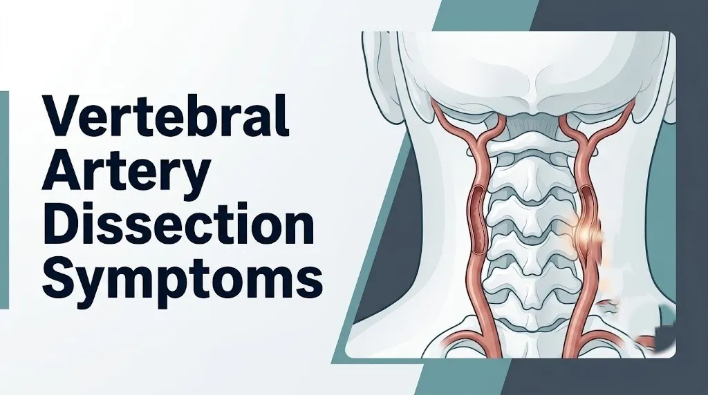
Vertebral artery dissection is a tear in the inner wall of one or both of the vertebral arteries, the paired vessels that run through the transverse foramina of the C6 through C1 cervical vertebrae and converge at the base of the brain to form the basilar artery.
The vertebral arteries are the main blood supply to the posterior circulation, which includes the brainstem, the cerebellum, the occipital cortex (the brain’s vision-processing center), and the inner ear’s blood supply. When the inner wall of a vertebral artery tears, blood forces its way between the arterial wall layers, creating what clinicians call a false lumen, a second channel running parallel to the normal vessel channel inside the arterial wall itself.
This is not like a garden hose splitting open. It is more like the layers of a thick rubber hose wall separating from each other while the hose stays intact on the outside. That separation creates two problems simultaneously: the normal channel narrows or blocks (reducing blood flow), and the exposed inner surface of the arterial wall triggers clot formation that can break off and travel downstream into the brain.
The condition is classified as either subintimal dissection (the false lumen forms between the tunica intima and tunica media, most often causing stenosis or occlusion) or subadventitial dissection (the false lumen forms between the tunica media and tunica adventitia, more likely to produce a bulging pseudoaneurysm rather than occlusion). The distinction matters clinically because the two subtypes carry different stroke risk profiles and different treatment considerations.
People over 50 should note that while VAD can occur at any age, the condition is most prevalent in adults aged 35 to 50. The left vertebral artery is the dominant vessel in approximately 75 percent of people and is slightly more frequently affected in many series.
Vertebral Artery Dissection Symptoms: The Complete Clinical Picture
The symptoms of vertebral artery dissection fall into two clinically distinct groups that every person with this condition and every clinician evaluating it needs to understand separately.
The first group is local symptoms, produced directly by the arterial wall injury, the intramural hematoma, and compression or irritation of adjacent structures. These include posterior neck pain, occipital headache, and occasionally tinnitus or a pulsatile sensation in the neck. These symptoms occur because pain-sensing C-fibers in the arterial adventitia are activated by the mechanical disruption and by local inflammatory mediators including bradykinin and prostaglandins released at the injury site.
The second group is ischemic symptoms, produced when the narrowed or occluded vessel reduces blood flow to the brainstem, cerebellum, or occipital cortex, or when a thrombus breaks free and travels into the posterior circulation. These symptoms include dizziness, visual disturbances, facial numbness, swallowing difficulty, and in the most severe presentations, the full picture of a posterior circulation stroke.
| Symptom Group | Mechanism | Examples | Clinical Significance |
|---|---|---|---|
| Local symptoms | Arterial wall tear, intramural hematoma, C-fiber activation | Posterior neck pain, occipital headache, pulsatile neck sensation | Present in most VAD cases; may precede ischemia by hours to days |
| Ischemic symptoms | Posterior circulation hypoperfusion or thromboembolism | Dizziness, ataxia, dysarthria, visual loss, facial numbness | Indicate neurological involvement; require immediate evaluation |
| Autonomic symptoms | Cervical sympathetic chain compression | Ptosis, miosis, anhidrosis (Horner syndrome) | Suggest subadventitial dissection with pseudoaneurysm |
| Cranial nerve symptoms | Brainstem nucleus ischemia | Dysphagia, hoarse voice, hiccups | Suggest lateral medullary involvement |
Older adults above 65 may present with a more insidious onset and a higher proportion of ischemic symptoms relative to local pain. Clinicians at Mayo Clinic note that the classic presentation (neck pain followed by posterior fossa ischemia) is recognized in only about 50 percent of confirmed VAD cases, making atypical presentations clinically challenging.
Neck Pain and Headache as First Symptoms of Vertebral Artery Dissection
Sudden, severe posterior neck pain combined with an occipital headache is the most common first symptom of vertebral artery dissection, present in an estimated 70 to 80 percent of confirmed cases based on clinical registry data reviewed by the American Heart Association/American Stroke Association.
What makes this headache and neck pain different from ordinary tension headache or cervicogenic pain? Three features. First, the onset is typically sudden and often described as the worst headache or neck pain the person has experienced, sometimes reaching maximum intensity within seconds to minutes. Second, the pain is almost exclusively posterior, centered at the base of the skull, the upper neck, or the suboccipital region, following the path of the vertebral artery itself. Third, it does not respond to the usual maneuvers that relieve tension headache, because the source is inside the artery wall, not in the cervical muscles or facet joints.
The anatomical reason is direct: the arterial adventitia is densely innervated by nociceptive fibers from the upper cervical nerve roots (C1 through C3) and from branches of the trigeminal nerve’s ophthalmic division. When the arterial wall tears, these fibers fire. Pain radiates from the suboccipital area forward toward the orbit or temple in a pattern that can closely mimic migraine, which is why VAD is one of the most underdiagnosed conditions in emergency and primary care settings.
People with a personal history of migraine are at particular risk of delayed diagnosis. Their characteristic migraine threshold may make them dismiss a new posterior headache as “just another migraine.” Any migraine patient who experiences a new headache with posterior neck pain that feels qualitatively different from their typical episode should treat it as a warning sign requiring immediate evaluation.
Quick Tip:
- Any new severe headache with posterior neck stiffness should be evaluated the same day, not monitored at home.
- Headache that peaks at maximum severity within 60 seconds (thunderclap pattern) is a neurological emergency regardless of prior migraine history.
- Neck pain that worsens with neck rotation toward the affected side sometimes, but not always, worsens with movement, which does not reliably distinguish VAD from musculoskeletal neck pain.
Dizziness and Balance Problems in Vertebral Artery Dissection
Dizziness and loss of balance in vertebral artery dissection are produced by ischemia to the vestibular nuclei in the lateral medulla or to the cerebellum, the brain structures responsible for detecting head position, processing spatial orientation, and coordinating movement.
The vestibular nuclei receive their blood supply primarily from the posterior inferior cerebellar artery (PICA), which is the first major branch of the vertebral artery after it enters the skull. When a dissection causes PICA territory ischemia, the result is a specific and recognizable pattern: true vertigo (the sensation that the room is spinning or the body is rotating), nystagmus (involuntary rhythmic eye movements), and ataxia (inability to coordinate movements, producing an unsteady, lurching gait).
This is not the vague lightheadedness of standing up too quickly or the dizziness from inner ear infection. The cerebellar ataxia from VAD produces a gait that clinicians describe as “drunken,” with the person consistently veering toward one side. They may be unable to walk a straight line. They may fall without warning. These features, particularly when combined with any degree of posterior neck pain or headache, make vertebral artery involvement the first concern until imaging proves otherwise.
According to a study published in Stroke (the journal of the American Stroke Association), isolated vertigo without other posterior circulation symptoms is present in a minority of VAD cases at onset but may be the only symptom for hours before ischemia becomes more extensive. This means a person who presents to the emergency department with severe vertigo, cannot walk steadily, and has a new posterior headache should not be assumed to have labyrinthitis or benign positional vertigo until posterior circulation imaging is completed.
People with pre-existing inner ear disorders (chronic benign paroxysmal positional vertigo, Meniere’s disease) are at higher risk of VAD-related dizziness being attributed to their known diagnosis. Any escalation in the severity, character, or pattern of their typical dizziness episodes warrants the same urgent neuroimaging evaluation.
Visual Symptoms of Vertebral Artery Dissection
Visual disturbances in vertebral artery dissection are produced by ischemia to the occipital cortex (served by the posterior cerebral arteries, the terminal branches of the basilar artery) or to the brainstem nuclei and pathways that coordinate eye movement.
The most commonly reported visual symptoms include sudden double vision (diplopia), blurred vision, loss of vision in one visual field (homonymous hemianopia), or a brief gray-out or blackening of vision. Double vision, specifically, occurs when ischemia affects the cranial nerve nuclei controlling eye movement, particularly the abducens nucleus (cranial nerve VI) in the pons or the oculomotor nucleus (cranial nerve III) in the midbrain, both of which receive their blood supply from the basilar artery and its perforating branches.
Homonymous hemianopia (loss of the same half of the visual field in both eyes) results from ischemia to the occipital cortex, specifically the calcarine cortex. Because both occipital lobes receive their blood supply from the posterior cerebral arteries through the basilar artery, a basilar artery occlusion can produce bilateral visual field loss, which may present as sudden cortical blindness.
Sudden double vision in an otherwise healthy young or middle-aged adult, particularly when combined with any element of neck pain, dizziness, or gait unsteadiness, is one of the clinical presentations that the American Stroke Association identifies as a warning sign of posterior circulation stroke and warrants emergency evaluation, not a next-day ophthalmology appointment.
Females taking combined hormonal contraceptives who experience any of these visual symptoms should specifically mention their contraceptive use when evaluated, as this information affects the clinical risk assessment. Visual aura associated with classic migraine can overlap with some of these symptoms, but migraine-related visual disturbances typically build gradually over 20 to 30 minutes and are not accompanied by gait instability or acute neck pain.
Vertebral Artery Dissection Stroke Symptoms
A posterior circulation stroke caused by vertebral artery dissection can produce a rapid constellation of symptoms that differ importantly from the better-publicized symptoms of anterior circulation stroke.
The American Heart Association/American Stroke Association BE-FAST acronym (Balance, Eyes, Face, Arm, Speech, Time) specifically added Balance and Eyes to the original FAST criteria because posterior circulation strokes, including those caused by VAD, were being missed when only facial droop, arm weakness, and speech slurring were used as screening criteria. Balance loss and eye movement problems are the signature posterior stroke signs.
Posterior circulation stroke symptoms from VAD include:
- Sudden severe imbalance or inability to walk
- Sudden double vision or vision loss in part of the visual field
- Sudden numbness or tingling affecting one side of the face or the opposite side of the body
- Sudden difficulty swallowing or a choking sensation without apparent cause
- Sudden hoarse or slurred speech
- Sudden severe headache at the back of the head with no known prior trigger
- Sudden loss of coordination in the hand or arm on one side
- Sudden drop in consciousness or profound confusion
The critical difference from anterior circulation stroke (the type most people recognize from FAST criteria) is that facial droop and unilateral arm weakness are less prominent or may be absent. A person experiencing a posterior circulation stroke from VAD may appear to have “only” dizziness and headache, which is exactly why this condition is misdiagnosed as migraine, labyrinthitis, or anxiety at initial presentation in a proportion of cases estimated at 35 to 40 percent in published retrospective series.
Older adults should note that confusion or sudden unexplained falls may be the most visible sign of posterior circulation stroke when other neurological symptoms are not immediately apparent to bystanders.
Key Takeaway: Posterior neck pain combined with sudden dizziness, visual change, or any difficulty swallowing or speaking is a posterior circulation stroke pattern until proven otherwise by emergency imaging.
Wallenberg Syndrome and Posterior Fossa Involvement
Wallenberg syndrome, also called lateral medullary syndrome, is the named clinical syndrome produced when vertebral artery dissection causes infarction of the lateral medulla, usually through PICA territory occlusion, and represents one of the most anatomically distinctive stroke presentations in all of clinical neurology.
The lateral medulla contains a tightly packed collection of neurological structures, and understanding which ones are injured explains every symptom in the Wallenberg syndrome picture. The nucleus ambiguus controls the muscles of swallowing and vocalization through cranial nerves IX and X: its ischemia produces dysphagia (difficulty swallowing), hoarse voice, and aspiration risk. The descending trigeminal spinal nucleus processes pain and temperature from the ipsilateral (same side) face: its ischemia produces loss of pain and temperature sensation on the same side of the face as the dissection.
At the same time, the lateral spinothalamic tract passing through the lateral medulla carries pain and temperature signals from the contralateral (opposite side) body: its ischemia produces loss of pain and temperature on the opposite side of the body from the dissection. This pattern, ipsilateral facial numbness with contralateral body numbness, is clinically called crossed sensory loss and is nearly pathognomonic for lateral medullary infarction.
According to a review published in JAMA Neurology, Wallenberg syndrome is recognizable in approximately 15 to 25 percent of confirmed PICA territory infarctions, but partial forms (incomplete Wallenberg syndrome with only some of the classic features) are considerably more common and more frequently missed.
The vestibular nuclei in the lateral medulla, when ischemic, produce the severe vertigo and nystagmus that many people with this syndrome describe as the most disabling symptom in the acute phase. Nausea is almost universal, driven by the same vestibular pathway dysregulation.
Horner Syndrome as a Sign of Vertebral Artery Dissection
Horner syndrome in the context of vertebral artery dissection is produced by compression or ischemia of the cervical sympathetic chain, the network of sympathetic nerve fibers that runs alongside the vertebral artery within the neck.
The clinical triad of Horner syndrome is precise: ptosis (partial drooping of the upper eyelid), miosis (constriction of the pupil on the same side), and anhidrosis (reduced sweating on the same side of the face). This triad occurs because the oculomotor sympathetic fibers, which normally maintain the upper eyelid’s position, dilate the pupil, and stimulate facial sweat glands, are interrupted by either direct compression from the arterial hematoma or by ischemia along the sympathetic chain’s course.
In clinical practice, ipsilateral Horner syndrome combined with posterior neck pain is a pairing that carries a high clinical suspicion for vertebral artery dissection until imaging proves otherwise. The Cleveland Clinic notes that Horner syndrome is present in a clinically significant proportion of VAD cases, particularly those involving a subadventitial dissection with pseudoaneurysm formation, because the pseudoaneurysm’s outward expansion compresses the adjacent sympathetic fibers.
| Feature | Horner Syndrome in VAD | Other Causes of Horner Syndrome |
|---|---|---|
| Onset | Acute, often within hours of neck pain onset | Gradual in lung apex tumor (Pancoast); variable in other causes |
| Associated neck pain | Present in majority of VAD-related Horner cases | Absent in most non-vascular causes |
| Imaging finding | Arterial wall hematoma or pseudoaneurysm on MRA | Apical lung mass, mediastinal mass, brachial plexus injury |
| Urgency | Emergency evaluation required | Depends on cause; urgent but not always emergency |
Anhidrosis (reduced sweating on the ipsilateral face) is the most underreported component of the Horner triad because many people do not notice asymmetric facial sweating without specifically testing for it. Clinicians use pharmacological pupil testing with apraclonidine or cocaine drops to confirm Horner syndrome when it is clinically suspected but not fully manifest.
Children and adolescents who present with new ptosis, miosis, and neck pain should have posterior neck imaging performed the same day, as VAD in pediatric populations carries a high risk of thromboembolic stroke if not identified and treated promptly.
Causes and Triggers of Vertebral Artery Dissection
Vertebral artery dissection occurs when mechanical stress on the arterial wall exceeds its structural tolerance, causing the inner layer to tear. The underlying cause determines whether the dissection is classified as traumatic (caused by identifiable physical force) or spontaneous (occurring without obvious external force, usually reflecting underlying structural fragility of the arterial wall).
The most clinically important causes and triggers include:
- Direct neck trauma: motor vehicle accidents (particularly rear-end impact with hyperextension-flexion of the cervical spine), direct blows to the neck in contact sports, falls with neck involvement, and strangulation
- Extreme neck rotation or hyperextension: sustained or forceful neck rotation beyond the normal physiological range, which mechanically stresses the vertebral artery as it traverses the transverse foramina and curves sharply at the C1-C2 level
- Chiropractic cervical manipulation: rapid high-velocity neck manipulation that produces a similar mechanical stress on the C1-C2 arterial segment
- Overhead sports and yoga: sustained hyperextension in positions such as deep backbends, overhead serves in tennis or volleyball, and swimming strokes requiring extreme neck rotation
- Trivial activities: turning the head to reverse a car, painting a ceiling, having hair washed in a salon with the neck hyperextended over the sink (called “beauty parlor syndrome” in the clinical literature)
- Spontaneous dissection: occurs in people with underlying arterial wall fragility from connective tissue disorders, fibromuscular dysplasia, or genetic conditions affecting collagen and elastin synthesis
According to the National Organization for Rare Disorders, a proportion of all cervical artery dissections classified as “spontaneous” may have been preceded by minor, unreported physical events that the patient did not consider relevant and did not report. A careful history at the time of presentation often reveals a triggering event.
Key Takeaway: Vertebral artery dissection can follow activities as trivial as a sudden head turn or a yoga backbend, which is why any new severe posterior headache after physical activity involving the neck should be evaluated urgently.
Vertebral Artery Dissection After Chiropractic Manipulation
Vertebral artery dissection following high-velocity, low-amplitude cervical spine manipulation by a chiropractor is one of the most clinically and medically significant, and most debated, associations in the field of spinal manual therapy.
The mechanical explanation is anatomically grounded. The vertebral artery is at its greatest mechanical vulnerability at the C1-C2 (atlantoaxial) segment, where it exits the C2 transverse foramen, curves sharply around the C1 lateral mass, and travels horizontally before entering the skull through the foramen magnum. High-velocity rotational manipulation of the upper cervical spine applies a stretching and torsional force directly to this segment. When the arterial wall has pre-existing fragility, from connective tissue disorder, fibromuscular dysplasia, or prior microtrauma, even a single manipulative thrust may be sufficient to initiate or extend a dissection.
The CADISS trial (Cervical Artery Dissection in Stroke Study), published in Lancet Neurology in 2015, did not examine the cause-and-effect relationship between cervical manipulation and VAD directly, but a systematic review published in Spine evaluated the epidemiological association and found that while causation cannot be confirmed from observational data, the temporal association between cervical manipulation and VAD presentation is clinically consistent across multiple case series.
The medical and chiropractic communities remain in active debate about the magnitude of this risk. What the clinical evidence does support: any person who experiences sudden posterior headache, neck pain, neurological symptoms, or dizziness during or immediately after cervical spine manipulation should be taken to an emergency department immediately for neuroimaging. The onset of symptoms within minutes of manipulation is the clinically significant pattern.
People with known connective tissue disorders (Ehlers-Danlos syndrome, Marfan syndrome, fibromuscular dysplasia) should specifically discuss their diagnosis with any manual therapy provider before any cervical manipulation is performed, and should consider this a relevant contraindication until cleared by a vascular neurologist.
Vertebral Artery Dissection Risk Factors
Several well-characterized factors increase the structural vulnerability of the vertebral artery wall or the likelihood of encountering the mechanical forces that initiate dissection.
| Risk Factor | Mechanism | Evidence Level |
|---|---|---|
| Connective tissue disorders (EDS vascular type, Marfan syndrome) | Defective collagen or fibrillin reduces arterial wall tensile strength | Well-established, multiple registry studies |
| Fibromuscular dysplasia (FMD) | Non-inflammatory, non-atherosclerotic arterial wall dysplasia causing structural weakness | Well-established, estimated in 15-20% of spontaneous cervical dissection cases |
| Hypertension | Elevated intraluminal pressure increases shear stress on arterial wall | Supported by clinical epidemiological data |
| Migraine with aura | Associated but mechanism not confirmed; possible shared vascular dysregulation | Observational association, causation not established |
| Recent upper respiratory infection | Inflammatory changes to arterial wall; possible immune-mediated weakening | Observational data from case series |
| Physical exertion with neck movement | Mechanical stress at vulnerable arterial segments | Clinical observation, consistent across registries |
| Prior cervical artery dissection | Underlying arterial wall fragility persists; contralateral dissection risk approximately 2% per year | Supported by longitudinal follow-up data |
| Hormonal contraceptive use in females | Possible prothrombotic effect amplifying stroke risk when dissection occurs | Observational association; mechanism under investigation |
According to a 2017 systematic review published in the European Journal of Neurology, fibromuscular dysplasia is found in approximately 15 to 20 percent of patients with spontaneous cervical artery dissection, making it the most commonly identified structural predisposition in this group.
Smoking history increases the risk of several vascular conditions and is considered a contributing factor in cervical artery dissection risk profiles, though its role is less well-characterized for VAD specifically than for intracranial aneurysm or atherosclerotic stroke.
Vertebral Artery Dissection in Young Adults and Females
Vertebral artery dissection is, relative to most stroke causes, a condition of younger people, and this population-specific pattern is one of the most clinically important features of the condition.
The American Heart Association reports that cervical artery dissection (which includes both vertebral and carotid artery dissection) accounts for up to 25 percent of strokes in adults under the age of 45. The peak incidence for vertebral artery dissection specifically falls in the 35 to 50 age range, though cases are reported in adolescents and in people over 70.
The sex-based picture is more nuanced than most consumer health content acknowledges. Data from European cervical artery dissection registries, including the CADISP (Cervical Artery Dissection and Ischemic Stroke Patients) study published in Stroke, showed that females represented approximately 40 to 45 percent of vertebral artery dissection cases in some registry cohorts but had a higher proportion of spontaneous (non-traumatic) dissections compared to males. Females under 45 specifically showed a different risk profile: the association with oral contraceptive use, migraine, and fibromuscular dysplasia was more prominent in this group than in age-matched males.
Pregnancy and the early postpartum period carry an elevated risk of cervical artery dissection, likely related to the hemodynamic, hormonal, and connective tissue changes of pregnancy. Any pregnant or postpartum person who develops sudden severe posterior headache, neck pain, or new neurological symptoms should be evaluated on an emergency basis, as this presentation must be differentiated from posterior reversible encephalopathy syndrome (PRES), cerebral venous sinus thrombosis, and eclampsia, all of which share some symptom features with VAD.
Male athletes in contact sports and combat sports (wrestling, football, ice hockey, martial arts) represent a separate high-risk subgroup for traumatic VAD, where direct neck impact or forceful rotational movement is the precipitating mechanism.
Key Takeaway: Vertebral artery dissection is not a condition of older adults with atherosclerosis; it is one of the most common causes of stroke in otherwise healthy people under 45, and females in this age group have a distinct risk profile that includes fibromuscular dysplasia, migraine, and hormonal factors.
Connective Tissue Disorders and Vertebral Artery Dissection
People with heritable connective tissue disorders face a substantially elevated lifetime risk of vertebral artery dissection because the structural proteins that give the arterial wall its tensile strength and elasticity are genetically abnormal.
The most clinically relevant disorders in this context are Ehlers-Danlos syndrome (EDS) vascular type (also called EDS type IV), caused by mutations in the COL3A1 gene that encodes type III collagen, a protein essential to the structural integrity of blood vessel walls. In people with vascular EDS, arterial dissection can occur with minimal or no mechanical provocation. The vascular type of EDS carries a risk of life-threatening arterial dissection or rupture that is not present in the more common hypermobility type (EDS type III). Any person with a confirmed or suspected EDS vascular type diagnosis should have their condition discussed with a vascular specialist before any manual therapy, vigorous physical activity, or elective procedure involving the neck.
Marfan syndrome, caused by mutations in the FBN1 gene encoding fibrillin-1, produces weakness in the tunica media of large and medium arteries. While aortic involvement is the most recognized vascular complication of Marfan syndrome, cervical artery dissection occurs at a higher rate than in the general population.
Fibromuscular dysplasia (FMD) is not a heritable collagen disorder but rather a non-inflammatory, non-atherosclerotic disease of the arterial media (smooth muscle layer) that produces arterial wall weakness. The FMD Society of America notes that FMD affects the vertebral arteries in a meaningful proportion of cases and is significantly more common in females than males. FMD is frequently not diagnosed until a dissection event occurs, because the condition produces no symptoms until arterial structural failure happens.
People from families with a history of early-onset arterial dissection, aortic aneurysm, or premature arterial rupture should discuss genetic connective tissue screening with a clinical geneticist or vascular specialist, even in the absence of a formal diagnosis.
How Vertebral Artery Dissection Is Diagnosed
Vertebral artery dissection is diagnosed through neuroimaging, and no clinical symptom pattern alone, however characteristic, is sufficient to confirm the diagnosis without imaging.
The current standard neuroimaging approach for suspected VAD includes:
- Computed tomography angiography (CTA) of the neck and brain: performed rapidly in the emergency setting, CTA can identify the narrowed lumen, the crescentic intramural hematoma, and the false lumen with high accuracy. CTA is typically the first-line imaging study in the emergency department because of its speed and wide availability.
- Magnetic resonance angiography (MRA) with fat-suppressed T1-weighted MRI: MRA provides excellent visualization of the arterial lumen without radiation exposure. Fat-suppressed T1 sequences of the neck are particularly valuable because they allow direct visualization of the intramural hematoma as a bright crescent surrounding the narrowed vessel lumen, a finding that is pathognomonic for dissection.
- Diffusion-weighted imaging (DWI) MRI of the brain: performed simultaneously to evaluate for acute ischemic infarction in the posterior fossa, cerebellum, or occipital cortex, DWI MRI detects cytotoxic edema within minutes of ischemic onset. This tells the clinical team whether a stroke has already occurred, which drives immediate treatment decisions.
- Digital subtraction angiography (DSA): the historical gold standard for arterial imaging, now used selectively when CTA and MRA findings are equivocal or when interventional treatment is under consideration. DSA carries a small procedural risk and is not the first-line emergency study.
- Doppler ultrasound of the vertebral arteries: can detect flow abnormalities and stenosis but has limited sensitivity for small dissections, particularly in the V3 (C2 to skull base) segment. Generally used as a follow-up or monitoring tool rather than for initial diagnosis.
According to the American Heart Association/American Stroke Association stroke guidelines, CTA or MRA is recommended for rapid evaluation of suspected cervical artery dissection in the emergency setting. Clinicians should also request a diffusion-weighted brain MRI concurrently to assess for concurrent ischemic stroke.
People with claustrophobia, implanted metal devices, or renal impairment (which affects contrast use for CTA) should inform the treating team immediately, as protocol modifications are available.
Vertebral Artery Dissection Prognosis and Recovery
The prognosis for vertebral artery dissection is meaningfully better than most people expect at the time of diagnosis, but the outcome depends heavily on whether a stroke occurred before the dissection was identified and treated.
For people in whom dissection is diagnosed before a stroke occurs, the majority of cases resolve without permanent neurological deficit. The dissected arterial segment heals through a process of reabsorption of the intramural hematoma and remodeling of the arterial wall. According to the CADISS trial investigators, arterial recanalization (restoration of normal blood flow through the vessel) occurs within three to six months in the majority of treated cases, regardless of whether antithrombotic or anticoagulant therapy was used, because the underlying arterial injury heals through natural mechanisms.
For people who have a posterior circulation stroke before diagnosis, the functional outcome depends on the extent and location of infarction. Lateral medullary infarction (Wallenberg syndrome) has a generally favorable long-term outcome in terms of survival, but the acute phase can be severely disabling, and dysphagia, ataxia, and vertigo may persist for months. Cerebellar infarction carries a risk of edema and mass effect requiring surgical decompression in a minority of cases.
The recurrence risk for vertebral artery dissection is estimated at approximately 1 to 2 percent per year for spontaneous cases, based on longitudinal data reviewed in the European Journal of Neurology systematic review. Most recurrences occur within the first year. People with underlying connective tissue disorders carry a higher lifetime recurrence risk and require long-term vascular surveillance.
Most clinicians advise avoiding high-risk neck maneuvers, contact sports, and cervical manipulation for at least three to six months after confirmed VAD while the arterial wall heals. Return to vigorous activity should be guided by repeat neuroimaging confirming arterial recanalization, not by symptom resolution alone.
Emergency Symptoms: When to Call 911 or Go to the ER
Certain symptoms associated with vertebral artery dissection require immediate emergency evaluation. Do not wait to see if these resolve on their own.
Call 911 or go to the nearest emergency room immediately if you experience:
- Sudden severe headache at the back of the head, described as the worst headache of your life, particularly with onset over seconds to minutes
- Sudden loss of balance or inability to walk, especially when combined with any other symptom listed here
- Sudden double vision or loss of vision in any part of your visual field
- Sudden difficulty swallowing, choking, or feeling that food or liquid is going down the wrong way for no clear reason
- Sudden slurred speech, hoarse voice, or loss of ability to speak normally
- Sudden numbness or tingling affecting one side of the face or one side of the body
- Sudden drooping of one eyelid with the pupil on that side appearing smaller than the other
- Sudden severe dizziness with nausea and inability to stand, particularly after a recent neck injury, physical exertion, or neck manipulation
- Any combination of neck pain plus new neurological symptom of any type, appearing within hours of each other
- Any new neurological symptom developing during or immediately after cervical spine manipulation
These presentations can indicate posterior circulation stroke, basilar artery thrombosis, or progressive vertebral artery occlusion, all of which require emergency neuroimaging and specialist evaluation within minutes, not an urgent care appointment or a next-day phone call.
If you are in a situation where you have any doubt about whether your symptoms are serious enough for 911, call 911. The window for effective stroke intervention is narrow, and the consequences of waiting are irreversible.
Frequently Asked Questions About Vertebral Artery Dissection Symptoms
What does vertebral artery dissection feel like?
Vertebral artery dissection most often feels like a sudden, severe pain at the back of the head and neck that people frequently describe as unlike any previous headache or neck pain.
The pain typically begins abruptly and may be accompanied by dizziness, nausea, or a sense of unsteadiness within minutes to hours.
Some people experience additional neurological symptoms including blurred or double vision, difficulty swallowing, or facial numbness, which reflect ischemia to the posterior brain circulation.
Can vertebral artery dissection cause a stroke?
Yes, vertebral artery dissection is one of the leading causes of ischemic stroke in adults under the age of 45, according to data reviewed by the American Heart Association.
The stroke occurs when a blood clot forms at the site of the arterial tear and travels into the basilar artery or its branches, blocking blood flow to the brainstem, cerebellum, or occipital cortex.
Rapid emergency evaluation and antithrombotic treatment are critical to reducing the risk of stroke or limiting its extent when dissection is identified.
What are the first signs of vertebral artery dissection?
The first signs of vertebral artery dissection are typically a sudden, severe posterior headache and posterior neck pain, present in an estimated 70 to 80 percent of cases based on clinical registry data.
These local symptoms may precede any neurological symptoms by hours or even days, which is why they should be taken seriously and not attributed to muscle strain or tension headache without evaluation.
Any person with a sudden severe occipital headache following physical activity, neck movement, or trauma should seek same-day emergency evaluation.
How long does it take to recover from vertebral artery dissection?
Recovery from vertebral artery dissection without stroke typically occurs over three to six months, during which the intramural hematoma reabsorbs and the arterial wall remodels.
The CADISS trial investigators reported that arterial recanalization occurs in the majority of treated patients within this timeframe regardless of whether anticoagulant or antiplatelet therapy was used.
Recovery after a posterior circulation stroke caused by VAD takes longer and depends on the size and location of the infarction, with some neurological symptoms such as dysphagia and ataxia persisting for months and requiring active rehabilitation.
Can vertebral artery dissection happen after chiropractic adjustment?
Vertebral artery dissection can occur in temporal association with high-velocity cervical spine manipulation, with multiple case series documenting onset of symptoms during or within hours of the procedure.
The vertebral artery is most mechanically vulnerable at the C1-C2 level during rotational neck manipulation, and this segment is a common site of dissection in post-manipulation cases.
Anyone who experiences sudden posterior headache, dizziness, visual changes, or neurological symptoms during or after neck manipulation should be transported to an emergency department immediately for neuroimaging.
Is vertebral artery dissection life-threatening?
Vertebral artery dissection can be life-threatening if it produces basilar artery occlusion or extensive posterior circulation stroke, conditions that carry mortality and severe disability risk.
Most isolated vertebral artery dissections diagnosed before significant stroke occurs are treated successfully with antithrombotic therapy and carry a favorable prognosis, with the majority of patients recovering without permanent neurological deficit.
The outcome depends critically on the speed of diagnosis, which is why recognizing the symptoms and calling 911 at the earliest warning signs is the most important factor within any individual’s control.
Closing
The single most important clinical point from everything in this article is that vertebral artery dissection symptoms follow a recognizable pattern: sudden posterior head and neck pain, followed by posterior fossa neurological symptoms. That sequence, in any combination, is a medical emergency until imaging proves otherwise. The window for effective intervention is short.
If you or someone near you develops sudden severe posterior headache combined with any neurological symptom, call 911 immediately. Do not drive yourself to the hospital. Do not wait to see if it improves. Do not take pain medication and lie down. These symptoms require neuroimaging within minutes, not hours.
Anyone who receives a diagnosis of vertebral artery dissection should request follow-up with a neurologist or vascular neurologist, ask about the timeline for repeat imaging to confirm arterial healing, and discuss when return to physical activity is safe based on their specific imaging findings. The condition heals in most cases, but that healing needs to be confirmed, not assumed.