Alcohol Gastritis Symptoms: What Your Stomach Is Telling You
Alcohol gastritis symptoms include burning pain in the upper abdomen, nausea, vomiting, bloating, loss of appetite, and in serious cases, vomiting blood or passing black tarry stools that indicate upper gastrointestinal bleeding requiring emergency medical attention. The severity ranges from a brief, self-limiting stomach upset after a single heavy drinking episode to a progressive, chronic inflammatory condition that can damage the gastric lining deeply enough to cause bleeding, anemia, and long-term structural changes.
According to the National Institute of Diabetes and Digestive and Kidney Diseases (NIDDK), gastritis is one of the most prevalent gastrointestinal conditions in the United States, with alcohol being among the most clinically recognized causes of both acute and chronic gastric mucosal inflammation. The underlying biology is specific: ethanol and its metabolite acetaldehyde disrupt the stomach’s protective barrier through multiple named biochemical pathways simultaneously.
This article covers every clinically recognized alcohol gastritis symptom with the physiological mechanism behind each one, how acute and chronic presentations differ, which symptoms signal a dangerous complication, and exactly when stomach symptoms after drinking require 911 or an emergency room rather than watchful waiting at home.
Alcohol Gastritis Symptoms
Alcohol gastritis symptoms are caused by ethanol-induced inflammation of the gastric mucosa, the innermost lining of the stomach, and range from mild epigastric discomfort after drinking to severe complications including upper gastrointestinal hemorrhage.
The symptom pattern you experience depends largely on whether you are dealing with acute gastritis from a single heavy drinking episode or chronic gastritis from sustained regular alcohol use. Acute symptoms often appear within hours of drinking and may resolve within one to three days with abstinence. Chronic symptoms develop insidiously over months of regular drinking and persist even during periods of reduced intake.
The full clinical picture of alcoholic gastritis includes symptoms from two overlapping sources: direct mucosal damage producing local stomach symptoms, and systemic effects of inflammation producing the fatigue, appetite loss, and malaise that many people attribute vaguely to “just feeling bad after drinking.”
Core alcohol gastritis symptoms, from earliest to most serious:
- Burning or gnawing pain in the upper abdomen, specifically the epigastric region between the navel and breastbone
- Nausea, with or without vomiting
- Feeling uncomfortably full after small amounts of food (early satiety)
- Bloating and a sensation of pressure or heaviness in the upper abdomen
- Loss of appetite or food aversion
- Belching and indigestion
- Hiccups from gastric distension stimulating the phrenic nerve
- Fatigue from chronic low-grade mucosal inflammation
- Vomiting blood (hematemesis) or coffee-ground material: this is a medical emergency
- Black, tarry, foul-smelling stool (melena): this is a medical emergency
People who take non-steroidal anti-inflammatory drugs (NSAIDs) such as ibuprofen, naproxen, or aspirin alongside regular alcohol consumption face substantially amplified gastric damage, because both agents suppress cyclooxygenase-1 (COX-1) and reduce prostaglandin E2 (PGE2) synthesis through different but additive mechanisms.
What Is Alcoholic Gastritis
Alcoholic gastritis (ICD-10 code K29.2) is an inflammatory condition of the gastric mucosa caused by the direct toxic effects of ethanol and its primary metabolite, acetaldehyde, on the stomach’s protective epithelial lining and underlying mucosal architecture. It is classified as either acute or chronic based on duration, drinking pattern, and histopathological findings.
The stomach’s inner surface is lined by a single layer of specialized surface mucous cells that produce a continuous mucus gel layer roughly 200 to 500 micrometers thick. This mucus gel, reinforced by bicarbonate ions secreted into it, forms the mucus-bicarbonate barrier: the primary physical and chemical defense separating the gastric epithelium from the highly acidic gastric lumen (pH 1.5 to 3.5). When this barrier is intact, the stomach can safely hold hydrochloric acid without self-digestion. When alcohol disrupts it, acid reaches the epithelium directly.
Alcoholic gastritis is distinct from other forms of gastritis in a clinically important way: the degree of mucosal damage is often disproportionate to the duration of symptoms. A single episode of heavy drinking can produce acute erosive gastritis (with visible erosions on endoscopy) that generates severe pain and nausea, while years of moderate drinking can produce chronic non-erosive gastritis with milder symptoms but deeper tissue changes including gastric atrophy and intestinal metaplasia that carry cancer risk.
According to the American College of Gastroenterology (ACG), alcoholic gastritis is typically confirmed by upper endoscopy with biopsy, which shows mucosal erythema, erosions, and neutrophilic infiltration of the gastric mucosa on histopathology. Clinical presentation alone is not sufficient for formal diagnosis.
People with alcohol use disorder have the highest risk of developing both forms, but clinically significant acute gastritis can occur in individuals who do not regularly drink heavily but consume a large amount of alcohol on a single occasion.
How Alcohol Damages the Stomach Lining
Alcohol damages the stomach lining through three simultaneous biochemical pathways: suppression of prostaglandin E2 (PGE2) synthesis that normally maintains the mucus-bicarbonate barrier; direct toxic effects of acetaldehyde on gastric epithelial tight junctions; and generation of reactive oxygen species (ROS) through CYP2E1-mediated oxidative stress. Each pathway removes a different layer of the stomach’s defenses.
Think of the gastric mucosa as a castle wall with three separate defense systems: a moat (the mucus-bicarbonate barrier), a fortified gate (the epithelial tight junctions), and a garrison of repair soldiers (the mucosal blood flow and cell turnover mechanism). Alcohol simultaneously lowers the moat, breaks the gate lock, and dismisses the garrison. The acid that was safely held in the courtyard now has direct access to the walls themselves.
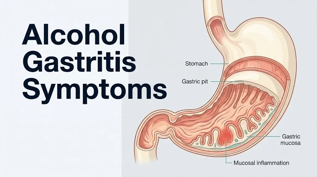
The first pathway begins with ethanol inhibiting COX-1 in gastric mucosal cells. COX-1 normally converts arachidonic acid into PGE2, which is the master regulator of all three mucosal defense systems: it stimulates surface mucous cell mucus secretion, drives bicarbonate secretion into the mucus layer, and maintains mucosal microvascular blood flow. When PGE2 falls, all three defenses decline simultaneously within hours of drinking.
The second pathway involves acetaldehyde, produced when alcohol dehydrogenase (ADH) and the microsomal enzyme CYP2E1 oxidize ethanol. Acetaldehyde forms protein adducts with gastric epithelial structural proteins, disrupting the claudin and occludin proteins that form tight junctions between cells. Once tight junctions are compromised, hydrochloric acid (HCl) from the gastric lumen back-diffuses through the epithelium into the submucosa, triggering mast cell degranulation, histamine release, and local inflammatory cascade amplification.
According to research reviewed in Gut, CYP2E1 activity is upregulated in chronic heavy alcohol users, meaning the oxidative stress pathway becomes more active and more damaging with prolonged drinking, not less.
Older adults metabolize acetaldehyde more slowly due to reduced aldehyde dehydrogenase (ALDH) activity, allowing it to accumulate to higher levels in gastric tissue from equivalent ethanol intake compared to younger adults.
Key Takeaway: Alcohol damages the stomach through three simultaneous pathways (PGE2 suppression, acetaldehyde epithelial toxicity, and CYP2E1-driven oxidative stress), meaning the mucosal damage compounds faster than a single protective mechanism can recover on its own.
Epigastric Pain and Burning from Alcoholic Gastritis
Epigastric pain and burning in alcoholic gastritis is produced when hydrochloric acid, no longer blocked by the compromised mucus-bicarbonate barrier, comes into direct contact with the gastric epithelium and the submucosal nerve plexus, stimulating nociceptive nerve endings in the stomach wall via the enteric nervous system and referred pain through the vagus nerve and thoracic splanchnic nerves. This pain is localized specifically to the upper abdomen between the navel and the lower breastbone.
The pain has characteristic features that distinguish it from other upper abdominal pain. It is typically a burning, gnawing, or aching sensation rather than a sharp stabbing pain. It often worsens on an empty stomach (when there is no food buffer between the gastric acid and the inflamed mucosa) and may temporarily improve after eating or drinking milk, because food neutralizes some gastric acid. It typically worsens again two to three hours after eating as acid secretion continues against the damaged epithelium.
The epigastric location reflects the anatomy of gastric innervation. Pain from the stomach is transmitted via the celiac plexus (a network of sympathetic ganglia at the T5-T9 spinal levels) and refereed to the mid-epigastric region. Some patients experience radiation to the back (particularly the left upper back) when gastric inflammation is severe or involves the posterior gastric wall adjacent to the pancreas.
According to the American Journal of Gastroenterology, epigastric pain is the most consistently reported symptom in patients with endoscopically confirmed gastritis, though pain intensity does not reliably correlate with the severity of mucosal damage seen on biopsy, meaning some patients with severe erosive gastritis have mild pain while others with minimal mucosal change have severe pain.
People with diabetes mellitus who develop gastroparesis (delayed gastric emptying from autonomic neuropathy) may experience epigastric pain patterns that overlap significantly with alcoholic gastritis, complicating self-assessment. Both alcohol and diabetic neuropathy can affect the same autonomic nerve pathways governing gastric motility.
Nausea and Vomiting from Alcoholic Gastritis
Nausea and vomiting in alcoholic gastritis are produced by two distinct mechanisms: direct stimulation of the area postrema (the brain’s chemoreceptor trigger zone, which lacks the normal blood-brain barrier and responds to blood-borne toxins including ethanol and acetaldehyde), and gastric mechanoreceptor activation from mucosal inflammation causing the stomach wall to signal the vagal nausea reflex. Both mechanisms can operate simultaneously.
The gastric component works through the enteric nervous system. Inflamed gastric mucosa generates abnormal electrical activity in the enteric nerve plexuses (Auerbach’s and Meissner’s plexuses) of the stomach wall. This activity is transmitted via the vagus nerve (cranial nerve X) to the nucleus tractus solitarius in the brainstem, which coordinates the vomiting reflex. Alcohol-induced gastric dysmotility, specifically delayed gastric emptying from ethanol’s direct depressant effect on gastric smooth muscle, amplifies this signal by keeping the distended, inflamed stomach in a state of prolonged mechanical stimulation.
Vomiting from acute alcoholic gastritis is typically non-bloody in early presentations. The vomited material is often acidic, bile-tinged, and may contain partially digested food or foam from gastric secretions. The onset of bloody vomiting, whether bright red blood or dark “coffee-ground” material (oxidized blood), signals a qualitative change in the underlying pathology: the gastric mucosa has eroded into a submucosal blood vessel. This is no longer simple gastritis vomiting.
Research published in the World Journal of Gastroenterology confirms that nausea is reported by the majority of patients with endoscopically confirmed alcoholic gastritis, with vomiting present in a substantial proportion, and that the nausea frequently begins within two to six hours of heavy drinking onset and may persist for 12 to 48 hours after cessation.
Pregnant women who drink alcohol and develop alcoholic gastritis face compounded nausea from overlapping pregnancy-induced nausea pathways (human chorionic gonadotropin-mediated hypothalamic stimulation) and the gastritis-driven vagal nausea reflex, making symptom severity assessment more difficult. Any stomach symptom in a pregnant person involving alcohol exposure warrants prompt evaluation by an obstetrician-gynecologist.
Bloating and Loss of Appetite in Alcoholic Gastritis
Bloating in alcoholic gastritis is produced by gastric dysmotility: ethanol directly impairs the contractility of gastric smooth muscle (the muscularis externa layer), reducing the coordinated peristaltic waves that normally move stomach contents into the duodenum, causing the stomach to retain gas and partially digested food longer than normal and producing the uncomfortable distension and pressure sensation patients describe as bloating. Loss of appetite follows from a combination of the distension itself and the cytokine-driven anorexia of systemic mucosal inflammation.
The gastric motility mechanism is important to understand because it explains why people with alcoholic gastritis feel full and distended even without eating much. Ethanol slows antral contractility (the muscular pump action of the pyloric antrum that propels food forward), delays gastric emptying of both solids and liquids, and impairs the migrating motor complex (MMC), the housekeeping contractions that clear the stomach between meals. The result is a stomach that retains its contents longer, generating gas from bacterial fermentation of undigested carbohydrates and producing the bloated, heavy sensation that persists for hours.
Appetite loss in alcoholic gastritis operates through two overlapping mechanisms. Locally, the gastric distension stimulates stretch receptors that signal satiety to the hypothalamus, suppressing hunger even when caloric intake is inadequate. Systemically, the elevated TNF-alpha and IL-6 produced by the inflamed gastric mucosa act on the hypothalamic arcuate nucleus (via the same pathway that drives anorexia during systemic infections), directly suppressing appetite drive independent of stomach fullness.
The National Institute on Alcohol Abuse and Alcoholism (NIAAA) notes that chronic alcohol use is associated with caloric substitution (calories from alcohol replacing nutritionally adequate food intake) and that appetite suppression from gastric inflammation compounds the nutritional deficiency pattern seen in alcohol use disorder.
People with chronic alcohol use disorder are frequently deficient in zinc, folate, and vitamin B12, all of which are required for gastric mucosal cell turnover and repair. These deficiencies impair the stomach’s capacity to regenerate damaged epithelial cells, prolonging gastritis episodes and slowing healing after abstinence.
Key Takeaway: Bloating after drinking is not just gas: it reflects ethanol’s direct impairment of gastric smooth muscle contractility and antral emptying, and in chronic alcohol users, this motility dysfunction can persist even during periods of reduced intake because the enteric nervous system has been recalibrated by sustained ethanol exposure.
Acute Alcoholic Gastritis Symptoms After Drinking
Acute alcoholic gastritis symptoms appear within two to six hours of a heavy drinking episode and are characterized by sudden-onset epigastric burning, nausea, retching, vomiting, and profound upper abdominal discomfort that reflects the rapid disruption of the gastric mucus-bicarbonate barrier by a large ethanol load. This form is typically the most symptomatically intense but often resolves within 24 to 72 hours with abstinence in otherwise healthy individuals.
The rapid onset reflects the speed of the PGE2 suppression pathway. Within two hours of ingesting alcohol at concentrations sufficient to suppress COX-1 activity in gastric mucosal cells, prostaglandin E2 levels in the gastric mucosa measurably decline. The mucus gel layer begins thinning immediately. HCl back-diffusion through the exposed epithelium triggers the pain and nausea cascade within the same time frame, which is why people often feel stomach symptoms while still drinking or within an hour of stopping.
Acute gastritis from a single episode is what most people colloquially call “stomach problems after drinking.” The distinction that matters clinically is whether the acute episode involves any bleeding (hematemesis, melena) or hemodynamic symptoms (dizziness, fainting, pallor, racing pulse), which indicate erosive gastritis or a Mallory-Weiss tear from repeated forceful vomiting.
| Symptom | Timing After Drinking | Duration Without Complications | Red Flag Version |
|---|---|---|---|
| Epigastric burning | 2 to 6 hours | Resolves in 24 to 72 hours | Severe, unrelenting, not food-responsive |
| Nausea | 2 to 6 hours | Resolves in 12 to 48 hours | Persistent beyond 48 hours |
| Vomiting | 3 to 8 hours | Resolves in 24 to 36 hours | Blood in vomit (any amount) |
| Bloating | 2 to 8 hours | Resolves in 24 to 48 hours | Increasing distension with pain |
| Appetite loss | 4 to 24 hours | Resolves in 24 to 72 hours | Persistent beyond 5 to 7 days |
Young adults (ages 18 to 25) experiencing acute alcoholic gastritis from binge drinking episodes are particularly vulnerable to Mallory-Weiss tears from forceful retching, because the intensity of the vomiting reflex in acute alcohol toxicity is often greater than the mucosal tissue can tolerate at the gastroesophageal junction.
Chronic Alcoholic Gastritis Symptoms
Chronic alcoholic gastritis symptoms are characterized by a more insidious presentation than the acute form: persistent low-grade epigastric discomfort, recurrent nausea, early satiety, fatigue, unintentional weight loss, and in advanced cases, symptoms of iron deficiency anemia from occult (hidden) gastrointestinal bleeding that has not produced visible blood in stool. These symptoms can develop over months to years of regular alcohol consumption.
The chronic form is driven by cumulative mucosal damage that progresses through stages. Repeated cycles of ethanol-induced PGE2 suppression, acetaldehyde toxicity, and oxidative stress produce inflammation that, over time, destroys the gastric glands. Parietal cells are lost (gastric atrophy), reducing acid secretion. Chief cells are replaced by intestinal-type cells (intestinal metaplasia). These histopathological changes represent a qualitative shift in the gastric mucosa’s nature, not just a quantitative increase in inflammation.
Chronic alcoholic gastritis is often clinically quieter than acute gastritis in terms of pain intensity, which is one reason it is frequently missed or attributed to other causes. A person with chronic gastritis may have persistent mild epigastric discomfort they consider normal, vague nausea most mornings, and a steadily worsening tolerance for food without recognizing these as indicators of progressive mucosal disease.
The association between chronic gastritis, intestinal metaplasia, and increased gastric adenocarcinoma risk is well-established in the gastroenterological literature. According to research in the American Journal of Gastroenterology, intestinal metaplasia of the gastric mucosa is considered a precancerous lesion, with progression risk that depends on the extent and location of metaplastic change.
People with iron deficiency anemia of unclear cause who drink regularly should be evaluated for occult GI blood loss from chronic erosive gastritis by a board-certified gastroenterologist, using upper endoscopy and fecal occult blood testing, before attributing the anemia to dietary causes alone.
Alcoholic Gastritis Symptoms the Morning After Drinking
Alcoholic gastritis symptoms the morning after drinking reflect the overnight persistence of mucosal inflammation combined with the physiological changes of alcohol withdrawal, producing a symptom complex that includes early-morning nausea, epigastric pain that is worse before eating, bile-tinged vomiting, headache, tremor, and pronounced appetite aversion.
The morning-after timing is clinically specific. Several mechanisms converge overnight. As blood alcohol concentration falls to zero, the rebound effect on gastric acid secretion begins: alcohol initially suppresses gastric acid output at very high concentrations, but as levels fall, a compensatory acid hypersecretion follows, driven by gastrin release from G cells in the pyloric antrum. This acid rebound hits a stomach whose protective PGE2-mediated barrier was depleted the night before, producing severe early-morning burning and pain on an empty stomach.
Bile reflux adds to the morning symptom picture. Alcohol reduces the tone of the pyloric sphincter, allowing duodenal contents including bile to reflux backwards into the stomach (duodenogastric reflux). Bile salts directly damage the gastric epithelium, amplifying the existing mucosal inflammation from ethanol. The bitter taste and early-morning nausea with bile-colored vomiting that many people report after heavy drinking is driven partly by this pyloric dysfunction.
Quick Tip:
- The urge to eat bland carbohydrates (toast, crackers) the morning after heavy drinking is physiologically appropriate: starchy food buffers gastric acid against the inflamed mucosa and stimulates mucin secretion from surface mucous cells.
- Avoid coffee and aspirin the morning after drinking. Coffee stimulates parietal cell acid secretion directly. Aspirin suppresses COX-1-derived PGE2 through the same pathway alcohol already activated overnight, compounding mucosal damage.
- Older adults who take low-dose aspirin for cardiovascular prevention should tell their primary care physician if they regularly experience morning epigastric symptoms after drinking, as the combination of alcohol and aspirin substantially elevates ulcer and hemorrhage risk.
Alcoholic Gastritis and H. Pylori
Helicobacter pylori co-infection with alcoholic gastritis produces additive and potentially synergistic mucosal damage because the two agents attack the gastric mucosal defense system through different but overlapping mechanisms, and the combined pathological effect on the gastric epithelium is substantially greater than either alone.
H. pylori is a gram-negative bacterium that colonizes the gastric mucus layer and the surface of gastric epithelial cells, particularly in the pyloric antrum. Its primary virulence factors include CagA (cytotoxin-associated gene A protein, which is injected directly into gastric epithelial cells and activates NF-kB, disrupting epithelial architecture and driving IL-8 production) and VacA (vacuolating cytotoxin, which creates vacuoles in epithelial cells and impairs the mucus barrier). H. pylori also produces urease, which converts urea into ammonia, raising local pH and creating its own barrier-disrupting chemical environment.
When H. pylori is present alongside chronic alcohol use, the NF-kB-driven inflammatory cascade from H. pylori’s CagA toxin and the NF-kB activation from alcohol’s CYP2E1-derived ROS converge on the same signaling pathway, producing higher levels of IL-8 (the primary neutrophil chemoattractant in gastric inflammation) and a more aggressive neutrophilic infiltrate in the gastric mucosa than either agent produces alone. The clinical result is a more symptomatic, faster-progressing, and harder-to-heal gastritis.
According to the American College of Gastroenterology, H. pylori testing is recommended for all patients with peptic ulcer disease, gastritis confirmed by endoscopy, and patients with iron deficiency anemia of unclear origin. For patients with alcoholic gastritis who drink regularly and have persistent symptoms, H. pylori testing should be part of the clinical workup rather than an afterthought.
People born outside of high-income countries, where H. pylori prevalence is substantially higher (over 70 percent in some regions, compared to approximately 30 to 40 percent in the United States per NIDDK data), who also drink regularly face a particularly elevated combined risk of symptomatic gastritis and peptic ulcer disease.
Key Takeaway: H. pylori co-infection multiplies the mucosal damage of alcoholic gastritis by activating the same NF-kB inflammatory pathway through a completely different mechanism, and testing for H. pylori is clinically warranted in any alcohol drinker with persistent gastritis symptoms.
Alcoholic Gastritis in Women and Older Adults
Women and older adults experience alcoholic gastritis through distinct physiological mechanisms that make them more vulnerable to mucosal damage from equivalent alcohol intake compared to young adult men, and these differences have direct implications for symptom severity, progression speed, and clinical management.
Women have measurably lower gastric alcohol dehydrogenase (ADH) activity than men, a sex difference documented in controlled clinical studies published in the New England Journal of Medicine. Gastric ADH is the enzyme that begins metabolizing ethanol in the stomach itself before it reaches the systemic circulation (first-pass gastric metabolism). When gastric ADH activity is lower, more unmetabolized ethanol passes through the gastric mucosa and into the bloodstream, and more acetaldehyde is produced per unit of ethanol consumed. The practical consequence: women reach equivalent blood acetaldehyde concentrations at substantially lower alcohol doses than men, exposing their gastric mucosa to greater acetaldehyde-mediated epithelial toxicity per drink.
This biological difference means that women develop alcohol-related gastric mucosal damage at lower drinking quantities and over shorter time periods than men, a phenomenon the NIAAA has specifically highlighted in its sex-differentiated alcohol use disorder guidance. A woman who drinks at levels considered moderate by standard definitions may be accumulating mucosal damage that would require heavier drinking to produce in a man.
Older adults face a different but equally important vulnerability. Baseline prostaglandin E2 synthesis in the gastric mucosa declines with age, independent of alcohol use, because COX-1 activity in mucosal cells decreases as part of normal aging. An older adult who drinks has therefore already started from a reduced PGE2 baseline before any alcohol suppression occurs, meaning the effective mucosal defense impairment from the same alcohol intake is greater than in a younger person.
Additionally, older adults are more likely to take NSAIDs, anticoagulants, and corticosteroids concurrently, each of which independently impairs mucosal defense or increases bleeding risk when gastric erosions develop. A 70-year-old taking naproxen for osteoarthritis pain who drinks wine regularly is at substantially elevated risk for erosive gastritis and upper GI hemorrhage compared to a 35-year-old drinker not on any medications.
Alcoholic Gastritis Bleeding and Upper GI Hemorrhage
Alcoholic gastritis bleeding occurs when mucosal erosions from sustained alcohol-induced inflammation reach the submucosal blood vessels of the stomach wall, producing upper gastrointestinal hemorrhage that manifests as hematemesis (vomiting blood) or melena (black tarry stool), and in the most severe cases, as hemodynamic instability from acute blood loss. This is the most dangerous complication of alcoholic gastritis.
The pathway from gastritis to hemorrhage involves the depth of tissue damage. Gastritis, by definition, involves inflammation of the mucosa and possibly the submucosa, without penetrating through the muscularis externa. Erosions are focal areas where the surface epithelium and underlying mucosa have been destroyed down to or through the muscularis mucosae. When erosions reach the rich submucosal vascular network, the gastric arterioles and venules in this layer are exposed to the gastric lumen, and bleeding begins.
A separate but related hemorrhagic complication is the Mallory-Weiss tear: a longitudinal laceration of the gastric mucosa at or just below the gastroesophageal junction, caused by the mechanical force of repeated, forceful vomiting. In acute alcohol toxicity with vigorous retching, the sudden pressure increase in the esophageal lumen tears the mucosal and submucosal layers at the gastroesophageal junction. These tears can involve gastric arterioles that branch from the left gastric artery, producing arterial-caliber bleeding that does not stop spontaneously.
In people with chronic alcohol use disorder who have developed alcoholic liver disease with portal hypertension, a third bleeding mechanism emerges: rupture of gastric varices. Portal hypertension from cirrhosis causes venous blood to be diverted through portosystemic collateral channels, including dilated submucosal veins in the stomach wall (gastric varices) and esophagus. When these varices rupture, the hemorrhage is typically massive, life-threatening, and requires emergent endoscopic and pharmacological intervention.
According to the Mayo Clinic, upper gastrointestinal bleeding is a medical emergency with mortality rates that increase significantly with delayed treatment, hemodynamic instability on presentation, and co-existing liver disease. People with alcoholic gastritis who develop any sign of GI bleeding should be treated as a medical emergency without exception.
When Alcoholic Gastritis Becomes a Medical Emergency
Certain symptoms associated with alcoholic gastritis require immediate emergency evaluation. Do not wait to see if these resolve on their own.
Call 911 or go to the nearest emergency room immediately if you experience:
- Vomiting blood (hematemesis): whether bright red blood or dark material resembling coffee grounds, indicating active upper gastrointestinal hemorrhage from erosive gastritis, Mallory-Weiss tear, or gastric varices
- Black, tarry, foul-smelling stool (melena): indicating that blood has passed through the gastrointestinal tract from an upper GI source, signaling active or recent hemorrhage
- Dizziness, fainting, or near-fainting with stomach symptoms: indicates hemodynamic instability from blood loss (hypovolemic shock), not simple intoxication
- Rapid, weak, or racing pulse combined with abdominal symptoms and pallor: cardiovascular signs of acute blood loss requiring immediate IV resuscitation
- Severe abdominal pain that worsens rapidly despite vomiting: may indicate gastric perforation, a surgical emergency distinct from gastritis
- Confusion or extreme drowsiness combined with stomach symptoms and heavy alcohol use: warrants emergency evaluation to distinguish alcohol toxicity from hemorrhage-related hemodynamic compromise
These presentations can indicate upper gastrointestinal hemorrhage, gastric perforation, or Mallory-Weiss syndrome with arterial bleeding, and require emergency medical assessment and potentially emergency endoscopy, not an urgent care appointment, a home remedy, or a wait-and-see approach.
If you are struggling with alcohol use and experiencing recurring gastric symptoms, contact the 988 Suicide and Crisis Lifeline by calling or texting 988, which also connects to substance use crisis counseling. This service is free, confidential, and available 24 hours a day, 7 days a week.
How Doctors Diagnose Alcoholic Gastritis
Alcoholic gastritis is formally diagnosed by a combination of clinical history, physical examination, and upper gastrointestinal endoscopy with gastric biopsy, with laboratory testing providing supportive evidence of mucosal inflammation and its complications. Clinical symptoms alone are not sufficient for a confirmed diagnosis because multiple conditions share the same symptom pattern.
The diagnostic process typically unfolds as follows:
- Clinical history: The physician will ask about alcohol consumption (frequency, quantity, duration), symptom timing in relation to drinking episodes, any prior gastrointestinal diagnoses, NSAID or medication use, and any family history of gastric disease or H. pylori infection.
- Physical examination: Abdominal palpation for epigastric tenderness, assessment of vital signs including blood pressure (low in hemorrhage), heart rate (elevated in acute blood loss), and evaluation for pallor or jaundice.
- Complete blood count (CBC): A low hemoglobin and hematocrit suggests either acute blood loss or chronic iron deficiency anemia from occult GI bleeding. Elevated white blood cell count suggests active infection or severe mucosal inflammation.
- H. pylori testing: The urea breath test (preferred for accuracy) or H. pylori stool antigen test are the standard non-invasive tests for active H. pylori infection, per ACG guidelines.
- Fecal occult blood test: Identifies microscopic blood in stool not visible to the naked eye, useful for detecting low-grade chronic mucosal bleeding.
- Upper endoscopy (esophagogastroduodenoscopy, EGD): Direct visualization of the gastric mucosa with a flexible camera. Allows the clinician to see mucosal erythema, erosions, varices, or active bleeding. Gastric biopsy at endoscopy provides histopathological confirmation: neutrophilic infiltration of the gastric mucosa confirms active gastritis; glandular atrophy and intestinal metaplasia confirm chronic gastritis.
- Serum ferritin and iron studies: Low serum ferritin in the context of alcoholic gastritis indicates iron deficiency from chronic occult bleeding.
How Long Does Alcoholic Gastritis Last
Acute alcoholic gastritis from a single heavy drinking episode typically resolves within 24 to 72 hours after abstinence from alcohol, while chronic alcoholic gastritis from regular sustained drinking can persist for weeks to months and may not fully resolve without medical treatment, extended abstinence, and management of co-existing factors like H. pylori infection. The healing timeline is directly tied to the severity of the underlying mucosal damage.
Mild acute gastritis, where mucosal inflammation is present but there are no erosions or structural changes, resolves relatively quickly because the gastric epithelium has a rapid cell turnover rate. Gastric surface mucous cells renew approximately every three to seven days under normal conditions. With abstinence removing the ongoing ethanol insult and adequate nutrition supporting cell repair, mild mucosal inflammation can resolve within a few days.
Erosive gastritis takes longer to heal because mucosal erosions require not just cell renewal but structural repair of the epithelial architecture and restoration of the mucosal vascular network. Healing of erosions typically takes one to three weeks with abstinence and appropriate medical management, though this varies by the depth and number of erosions present.
Chronic alcoholic gastritis with established gastric atrophy or intestinal metaplasia does not reverse fully with abstinence alone. According to research reviewed in the World Journal of Gastroenterology, gastric atrophy can partially recover over months to years with sustained abstinence and H. pylori eradication if co-infection is present, but established intestinal metaplasia tends to persist and requires endoscopic surveillance.
People with active alcohol use disorder who return to drinking after any period of abstinence will restart the mucosal damage cascade, making sustained abstinence the only pathway to durable gastric recovery rather than cyclical partial healing.
When to See a Doctor for Alcohol Gastritis Symptoms
You should contact a primary care physician within 24 to 72 hours for any alcohol gastritis symptoms that do not improve with 48 hours of abstinence, that are recurring with each drinking episode, that include unintentional weight loss, or that cause any change in stool color or consistency. Waiting longer substantially increases the risk that a treatable early-stage problem becomes a more advanced one.
The right provider for each clinical scenario:
- Primary care physician: First contact for persistent symptoms, request for H. pylori testing, prescription-strength acid suppression therapy, or alcohol use counseling referral. Bring a specific alcohol use history including quantity, frequency, and duration, all current medications, and a description of your stool color and any changes.
- Board-certified gastroenterologist: Required for upper endoscopy evaluation if symptoms are persistent despite treatment, if iron deficiency anemia is present without explanation, if there is any history of GI bleeding, or if chronic gastritis is suspected based on symptom duration and drinking history.
- Emergency medicine physician / emergency room: Required immediately for any hematemesis, melena, syncope, rapid weak pulse, or severe rapidly worsening abdominal pain.
What to bring to your appointment:
- Accurate and honest alcohol use history (quantity per day or week, type of alcohol, duration of current drinking pattern)
- List of all medications including NSAIDs, aspirin, antacids, and any supplements
- Description of stool appearance over the past two weeks
- Any prior diagnosis of H. pylori, peptic ulcer, or liver disease
- Any history of anemia or low hemoglobin on prior blood tests
People who are concerned about their alcohol use alongside their gastritis symptoms can ask their primary care physician about completing the Alcohol Use Disorder Identification Test (AUDIT), a validated screening tool, to better understand their consumption level and associated health risks.
Frequently Asked Questions About Alcohol Gastritis Symptoms
What does alcoholic gastritis feel like?
Alcoholic gastritis typically feels like a burning or gnawing pain in the upper center of the abdomen (the epigastric region), often accompanied by nausea, a heavy bloated sensation, and loss of appetite.
The pain tends to worsen on an empty stomach and may briefly improve after eating, then worsen again two to three hours later as acid production continues against the inflamed gastric lining.
The burning sensation is caused by hydrochloric acid contacting the gastric epithelium directly after alcohol has disrupted the mucus-bicarbonate barrier that normally separates the acid from the stomach wall.
How do I know if my stomach pain is from alcohol gastritis?
Stomach pain from alcoholic gastritis is typically located in the upper abdomen between the navel and breastbone, begins two to six hours after drinking, and is accompanied by nausea, bloating, or appetite loss.
The timing connection to alcohol consumption is the most useful distinguishing feature: pain that consistently appears after drinking and improves during periods of abstinence fits the alcoholic gastritis pattern.
A confirmed diagnosis requires a physician’s evaluation and, ideally, upper endoscopy with biopsy, because several other conditions including peptic ulcer disease and H. pylori gastritis produce nearly identical symptoms.
Can alcoholic gastritis cause bleeding?
Yes, alcoholic gastritis can cause upper gastrointestinal bleeding when mucosal erosions reach submucosal blood vessels, producing hematemesis (vomiting blood) or melena (black tarry stool).
Forceful vomiting associated with acute alcohol toxicity can also cause Mallory-Weiss tears at the gastroesophageal junction, producing arterial-caliber bleeding that requires emergency endoscopy.
Any blood in vomit or black stool in the context of alcohol use requires immediate emergency room evaluation, not a wait-and-see approach.
How long does alcoholic gastritis take to heal?
Mild acute alcoholic gastritis typically improves within 24 to 72 hours of abstinence from alcohol, with full symptomatic resolution within three to seven days in people without erosions or structural damage.
Erosive gastritis takes one to three weeks to heal with abstinence and medical management, while chronic alcoholic gastritis with gastric atrophy or intestinal metaplasia may not fully reverse even with extended sobriety.
The single most effective step for healing is complete abstinence from alcohol, as continued drinking restarts the mucosal damage cascade before the previous damage can repair.
What are the warning signs that alcohol gastritis is serious?
The most serious warning signs of alcoholic gastritis are vomiting blood (bright red or coffee-ground material), black tarry stool (melena), dizziness or fainting with stomach symptoms, and a rapid or weak pulse accompanying abdominal pain.
These signs indicate upper gastrointestinal hemorrhage from erosive gastritis, a Mallory-Weiss tear, or gastric variceal rupture, all of which are life-threatening without emergency medical treatment.
Any single one of these signs requires calling 911 or going to the nearest emergency room immediately, not waiting to see if symptoms improve.
Can alcoholic gastritis go away if I stop drinking?
Mild to moderate alcoholic gastritis can resolve with complete abstinence from alcohol, typically within days to weeks depending on the severity of mucosal damage, but resolution is not guaranteed without medical assessment and management of co-existing factors.
If H. pylori co-infection is present, abstinence alone will not fully resolve the gastritis because H. pylori continues its own mucosal damage independently; H. pylori eradication therapy prescribed by a physician is also required.
Chronic alcoholic gastritis with gastric atrophy does not fully reverse with abstinence but can stabilize and partially improve, making sustained sobriety the most important step toward limiting further progression.
Closing
Alcohol gastritis is not just a “bad stomach” after drinking. It is a specific pattern of mucosal damage with a clear biochemical mechanism, a predictable symptom progression, and a serious complication pathway that can become life-threatening without warning. The most important thing you can do with the symptoms you are experiencing right now is to take the bleeding signs seriously.
If your symptoms include any blood in vomit or black stool, do not wait. Go to an emergency room. For symptoms that are uncomfortable but not including any bleeding, contact a primary care physician within 48 to 72 hours if they are not improving with abstinence, and ask specifically about H. pylori testing and whether an endoscopy referral to a gastroenterologist is appropriate.
Your gastric mucosa can heal. The timeline and the extent of that healing depend on what you do next.