Pseudogout Symptoms: Full Clinical Guide and Red Flags 2026
Pseudogout symptoms appear suddenly, typically in a single large joint that becomes intensely painful, swollen, warm, and stiff within hours, a presentation so striking that many people experiencing their first attack genuinely believe they have broken a bone or developed a severe infection. The condition is caused by calcium pyrophosphate dihydrate (CPPD) crystals depositing in joint cartilage and shedding into the joint space, triggering an acute inflammatory cascade that can rival the intensity of gout.
According to the National Institute of Arthritis and Musculoskeletal and Skin Diseases (NIAMS), CPPD disease becomes increasingly prevalent with age, with radiographic evidence of calcium deposits present in the cartilage of approximately 7% of adults in their 60s, rising to approximately 50% of adults in their 90s. This makes pseudogout one of the most common causes of acute joint inflammation in older adults, yet it remains underdiagnosed because its symptoms closely mimic gout, rheumatoid arthritis, and, in the most clinically dangerous scenario, septic joint infection.
This guide covers the full symptom picture of pseudogout, organized by body system, joint location, and clinical subtype, with the biochemical mechanism behind each symptom, clear severity guidance, the critical distinction from septic arthritis, and the specific thresholds at which symptoms require emergency evaluation.
Pseudogout Symptoms: The Full Clinical Picture
Pseudogout symptoms consist of acute joint pain, swelling, warmth, stiffness, and sometimes fever, arising from CPPD crystal-induced inflammation within one or more joints, most commonly the knee.
The symptom pattern typically develops over hours rather than days. The joint becomes visibly swollen, hot to the touch, and exquisitely tender, with pain that worsens sharply on any movement or palpation of the joint. Unlike the chronic, insidious joint pain of osteoarthritis, a pseudogout attack is rapid, intense, and often disabling at its peak.
The full clinical symptom inventory of an acute pseudogout attack includes:
- Severe joint pain, typically rated 7 to 10 on the Visual Analog Scale for pain, arising within 6 to 24 hours
- Joint swelling from synovial fluid accumulation (effusion) in the joint space
- Joint warmth and redness of the overlying skin, from increased local blood flow driven by inflammatory mediators
- Restricted range of motion, caused by joint effusion volume and pain-inhibited movement
- Morning stiffness lasting more than 30 minutes in people with chronic CPPD inflammatory arthritis
- Fever in approximately 30% to 50% of acute attacks, ranging from low-grade to 102°F or higher
- Systemic malaise and fatigue, from elevated interleukin-6 driving the acute phase response
People with diabetes mellitus should be aware that their typical inflammatory response may be altered, potentially blunting fever and masking the full local inflammatory picture even when CPPD crystal-induced synovitis is active. A board-certified rheumatologist or primary care physician should evaluate any acute joint swelling in a person with diabetes, regardless of whether fever is present.
What Is Pseudogout and How Does It Differ from Gout?
Pseudogout is an acute or chronic inflammatory arthritis caused by the deposition of calcium pyrophosphate dihydrate (CPPD) crystals in articular cartilage and synovial fluid, while gout is caused by monosodium urate (MSU) crystals forming from elevated serum uric acid.
The two conditions share the same dramatic clinical presentation: sudden, severe, incapacitating joint pain that arrives without warning. Both involve crystal-triggered NLRP3 inflammasome activation and IL-1 beta-mediated synovitis. The clinical experience of a severe attack is nearly identical. The distinction lies in the crystal type, the metabolic cause, the joints typically affected, and the patient demographics.
| Feature | Pseudogout (CPPD) | Gout (MSU) |
|---|---|---|
| Crystal type | Calcium pyrophosphate dihydrate | Monosodium urate |
| Crystal shape under polarized light | Rhomboid, weakly positively birefringent | Needle-shaped, strongly negatively birefringent |
| Most common joint | Knee (tibiofemoral joint) | First metatarsophalangeal joint (great toe) |
| Typical age of onset | Over 60 years (idiopathic); any age with metabolic disease | Any adult age; peak in men 40s to 60s |
| Sex distribution | Slight female predominance in older adults | Strong male predominance (ratio approximately 4:1) |
| Serum marker | No reliable serum crystal marker | Serum uric acid (often elevated, not diagnostic alone) |
| Associated metabolic conditions | Hyperparathyroidism, hemochromatosis, hypomagnesemia, hypothyroidism | Chronic kidney disease, metabolic syndrome, alcohol use, certain medications |
| Radiographic finding | Chondrocalcinosis (calcium in cartilage) | Punched-out bony erosions with overhanging edges (late gout) |
The critical clinical point is that these two diagnoses cannot be reliably distinguished from each other on clinical presentation alone. Synovial fluid analysis with compensated polarized light microscopy is the only way to confirm the crystal type and thus the diagnosis.
How Calcium Pyrophosphate Crystals Form in Joints
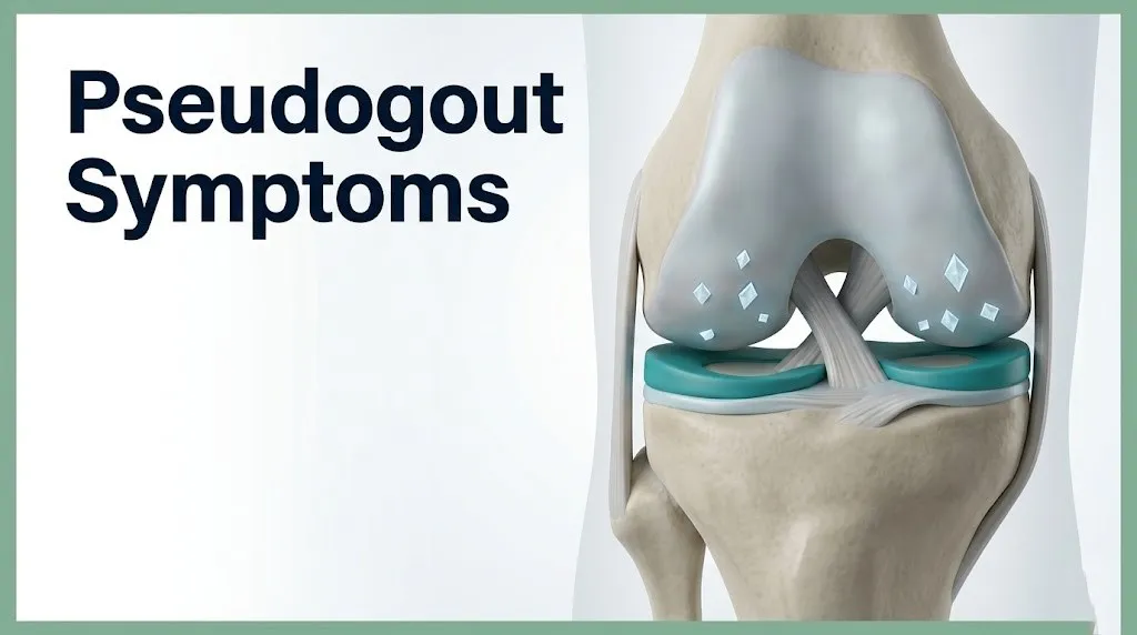
CPPD crystals form in articular cartilage when inorganic pyrophosphate (PPi) accumulates in the cartilage extracellular matrix and combines with calcium ions to form calcium pyrophosphate dihydrate crystals.
The biochemical pathway begins in chondrocytes, the cells that maintain articular cartilage. Chondrocytes produce PPi as a byproduct of ATP hydrolysis via the ENPP1 (nucleotide pyrophosphatase/phosphodiesterase 1) enzyme. PPi is then transported from the intracellular space into the cartilage extracellular matrix by the ANKH transmembrane protein. Normally, an enzyme called tissue-nonspecific alkaline phosphatase (TNAP) breaks down PPi into harmless inorganic phosphate, keeping PPi levels in the extracellular matrix low enough to prevent crystal formation.
Think of this system like a water treatment plant. TNAP is the filtration stage that clears excess PPi before it can accumulate. When TNAP activity is reduced (as in magnesium deficiency, where magnesium is a required cofactor for TNAP), or when PPi production overwhelms TNAP capacity (as in hyperparathyroidism, where increased calcium turnover stimulates chondrocyte activity), PPi builds up and crystallizes with calcium.
Once crystals form in cartilage, they can be stable for years. The trigger for an acute attack is crystal shedding: when crystals break free from the cartilage surface into the synovial joint fluid. According to research published in Annals of the Rheumatic Diseases, crystal shedding can be provoked by mechanical joint trauma, surgery, acute illness, or rapid shifts in serum calcium or magnesium levels.
Younger adults with the rare genetic condition hypophosphatasia have markedly reduced TNAP activity from birth, making them susceptible to premature CPPD crystal formation decades before the typical age of onset. A board-certified rheumatologist should evaluate any person under 50 with confirmed chondrocalcinosis to screen for an underlying metabolic or genetic cause.
Key Takeaway: CPPD crystals form when inorganic pyrophosphate accumulates in joint cartilage because TNAP enzyme activity is insufficient to clear it, which is why metabolic diseases that reduce TNAP function or increase calcium and PPi production all raise pseudogout risk.
Pseudogout Attack Symptoms: The Acute Flare
An acute pseudogout attack, classified by the European League Against Rheumatism (EULAR) 2023 recommendations as “acute CPP crystal arthritis,” begins with rapid-onset severe pain in a single joint that typically reaches maximum intensity within 6 to 24 hours and, without treatment, resolves spontaneously over 1 to 3 weeks.
The pain arises through a specific sequence. When CPPD crystals shed into the synovial fluid, synovial macrophages and neutrophils phagocytose the crystals. Inside these immune cells, crystals rupture the phagosome membrane, releasing lysosomal contents into the cell cytoplasm and activating the NLRP3 inflammasome. The NLRP3 complex then activates caspase-1, which cleaves the inactive precursor pro-IL-1 beta into active interleukin-1 beta (IL-1 beta). Massive IL-1 beta release into the joint space drives prostaglandin E2 production, bradykinin sensitization of joint nociceptors, and a neutrophil flood into the synovial fluid.
The result is joint pain that is not just aching but deeply throbbing, with exquisite sensitivity to even light touch of the joint or overlying skin. Patients frequently describe it as feeling like the joint is being stabbed or crushed from the inside.
The attack phase typically follows this timeline:
| Phase | Timing | Dominant Symptoms |
|---|---|---|
| Prodromal | 0 to 6 hours | Mild joint ache, early warmth, vague discomfort |
| Peak inflammation | 6 to 24 hours | Severe pain, maximal swelling, fever, immobility |
| Plateau | 1 to 7 days (untreated) | Sustained severe symptoms, possible fatigue and malaise |
| Resolution | 1 to 3 weeks (untreated) | Gradual subsiding of pain and swelling; residual stiffness |
People taking immunosuppressive medications such as corticosteroids for transplant rejection or autoimmune conditions may experience a blunted or delayed attack timeline, as their suppressed neutrophil response reduces the peak inflammatory intensity. This population should be evaluated by their prescribing specialist when any joint swelling develops, because the clinical picture may be less typical.
Pseudogout Joint Pain and Swelling
The joint pain and swelling of a pseudogout attack are driven by two distinct physiological processes: the direct nociceptive sensitization of joint capsule and synovial pain receptors by inflammatory mediators, and the mechanical distension of the joint capsule by synovial fluid effusion.
Joint effusion in acute CPP crystal arthritis develops because IL-1 beta and prostaglandin E2 increase the permeability of synovial blood vessels, allowing plasma proteins and fluid to leak into the joint space. The joint capsule, which encloses the synovial space, has a limited capacity to expand. As fluid volume increases, the capsule is stretched, activating mechanoreceptors and nociceptors within the capsular tissue. This stretch pain adds to the inflammatory pain from prostaglandin E2 sensitization of free nerve endings in the synovium, producing the characteristic double-component pain: deep aching from inflammation plus sharp capsular pain from distension.
The swelling is typically visible and palpable within hours. In the knee, a large effusion can be detected clinically by pressing on the medial side of the joint and observing a fluid wave on the lateral side, a finding clinicians call the bulge sign or patella tap test. In the wrist, swelling may be more diffuse, following the joint capsule contours and limiting flexion and extension symmetrically.
People with pre-existing osteoarthritis in the affected joint may find that the superimposed pseudogout swelling is dramatically larger than any prior osteoarthritis-related effusion, because the crystal-triggered inflammatory response generates far more synovial fluid than mechanical osteoarthritis alone. A primary care physician or emergency medicine physician should evaluate any person with known osteoarthritis who develops a disproportionately large, acutely painful joint effusion to exclude crystal arthritis and infection.
Pseudogout and Fever: What It Means Clinically
Fever in pseudogout occurs when systemic levels of interleukin-6 (IL-6) and prostaglandin E2 (PGE2) released during the joint inflammation cascade reach the hypothalamic thermoregulatory center and shift the temperature set point upward.
Fever is present in approximately 30% to 50% of acute pseudogout attacks according to clinical observation data, with temperatures typically ranging from 100°F to 102°F (37.8°C to 38.9°C). This temperature range is clinically important but not diagnostic: fever at these levels occurs in both pseudogout and in the condition that must be urgently excluded, septic arthritis (bacterial joint infection).
The critical clinical problem is that fever, joint swelling, and elevated inflammatory markers (elevated CRP, elevated ESR, elevated synovial fluid white blood cell count) are present in both pseudogout and septic arthritis. Clinically, these two conditions are indistinguishable at the bedside. The American College of Rheumatology states that joint aspiration with synovial fluid analysis is required whenever an acutely inflamed joint with fever is present, regardless of how confident a clinician or patient may be that the diagnosis is pseudogout.
The presence of fever should never be used to reassure a patient that the inflammation is “just pseudogout.” Fever above 101°F in someone with an acutely swollen joint is a signal to seek same-day evaluation by an emergency medicine physician or primary care physician who can perform or arrange arthrocentesis, not a signal to manage the attack at home.
People who are immunocompromised (from chemotherapy, biologic agents, or HIV) may have a blunted fever response even during septic arthritis, making the absence of high fever in this population unreliable as a sign that infection is excluded. Any acutely swollen joint in an immunocompromised person requires same-day emergency evaluation.
Key Takeaway: Fever alongside a swollen joint in a pseudogout attack is clinically indistinguishable from septic arthritis without synovial fluid analysis, and every such presentation requires same-day medical evaluation rather than home management.
Pseudogout in the Knee
Pseudogout affects the knee joint more frequently than any other joint, with clinical observation data reviewed by the NIAMS indicating that the knee is involved in approximately 50% of all acute pseudogout attacks.
The knee’s predisposition to CPPD crystal deposition relates to its fibrocartilaginous menisci. The medial and lateral menisci, the C-shaped fibrocartilaginous shock absorbers within the tibiofemoral joint, are composed of the type of fibrocartilage that preferentially accumulates CPPD crystals. The triangular fibrocartilage complex of the wrist is the other primary fibrocartilaginous site; the knee simply has a larger volume of susceptible tissue.
Symptoms of pseudogout in the knee include:
- Severe pain rated 8 to 10 out of 10 on the Visual Analog Scale at peak attack, typically within 12 to 24 hours of onset
- Visible swelling of the knee extending above and below the kneecap (patella), caused by joint effusion filling the suprapatellar bursa and joint recesses
- Warmth of the knee skin palpable to touch, often extending 5 to 10 centimeters above and below the joint line
- Inability to fully straighten or bend the knee, from both effusion volume and pain-inhibited movement
- Difficulty weight-bearing, with most patients describing inability to walk normally at the peak of the attack
Radiographic chondrocalcinosis (calcium deposits visible on plain X-ray) in the menisci of the knee is found incidentally on knee X-rays in a substantial proportion of older adults and is itself not a symptom. The acute attack occurs when crystals shed from the menisci into the joint fluid, not from the presence of the calcification itself.
Older adults with a painful, swollen knee should not assume that any knee pain is from their pre-existing osteoarthritis. A superimposed acute pseudogout attack on an osteoarthritic knee is a distinct event requiring different management than routine osteoarthritis care, and a primary care physician should be contacted the same day it develops.
Pseudogout in the Wrist and Other Joints
Pseudogout in the wrist is the second most frequently affected site, occurring in approximately 20% of acute pseudogout attacks, with crystal deposition concentrated in the triangular fibrocartilage complex (TFCC) at the ulnar side of the wrist.
The TFCC is a fibrocartilaginous structure that stabilizes the distal radioulnar joint and cushions the ulnar side of the radiocarpal joint. Its fibrocartilaginous composition makes it a preferred crystal deposition site. An acute attack in the wrist produces severe pain and swelling across the dorsal and ulnar wrist, with dramatically reduced wrist flexion, extension, and forearm rotation.
Beyond the knee and wrist, pseudogout can affect:
- Glenohumeral joint (shoulder): Acute shoulder pain mimicking rotator cuff pathology, with restricted range of motion and effusion visible on ultrasound
- Ankle joint (talocrural joint): Acute ankle swelling that may be misattributed to a sprain
- Pubic symphysis: A fibrocartilaginous structure that can develop CPPD crystals; attacks here produce severe groin and lower abdominal pain that mimics urological, gynecological, or gastrointestinal pathology
- Metacarpophalangeal joints (knuckles): Particularly in people with hemochromatosis, where iron overload preferentially drives CPPD deposition in these joints, producing a pattern that closely mimics early rheumatoid arthritis
- Spinal facet joints and intervertebral disc annulus: Rare but documented, causing acute neck or back pain with fever in a presentation called crowned dens syndrome when crystals deposit around the odontoid process of C2
Women over 60 are more likely than men to present with wrist pseudogout as a first attack, whereas men with hemochromatosis characteristically present with metacarpophalangeal joint involvement as an early manifestation. A board-certified rheumatologist should evaluate any man presenting with bilateral metacarpophalangeal joint swelling, particularly if accompanied by elevated serum ferritin, to screen for hemochromatosis-associated CPPD disease.
Chronic Pseudogout Symptoms: The Three CPPD Subtypes
The EULAR 2023 recommendations for CPPD disease, published in the Annals of the Rheumatic Diseases, classify CPPD disease into three distinct clinical subtypes that have different symptom patterns, natural histories, and diagnostic implications.
Subtype 1: Acute CPP Crystal Arthritis (the classic pseudogout attack): This is the explosive, episodic inflammatory attack described throughout this article. Attacks are separated by periods of complete or near-complete symptom resolution. Between attacks, the affected joint may feel entirely normal. The attack duration without treatment is typically 1 to 3 weeks, and attacks may recur months to years apart or in rapid succession.
Subtype 2: Chronic CPP Crystal Inflammatory Arthritis: This subtype produces persistent joint inflammation lasting more than 6 weeks at a time, affecting multiple joints simultaneously, and closely mimics rheumatoid arthritis (RA) in its presentation. Symptoms include prolonged morning stiffness (more than 45 minutes), symmetric joint swelling of the wrists and metacarpophalangeal joints, fatigue, and elevated inflammatory markers (CRP, ESR). It is distinguished from RA by the absence of anti-cyclic citrullinated peptide (anti-CCP) antibodies and rheumatoid factor in most cases, and by the presence of chondrocalcinosis on imaging.
Subtype 3: CPPD Associated with Osteoarthritis: This subtype produces chronic low-grade joint pain, stiffness, and progressive joint damage in joints with underlying osteoarthritis. It does not typically produce the dramatic acute flare of subtype 1. Symptoms are chronic, slowly progressive, and often attributed entirely to osteoarthritis, leading to delayed recognition of the CPPD component.
People with a new diagnosis of what appears to be rheumatoid arthritis who are over 65 should specifically request serum testing for anti-CCP antibodies and imaging for chondrocalcinosis before accepting an RA diagnosis, because chronic CPP crystal inflammatory arthritis is a distinct condition requiring different long-term management than RA.
Key Takeaway: The classic explosive pseudogout attack (acute CPP crystal arthritis) is just one of three distinct clinical subtypes of CPPD disease; the chronic inflammatory subtype mimics rheumatoid arthritis and is frequently misdiagnosed, particularly in older adults.
Pseudogout Flare Triggers
Pseudogout flares are triggered by events that cause rapid shifts in joint calcium levels, mechanical disruption of crystal-laden cartilage, or acute physiological stress that alters the equilibrium between crystal stability and shedding.
The most clinically recognized pseudogout flare triggers include:
- Acute illness or hospitalization: Serious infections, myocardial infarction, pneumonia, or any acute medical event can trigger pseudogout by causing rapid electrolyte shifts (particularly calcium and magnesium) that destabilize crystal deposits. Pseudogout is one of the most common causes of acute monoarthritis in hospitalized patients, according to clinical data reviewed by the Mayo Clinic.
- Surgery, particularly parathyroid or thyroid surgery: Parathyroid surgery produces an acute drop in serum parathyroid hormone (PTH), which rapidly shifts calcium equilibrium in cartilage and can trigger crystal shedding within 24 to 72 hours post-operatively.
- Joint trauma or injury: Direct mechanical impact on a crystal-laden joint can physically dislodge crystals into the synovial space.
- Blood transfusion: Transfused blood contains citrate as an anticoagulant; citrate chelates calcium, causing a transient drop in ionized serum calcium that can destabilize cartilage crystal deposits.
- Intravenous fluid administration: Large volumes of calcium-free IV fluids dilute serum calcium acutely, producing a similar destabilizing effect on crystal equilibrium.
- Physical overexertion of a crystal-laden joint: Prolonged walking or exercise involving a knee with chondrocalcinosis can mechanically provoke crystal shedding.
People scheduled for elective parathyroid or thyroid surgery should be informed by their surgical team that post-operative pseudogout attacks are a recognized complication. Awareness of this risk allows patients and their primary care physicians to recognize a post-operative joint attack as pseudogout rather than a surgical site infection.
Pseudogout Risk Factors and Who Gets It
The primary risk factors for pseudogout are age, genetic predisposition, prior joint damage, and a set of specific metabolic conditions that increase PPi production or reduce PPi clearance in joint cartilage.
Risk factors organized by clinical category:
- Age: The strongest non-modifiable risk factor. CPPD crystal prevalence in joint cartilage rises steeply after age 60.
- Prior joint trauma or surgery: Any joint that has sustained significant trauma accumulates CPPD crystals at an accelerated rate at that specific site.
- Hyperparathyroidism: Elevated PTH increases osteoclast bone resorption, releasing calcium into the circulation and increasing cartilage calcium availability, accelerating CPPD crystal formation.
- Hemochromatosis: Excess iron deposition in cartilage tissue directly inhibits pyrophosphatase enzyme activity, reducing PPi clearance and allowing crystal accumulation. Men with hemochromatosis may develop CPPD disease in their 40s and 50s, decades earlier than the typical idiopathic form.
- Hypomagnesemia: Magnesium is a required cofactor for pyrophosphatase (TNAP) function. Chronically low magnesium, from loop diuretic use (furosemide), chronic alcoholism, or malabsorption syndromes, reduces PPi clearance.
- Hypothyroidism: Reduces the metabolic rate of PPi clearance in cartilage, allowing accumulation.
- Hypophosphatasia: A rare genetic disorder in which TNAP activity is severely reduced, causing PPi accumulation and premature CPPD crystal formation, often presenting in adults in their 40s.
- Familial CPPD disease: Mutations in the ANKH gene that increase PPi transport out of chondrocytes cause autosomal dominant familial CPPD disease, typically presenting at a younger age than idiopathic CPPD.
People on long-term loop diuretics for heart failure or hypertension should ask their prescribing cardiologist or primary care physician about monitoring serum magnesium levels, as chronic loop diuretic use is one of the most common reversible risk factors for hypomagnesemia-related pseudogout.
Pseudogout in Older Adults
Pseudogout in older adults represents the most clinically complex presentation of the condition, because the combination of age-related cartilage changes, polypharmacy, comorbidities, and baseline joint disease makes both accurate diagnosis and appropriate management more challenging than in younger patients.
Age-related cartilage changes increase CPPD crystal susceptibility through multiple pathways: chondrocytes in older cartilage show increased ENPP1 activity and increased ANKH expression, both driving higher PPi production and export. Simultaneously, TNAP activity in older cartilage declines, reducing the enzymatic clearance of PPi. The net result is that virtually all articular cartilage in very elderly people contains some degree of CPPD crystal deposition, which is why the radiographic prevalence approaches 50% in people in their 90s according to NIAMS data.
Clinically, pseudogout in older adults presents several specific diagnostic challenges:
- Fever may be lower or absent: Older adults often have a blunted febrile response even to severe joint infection, making the absence of fever a less reliable indicator that septic arthritis is excluded.
- Symptoms may involve multiple joints simultaneously: Older adults with the chronic CPP inflammatory subtype may present with polyarthritis affecting multiple joints at once, mimicking late-onset rheumatoid arthritis or polymyalgia rheumatica.
- Cognitive impairment may impede symptom reporting: In older adults with dementia or delirium, the presenting sign of a pseudogout attack may be behavioral change, agitation, or unexplained refusal to bear weight rather than a verbal complaint of joint pain.
- Polypharmacy interactions: Older adults are frequently on diuretics (increasing hypomagnesemia risk), nonsteroidal anti-inflammatory drugs (renal and GI risk), and anticoagulants (complicating joint aspiration planning).
A board-certified geriatrician working in coordination with a board-certified rheumatologist provides the most appropriate specialist care for older adults with recurrent or chronic pseudogout who have multiple comorbidities affecting both diagnosis and treatment options.
Pseudogout vs Septic Arthritis: The Critical Distinction
Pseudogout and septic arthritis (bacterial joint infection) can be clinically indistinguishable at the bedside, and this distinction is one of the most clinically urgent differentiations in all of acute joint medicine because the consequences of missing septic arthritis are catastrophic.
Septic arthritis occurs when bacteria (most commonly Staphylococcus aureus, Streptococcal species, or gram-negative organisms in immunocompromised patients) seed the synovial joint space and establish infection. Articular cartilage destruction begins within 24 to 72 hours of untreated septic arthritis, and without antibiotic treatment plus joint drainage, permanent joint destruction and systemic sepsis with potential mortality follow.
Both conditions can produce:
- Acute monoarticular (single joint) severe pain, swelling, and warmth
- Fever ranging from 100°F to 103°F
- Elevated CRP and ESR
- Elevated synovial fluid WBC count (often 30,000 to 100,000 cells/microliter in both)
- Peripheral blood leukocytosis (elevated WBC on complete blood count)
The only reliable way to distinguish them is arthrocentesis (joint aspiration) with synovial fluid sent for:
- Crystal identification under compensated polarized light microscopy (rhomboid, weakly positively birefringent CPPD crystals confirm crystal arthritis)
- Gram stain and culture and sensitivity (bacterial growth confirms septic arthritis)
- Synovial fluid WBC count and differential (extremely high WBC, particularly above 100,000 cells/microliter, raises suspicion for infection even if crystals are present)
Critically, pseudogout and septic arthritis can coexist in the same joint simultaneously. A joint can have both CPPD crystals and bacterial infection at the same time. The presence of crystals on polarized light microscopy does not exclude co-existing septic arthritis. All culture results must be awaited before concluding that infection is absent.
People with prosthetic (artificial) joint replacements who develop acute pain and swelling in or around the prosthetic joint require emergency evaluation regardless of a prior pseudogout history, because prosthetic joint infection is a surgical emergency requiring different management from native joint septic arthritis.
Key Takeaway: Crystal identification in joint fluid confirms CPPD disease but does not exclude coexisting septic arthritis, which is why synovial fluid culture must be completed alongside crystal analysis in every acutely inflamed joint evaluation.
How Pseudogout Is Diagnosed
Pseudogout is definitively diagnosed by identifying CPPD crystals in synovial fluid from the affected joint using compensated polarized light microscopy, a laboratory technique that reveals the characteristic rhomboid shape and weak positive birefringence of CPPD crystals.
The diagnostic process typically unfolds as follows:
- Clinical history and physical examination: A physician documents the onset, affected joint(s), symptom character, associated fever, and prior attack history. Examination assesses joint swelling, warmth, range of motion, and tenderness.
- Arthrocentesis (joint aspiration): A physician inserts a needle into the joint space under sterile conditions and withdraws synovial fluid. This procedure also provides therapeutic benefit in the knee by relieving pain from effusion pressure.
- Synovial fluid analysis: The aspirated fluid is analyzed for crystal morphology under compensated polarized light microscopy, white blood cell count and differential, and Gram stain with culture and sensitivity.
- Plain X-ray of the affected joint: Radiographs may show chondrocalcinosis (linear calcium deposits in cartilage), particularly in the menisci of the knee or the TFCC of the wrist. Chondrocalcinosis supports the diagnosis but is not definitive; crystals must be seen in fluid to confirm active disease.
- Blood tests: These do not diagnose pseudogout directly but identify metabolic conditions that increase risk and guide further investigation. Relevant tests include serum calcium, serum PTH, serum ferritin and transferrin saturation, serum magnesium, and thyroid-stimulating hormone (TSH).
- Ultrasound of the joint: Joint ultrasound can identify CPPD crystal deposits in cartilage as hyperechoic (bright) deposits within the cartilage thickness, a finding distinct from the surface deposits seen with gout crystals.
- CT scan of the joint: High-resolution CT is more sensitive than plain X-ray for detecting chondrocalcinosis in deep joint structures, such as the facet joints of the spine.
People whose pseudogout diagnosis has not prompted a metabolic workup (calcium, PTH, ferritin, magnesium, TSH) should ask their primary care physician or board-certified rheumatologist for this panel at the next visit, because identifying and treating an underlying metabolic condition can reduce future attack frequency.
Pseudogout Symptoms in People with Metabolic Conditions
Pseudogout symptoms in people with underlying metabolic conditions often present at a younger age, affect atypical joints, and may recur more frequently than idiopathic age-related CPPD disease, because the metabolic driver continuously promotes crystal deposition rather than allowing the gradual accumulation of normal aging.
The specific symptom patterns by metabolic condition:
| Metabolic Condition | CPPD Symptom Pattern | Typical Age of Onset | Key Differentiating Feature |
|---|---|---|---|
| Hyperparathyroidism | Acute attacks in multiple joints; may recur rapidly | Any adult age; often 30s to 60s | Elevated serum calcium and PTH |
| Hemochromatosis | Metacarpophalangeal joint involvement; chronic progressive | Men in 40s to 50s | Elevated serum ferritin; skin bronzing; liver disease |
| Hypomagnesemia (loop diuretics, alcoholism) | Acute attacks; exacerbated by intercurrent illness | Any adult age on loop diuretics | Low serum magnesium |
| Hypothyroidism | Indolent chronic CPPD; fewer acute attacks | Older adults; postmenopausal women | Elevated TSH; fatigue; weight gain |
| Hypophosphatasia | Early-onset CPPD; multiple joint attacks | Adults in 40s to 50s | Markedly reduced serum alkaline phosphatase |
| Familial CPPD (ANKH mutation) | Early-onset; positive family history | Adults in 30s to 50s | Autosomal dominant family history of early arthritis |
According to the EULAR 2023 recommendations, all patients under 55 years of age with confirmed CPPD disease should undergo systematic metabolic screening for hyperparathyroidism, hemochromatosis, hypomagnesemia, hypothyroidism, and hypophosphatasia, because a treatable metabolic condition is identified in a substantial minority of younger CPPD patients.
Postmenopausal women with hypothyroidism on inadequate levothyroxine replacement (resulting in persistently elevated TSH) may experience more frequent pseudogout attacks than euthyroid women. A primary care physician or endocrinologist should optimize thyroid hormone replacement in this group, as correcting hypothyroidism may reduce attack frequency.
When to Seek Urgent Care for Pseudogout Symptoms
The urgency of care for pseudogout symptoms depends on the presence of fever, the rate of symptom progression, joint involvement type (native vs. prosthetic), and the patient’s underlying immune status.
How to determine the right level of care based on symptom pattern:
- Home monitoring is appropriate only if: The joint swelling is mild to moderate, there is no fever above 100.4°F (38°C), the patient has had prior identical attacks confirmed as pseudogout by joint aspiration, symptoms are not rapidly worsening within the first 6 hours, and the patient has a same-day follow-up option available.
- Contact a primary care physician the same day if: The joint is more swollen and painful than any prior pseudogout attack, fever between 100.4°F and 101°F is present, or the patient has not had a prior attack confirmed by crystal analysis.
- Go to urgent care or emergency department the same day if: Fever is above 101°F, the patient is immunocompromised (from medications, HIV, or chronic disease), symptoms are rapidly worsening within hours of onset, or the patient cannot bear weight on a lower extremity joint.
- Go to the emergency room immediately if: Fever exceeds 103°F with joint swelling, the patient shows signs of systemic sepsis (rigors, confusion, rapid heart rate, low blood pressure), a prosthetic joint is acutely swollen and painful, or the patient has had prior bacteremia or skin infection in the preceding weeks.
- Call 911 if: The patient with joint symptoms also shows altered consciousness, extreme hypotension, or other signs of systemic septic shock.
People with rheumatoid arthritis who are on biologic agents (such as TNF inhibitors or IL-6 inhibitors) are at elevated risk for septic arthritis because of their immunosuppressive medication. Any acutely swollen joint in a person on biologic therapy must be evaluated as a potential septic arthritis until proven otherwise, regardless of a prior CPPD diagnosis.
Emergency Symptoms: When to Call 911 or Go to the ER
Certain symptoms associated with pseudogout require immediate emergency evaluation. Do not wait to see if these resolve on their own.
Call 911 or go to the nearest emergency room immediately if you experience:
- Fever above 103°F (39.4°C) with an acutely swollen, painful joint, which cannot be distinguished from septic arthritis without emergency synovial fluid analysis and requires immediate joint aspiration and empiric antibiotics until culture results are available
- Signs of systemic sepsis alongside joint swelling: rigors (uncontrollable shaking chills), confusion or altered consciousness, rapid heart rate above 120 beats per minute, blood pressure below 90/60 mmHg
- Acute joint pain and swelling in a prosthetic (artificial) joint, which represents a surgical emergency because prosthetic joint infection carries high morbidity and requires urgent orthopedic evaluation
- Rapid progression from mild to severe joint symptoms within 4 to 6 hours in an immunocompromised person, including those on chemotherapy, biologic agents for rheumatoid arthritis, or long-term corticosteroids
- Acute neck pain with fever and new neurological symptoms (arm weakness, sensory changes, difficulty swallowing) in an older adult, which can represent crowned dens syndrome from CPPD crystal deposition around the odontoid process of C2, a rare but potentially serious spinal complication
- Complete inability to bear weight combined with fever above 101°F in a lower extremity joint, requiring emergency evaluation to exclude septic joint destruction
These presentations can indicate septic arthritis, prosthetic joint infection, or spinal CPPD disease with neurological involvement and require emergency medical assessment, not an urgent care appointment or a phone call.
Frequently Asked Questions About Pseudogout Symptoms
What does pseudogout feel like when it first starts?
Pseudogout typically begins with a vague ache or mild discomfort in a single joint that rapidly intensifies to severe, throbbing pain within 6 to 24 hours.
The joint becomes hot, swollen, and so tender that even light contact with bedsheets or clothing can be unbearable at the peak of the attack.
Most people describe the pain as 8 to 10 out of 10 in severity at peak intensity, often preventing any use of the affected joint.
How do you tell the difference between pseudogout and gout?
Pseudogout and gout cannot be reliably distinguished by symptoms alone, because both cause sudden, severe joint pain and swelling through crystal-triggered inflammation.
The definitive distinction requires synovial fluid aspiration and analysis under compensated polarized light microscopy: CPPD crystals appear rhomboid-shaped and weakly positively birefringent, while gout crystals (monosodium urate) appear needle-shaped and strongly negatively birefringent.
The knee is the most commonly affected joint in pseudogout, while gout most frequently attacks the first metatarsophalangeal joint (great toe), but neither pattern is specific enough to confirm the diagnosis without crystal analysis.
Can pseudogout cause a fever?
Yes, pseudogout can cause fever in approximately 30% to 50% of acute attacks, typically ranging from 100°F to 102°F.
Fever occurs because interleukin-6 and prostaglandin E2 released during the joint inflammation reach the hypothalamus and shift the body’s temperature set point upward.
Any fever above 101°F alongside an acutely swollen joint must be evaluated by a physician the same day, because this presentation is clinically indistinguishable from septic arthritis without synovial fluid analysis.
How long does a pseudogout attack last?
An untreated acute pseudogout attack typically lasts 1 to 3 weeks before resolving spontaneously.
With appropriate anti-inflammatory treatment, the attack duration is often shortened to several days.
Between attacks, affected joints may feel entirely normal (in the acute episodic subtype), though people with the chronic CPPD inflammatory subtype experience persistent joint symptoms lasting more than 6 weeks at a time.
What joints does pseudogout most commonly affect?
The knee is the most commonly affected joint in pseudogout, involved in approximately 50% of acute attacks, followed by the wrist (approximately 20% of attacks).
Other joints that can be affected include the shoulder, ankle, elbow, hip, and metacarpophalangeal joints (knuckles).
The specific joint affected often provides a diagnostic clue: metacarpophalangeal joint involvement in a middle-aged man warrants screening for hemochromatosis, while knee and wrist involvement in an older adult is more consistent with age-related idiopathic CPPD disease.
What blood tests are done for pseudogout?
Blood tests for pseudogout do not diagnose the condition directly but identify metabolic conditions that drive crystal formation, including serum calcium, parathyroid hormone (PTH), serum ferritin, serum magnesium, and thyroid-stimulating hormone (TSH).
Inflammatory markers including C-reactive protein (CRP) and erythrocyte sedimentation rate (ESR) are typically elevated during an acute attack but are nonspecific.
According to the EULAR 2023 recommendations, all patients under 55 with confirmed CPPD disease should undergo this full metabolic panel to screen for a treatable underlying condition driving crystal formation.
Closing
The most clinically actionable takeaway from this article is the fever rule: an acutely swollen joint with fever above 101°F is a septic arthritis until proven otherwise, regardless of how characteristic the presentation looks for pseudogout and regardless of a prior pseudogout history. That distinction requires synovial fluid analysis, and it requires a physician, not a home assessment.
If you are experiencing an acute joint attack for the first time, contact a primary care physician or go to urgent care or the emergency room the same day, particularly if fever is present. Bring a list of your current medications, any prior joint diagnoses, and any metabolic conditions you have been told about, because the metabolic workup that follows a confirmed pseudogout diagnosis is as important as the acute treatment.
You now know the biochemical mechanism behind your symptoms, the three distinct subtypes of CPPD disease, and exactly where the line sits between watchful waiting and emergency care. Use that knowledge to act with the right level of urgency.