Vagus Nerve Compression Symptoms: Full Clinical Guide 2026
Vagus nerve compression symptoms can span the heart, throat, ears, stomach, and lungs simultaneously because the vagus nerve is the longest cranial nerve in the body and governs parasympathetic control of nearly every major organ in the thorax and abdomen. When this nerve is mechanically compressed at any point along its extensive anatomical course, the resulting symptom pattern depends precisely on where the compression occurs and which of the nerve’s fiber types are affected first.
The National Institute of Neurological Disorders and Stroke (NINDS) identifies the vagus nerve as the primary carrier of parasympathetic information to the heart, lungs, and gastrointestinal tract, with approximately 80% of its fibers carrying sensory information upward to the brainstem rather than motor signals downward. This anatomical fact explains why sensory symptoms such as chronic cough, ear pain, nausea, and palpitations often appear before the more recognizable motor symptoms such as hoarseness and difficulty swallowing.
This guide covers the complete symptom inventory of vagus nerve compression, organized by body system and anatomical site, with specific physiological explanations for each symptom, clear guidance on severity and urgency, and the exact clinical threshold at which each symptom pattern requires professional evaluation.
Vagus Nerve Compression Symptoms: The Clinical Picture
Vagus nerve compression symptoms produce a clinically confusing pattern because they involve multiple body systems simultaneously, including the heart, digestive tract, throat, lungs, and ears, making them easy to misattribute to anxiety, acid reflux, or cardiac arrhythmia.
The symptom picture varies by the site of compression and the severity of nerve impingement. Mild compression typically irritates the nerve, producing excessive parasympathetic output before any deficiency develops. This irritation phase produces symptoms like bradycardia, nausea, increased gut motility, and cough through upregulated afferent signaling. Severe or prolonged compression ultimately leads to fiber damage and signal dropout, producing the opposite: gastroparesis, dysphagia, vocal cord weakness, and autonomic instability.
Key symptoms that span multiple body systems in vagus nerve compression:
- Bradycardia (heart rate below 60 beats per minute at rest, particularly if new-onset)
- Heart palpitations caused by disrupted sinoatrial node regulation
- Chronic cough not explained by respiratory infection or reflux
- Dysphagia (difficulty swallowing, particularly with solids)
- Dysphonia (hoarseness or voice changes)
- Nausea and gastroparesis (delayed gastric emptying)
- Referred ear pain (otalgia) without ear infection or structural ear pathology
- Dizziness and syncope (fainting) from impaired cardiovascular baroreflex
- Throat tightness or globus sensation (the feeling of something stuck in the throat)
- Shortness of breath from impaired bronchomotor tone regulation
People taking beta-blocker medications for blood pressure or cardiac conditions face compounded bradycardia risk if vagal compression is also reducing heart rate, because the mechanisms are additive. Any beta-blocker user who develops new resting bradycardia should report this to their prescribing cardiologist before attributing it to medication.
What Is Vagus Nerve Compression?
Vagus nerve compression is a condition in which mechanical pressure applied to the vagus nerve at one or more points along its anatomical course disrupts normal nerve signal transmission, producing parasympathetic dysfunction across the organ systems it controls.
The term “compression” describes physical impingement by adjacent structures: bone, disc material, a tumor, a cyst, enlarged lymph nodes, fibrous scar tissue, or adjacent vascular structures. The compression can be partial or complete, unilateral or bilateral, fixed or positional. Each of these variables changes the clinical symptom picture in predictable ways.
The vagus nerve is distinct from purely motor or purely sensory cranial nerves because it carries four types of nerve fibers simultaneously: general visceral efferent (parasympathetic motor to thoracic and abdominal organs), general visceral afferent (sensory from the same organs), general somatic efferent (motor to pharyngeal and laryngeal muscles), and general somatic afferent (sensory from a small area of external ear skin). This mixed fiber composition means that virtually no other single nerve, when compressed, can produce as wide a symptom variety.
The Mayo Clinic notes that vagus nerve disorders can produce symptoms affecting cardiovascular, gastrointestinal, and respiratory function simultaneously, a clinical presentation that often leads to multiple specialist evaluations before the underlying neurological cause is identified.
Vagus nerve compression is not the same as vagal nerve stimulation therapy, a clinical treatment in which electrical pulses are intentionally delivered to the nerve. Compression is an uncontrolled mechanical event, not a therapeutic intervention. The distinction matters because some readers confuse the two when researching vagal symptoms online.
Vagus Nerve Anatomy: Course and Fiber Types
The vagus nerve originates in the lateral medulla oblongata of the brainstem, with two key nuclei: the dorsal motor nucleus of the vagus (DMV), which sends parasympathetic efferent fibers to thoracic and abdominal organs, and the nucleus ambiguus, which sends motor fibers to pharyngeal and laryngeal muscles. Sensory information from vagal afferent fibers terminates in the nucleus tractus solitarius (NTS).
From the brainstem, the nerve exits the skull through the jugular foramen, a narrow opening at the skull base where cranial nerves IX (glossopharyngeal), X (vagus), and XI (accessory) pass in close proximity. Below the skull, the vagus nerve descends within the carotid sheath alongside the internal and common carotid artery and the internal jugular vein. It then passes through the thoracic inlet, courses alongside the trachea and esophagus within the tracheoesophageal groove, loops under the aortic arch on the left side (giving off the recurrent laryngeal nerve), passes through the diaphragm via the esophageal hiatus, and terminates in anterior and posterior vagal trunks supplying the stomach, liver (via the hepatic vagal branch), and intestines (via the celiac vagal branch).
Think of the vagus nerve like a telephone trunk cable running from the central exchange (the brainstem) down through a building’s infrastructure conduit (the neck, chest, and abdomen). Compression at any segment of that conduit disrupts signals to every endpoint downstream of that point.
According to established neuroanatomical data referenced in Gray’s Anatomy and confirmed by multiple neurophysiology studies, approximately 80% of vagal fibers are afferent (sensory, ascending to the brainstem) and approximately 20% are efferent (descending motor and parasympathetic fibers). This fiber ratio is the anatomical reason why sensory symptoms dominate the early clinical picture.
Key Takeaway: The vagus nerve carries approximately 80% sensory (afferent) fibers and 20% motor/parasympathetic (efferent) fibers, which is why pain, cough, nausea, and ear symptoms appear before hoarseness and swallowing problems in early vagus nerve compression.
Early Signs of Vagus Nerve Compression
The earliest signs of vagus nerve compression are predominantly sensory and autonomic, arising from afferent fiber irritation before any motor fiber damage occurs.
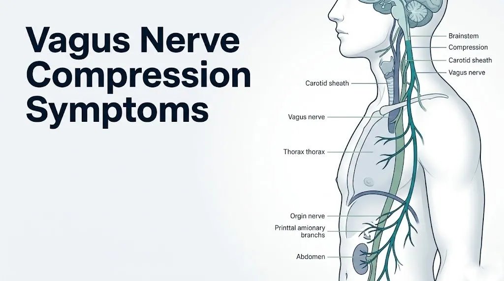
Early signs typically include:
- Unexplained chronic cough: Irritation of the vagal afferent fibers in the larynx and trachea triggers the Arnold’s nerve cough reflex even without infection or aspiration.
- Throat globus sensation: A persistent feeling of a lump or tightness in the throat at the level of the thyroid cartilage, produced by abnormal efferent motor signaling to the pharyngeal constrictor muscles.
- Intermittent nausea without vomiting: Afferent signal disruption from the gastric branches creates abnormal sensory input to the NTS, which the brainstem interprets as nausea.
- Episodes of transient dizziness: Impaired baroreflex afferent signaling produces brief drops in blood pressure on position change.
- Ear discomfort or aching: Irritation of Arnold’s nerve (the auricular branch) causes referred pain to the external auditory meatus and posterior auricle.
Clinical observation consistently shows that early vagus nerve compression is frequently misattributed to gastroesophageal reflux disease (GERD), anxiety, or viral post-nasal drip cough, delaying accurate diagnosis by months to years. The distinguishing feature of early vagal compression is that these symptoms cluster across multiple systems simultaneously without a single unifying diagnosis explaining all of them.
People over 60 may present with early vagal compression symptoms that overlap significantly with age-related autonomic decline, making the distinction harder. A board-certified neurologist specializing in autonomic disorders is the appropriate provider to evaluate whether a symptom cluster in an older adult represents age-related changes or structural nerve compression.
Vagus Nerve Compression Heart Symptoms
Vagus nerve compression heart symptoms arise from disrupted parasympathetic control of the sinoatrial (SA) node, the heart’s primary pacemaker located in the posterior wall of the right atrium.
The vagus nerve’s superior and inferior cervical cardiac branches and thoracic cardiac branches deliver parasympathetic signals directly to the SA node and, to a lesser degree, the atrioventricular (AV) node. Under normal conditions, these branches slow heart rate at rest, producing the physiological resting heart rate of 60 to 100 beats per minute. When the vagus nerve is compressed and initially irritated, paradoxical upregulation of these signals can produce bradycardia (heart rate below 60 beats per minute at rest), and in severe cases, AV conduction delays.
Conversely, when chronic compression damages vagal fibers, the loss of parasympathetic tone allows sympathetic drive to dominate, which can produce inappropriate resting tachycardia (heart rate above 100 beats per minute). This switch from bradycardia to tachycardia as compression progresses is a clinically important pattern that distinguishes vagal compression from intrinsic cardiac arrhythmia.
| Cardiac Symptom | Mechanism | Clinical Stage |
|---|---|---|
| Bradycardia at rest | SA node over-suppression from irritated vagal branches | Early/irritation phase |
| Heart palpitations | Disrupted SA node rhythm regulation | Early to intermediate |
| AV conduction delay | Vagal efferent excess to AV node | Intermediate |
| Inappropriate resting tachycardia | Loss of parasympathetic tone; sympathetic dominance | Advanced/fiber damage phase |
| Orthostatic hypotension | Impaired baroreflex afferent arc | Intermediate to advanced |
People with pre-existing first-degree AV block who then develop vagal compression face an elevated risk of more advanced AV conduction slowing. A board-certified cardiologist or electrophysiologist should evaluate any patient with known conduction abnormalities who develops new bradycardia or syncope.
Vagus Nerve Compression Digestive Symptoms
Vagus nerve compression digestive symptoms result from impaired parasympathetic control of the esophageal plexus, gastric branches, and celiac branch of the vagus nerve, affecting motility and secretion throughout the gastrointestinal tract from the esophagus to the proximal colon.
The vagus nerve accounts for approximately 75% of all parasympathetic outflow to the gastrointestinal tract, according to data reviewed by the National Institute of Neurological Disorders and Stroke (NINDS). This level of parasympathetic dependency means that even partial vagal compression produces measurable changes in gut function. Under irritation conditions, increased afferent signaling from the gut produces nausea and accelerated gastric motility, sometimes presenting as diarrhea. As compression progresses and efferent motor fibers are damaged, the opposite occurs: gastroparesis (delayed gastric emptying) and constipation.
Digestive symptoms associated with vagus nerve compression include:
- Nausea, particularly after meals, not explained by food intake or other GI pathology
- Early satiety, caused by impaired gastric fundic relaxation from disrupted vagal efferents
- Bloating and abdominal distension, from reduced gastric and intestinal motility
- Dysphagia (difficulty swallowing), from impaired esophageal peristalsis via the esophageal plexus
- Unexplained weight loss, a downstream effect of inadequate caloric intake from early satiety and nausea
- Alternating diarrhea and constipation, reflecting the transitional phase between irritation-driven hypermotility and damage-driven hypomotility
People with diabetes mellitus have a compounding risk factor: diabetic autonomic neuropathy can independently damage vagal fibers supplying the gut, producing gastroparesis that may be mistaken for compression-related symptoms. In diabetic patients, a board-certified gastroenterologist should evaluate digestive symptoms with a gastric emptying study before attributing them to compression alone.
Vagus Nerve Compression Throat and Voice Symptoms
Vagus nerve compression throat and voice symptoms arise specifically from involvement of the recurrent laryngeal nerve and the superior laryngeal nerve, both branches of the vagus that supply the intrinsic muscles of the larynx and the sensory innervation of the supraglottic and infraglottic larynx.
The recurrent laryngeal nerve (RLN) is particularly vulnerable to compression because of its unusual anatomical course. On the left side, the RLN loops under the aortic arch before ascending in the tracheoesophageal groove to reach the larynx, making it the longest path of any laryngeal nerve and exposing it to compression from mediastinal masses, aortic arch aneurysm, and enlarged mediastinal lymph nodes. On the right side, the RLN loops under the right subclavian artery, a shorter but still surgically relevant course.
Throat and voice symptoms of vagus nerve compression include:
- Dysphonia (hoarseness or a breathy, weak voice quality), from paralysis or paresis of the vocalis muscle and posterior cricoarytenoid muscle, the only abductor of the vocal cord
- Dysphagia, particularly for liquids (aspiration risk), from pharyngeal constrictor weakness via nucleus ambiguus fiber involvement
- Globus pharyngeus (the sensation of a lump in the throat), from abnormal sensory signaling in the hypopharyngeal afferent fibers
- Chronic throat clearing, from irritated laryngeal afferents signaling to the NTS
- Aspiration of food or liquid into the trachea, a serious complication of complete unilateral vocal cord paralysis
A research review published in the Journal of Neurology, Neurosurgery and Psychiatry noted that new-onset hoarseness lasting more than three weeks without a clear upper respiratory tract infection as the cause warrants direct laryngoscopy to assess vocal cord mobility before any imaging is ordered. This sequence matters because laryngoscopy directly visualizes whether unilateral cord immobility is present, which then guides imaging decisions.
Postmenopausal women have a modestly higher prevalence of voice changes for multiple independent reasons including hormonal changes to laryngeal mucosa, which can complicate attribution of dysphonia to vagal compression versus hormonal effects. An otolaryngologist (ENT physician) with laryngeal expertise should assess persistent dysphonia in this population.
Key Takeaway: New-onset hoarseness lasting more than three weeks in an adult is a red flag that requires laryngoscopy to assess vocal cord mobility, because unilateral cord paralysis from vagal or recurrent laryngeal nerve compression can be the presenting sign of a compressive mediastinal or neck mass.
Vagus Nerve Compression and Dizziness and Syncope
Dizziness and syncope (fainting) from vagus nerve compression result from disruption of the baroreflex arc, the feedback loop that continuously adjusts heart rate and vascular tone to maintain adequate blood pressure and cerebral perfusion.
The afferent limb of the baroreflex carries blood pressure information from baroreceptors in the carotid sinus and aortic arch to the nucleus tractus solitarius (NTS) via vagal afferent fibers. The NTS then modulates vagal efferent output to the SA node and sympathetic tone to peripheral blood vessels. When vagal afferent fibers are compressed and signal transmission is disrupted, the NTS receives inaccurate or absent blood pressure data. The cardiovascular system responds by failing to adjust appropriately to position changes or exertion, producing orthostatic hypotension (blood pressure drop on standing) and vasovagal syncope.
Vasovagal syncope is a specific form of fainting in which paradoxical vagal activation (often triggered by pain, emotional stress, or prolonged standing) causes simultaneous extreme bradycardia and peripheral vasodilation, dropping cerebral perfusion pressure below the threshold required for consciousness. In people with pre-existing vagal compression, the threshold for triggering this response is lower.
According to the Mayo Clinic, vasovagal syncope accounts for approximately one-third of all syncopal episodes evaluated in clinical practice. When syncope occurs alongside other vagal symptoms such as chronic cough, dysphagia, or bradycardia, the clinical picture becomes more specific to vagal pathology rather than isolated reflex syncope.
To accurately describe dizziness to a neurologist for evaluation:
- Note whether dizziness is rotational (vertigo) or a sense of lightheadedness (presyncope) without spinning.
- Record whether it occurs on standing up, during specific head positions, or without positional trigger.
- Track whether the heart rate is perceptibly slow (bradycardia) or racing (compensatory tachycardia) at the time of dizziness.
- Note any preceding nausea, throat symptoms, or cough before the dizziness episode.
- Bring this documented pattern to an evaluation by a board-certified neurologist with autonomic disorder expertise.
Vagus Nerve Compression Ear Symptoms
Vagus nerve compression ear symptoms arise from irritation or compression of the auricular branch of the vagus nerve, also called Arnold’s nerve, which supplies sensory innervation to the skin of the posterior external auditory meatus (ear canal), the concha of the auricle, and the posterior surface of the eardrum (tympanic membrane).
Arnold’s nerve exits the main vagal trunk at the level of the jugular ganglion (superior ganglion of the vagus) and passes through the mastoid canaliculus in the temporal bone to reach the ear. When the vagus nerve is compressed at or near the jugular foramen or in the upper carotid sheath, Arnold’s nerve afferents are irritated, producing referred pain, aching, or strange sensations in the ear without any pathology of the ear itself.
This presentation is clinically important because ear pain in the absence of middle ear infection, ear canal pathology, or temporomandibular joint disorder is a known presentation of vagal and glossopharyngeal nerve irritation. According to clinical case series published in Annals of Neurology, referred otalgia (ear pain) from vagal branch irritation is underrecognized in primary care, where it is frequently attributed to eustachian tube dysfunction or atypical otitis media.
Ear symptoms associated with vagal compression include:
- Referred otalgia, a deep aching in the ear canal or concha without visible ear pathology
- Auricular paresthesia, tingling or altered sensation in the posterior ear and mastoid area
- Arnold’s nerve cough reflex, in which stimulation of the external ear canal (for example, during ear cleaning or ear examination) triggers an immediate cough response
- Tinnitus (ringing or buzzing in the ear) in some cases, though this symptom is more directly associated with the adjacent glossopharyngeal nerve and cochlear nerve than the vagus itself
People who notice that touching or cleaning their ear canal consistently triggers a cough should specifically mention this to their primary care physician or otolaryngologist, because Arnold’s nerve cough reflex is a clinically recognized diagnostic clue pointing to vagal involvement in the neck or skull base.
Vagus Nerve Compression Breathing Symptoms
Vagus nerve compression breathing symptoms arise from disrupted vagal control of the pulmonary plexus, the network of vagal fibers that regulates bronchomotor tone, controls the cough reflex from bronchial mucosa, and modulates respiratory rate via the Hering-Breuer reflex arc.
The vagus nerve’s pulmonary branches, traveling within the pulmonary plexus at the lung hilum, carry parasympathetic efferent fibers that cause bronchoconstriction and mucus secretion. They also carry afferent fibers that detect lung stretch and airway irritants, feeding this information to the NTS. Compression of these fibers can produce paradoxical bronchoconstriction mimicking asthma, or alternatively a blunted cough reflex that impairs airway protection.
According to research published in the journal Neurology, vagal afferent fiber irritation in the airway is a recognized mechanism for chronic refractory cough, a cough that persists despite standard treatment for the three most common causes (upper airway cough syndrome, asthma, and gastroesophageal reflux disease). Clinicians now recognize vagal hypersensitivity or nerve irritation as a distinct cause category in refractory cough guidelines.
Breathing symptoms associated with vagus nerve compression include:
- Chronic nonproductive cough, particularly triggered by speaking, laughing, cold air, or position changes rather than by respiratory illness
- Throat-centered breathing difficulty (laryngeal dyspnea), caused by impaired vocal cord abductor function
- Paradoxical vocal cord motion, a condition where the vocal cords adduct (close) on inhalation rather than abducting, causing inspiratory stridor
- Exercise intolerance from impaired bronchomotor regulation and blunted cardiac acceleration
- Hiccups (singultus), caused by irritation of vagal afferents at the diaphragmatic level, triggering uncontrolled diaphragmatic spasm
People with pre-existing asthma may find their asthma control worsening if vagal compression is adding bronchomotor instability on top of their pre-existing airway hyperresponsiveness. A board-certified pulmonologist should evaluate any asthmatic patient whose disease becomes unexpectedly difficult to control, to rule out a vagal contribution.
Key Takeaway: Chronic cough that persists for more than eight weeks despite standard treatment for reflux, post-nasal drip, and asthma should prompt evaluation of vagal nerve irritation or compression as a distinct diagnostic category.
Vagus Nerve Compression Neck Pain and Referred Pain
Vagus nerve compression neck pain is typically not the primary presenting complaint, but somatic pain in the neck and referred pain to the jaw, ear, or shoulder can arise when compression involves the upper carotid sheath or the jugular foramen region where adjacent somatic structures are also affected.
The vagus nerve itself carries limited somatic afferent input compared to its visceral afferent load. However, the close anatomical relationship within the carotid sheath between the vagus nerve and the carotid artery and internal jugular vein means that any compressive pathology in this region often affects all three structures. Compression from a cervical lymph node mass, carotid body tumor (paraganglioma), or cervical disc herniation at levels C3-C6 can produce deep cervical aching that patients describe as neck pain, though the primary neural injury is vagal.
Referred pain patterns from upper vagal compression can include:
- Deep aching in the lateral neck below the jaw angle, at the level of the thyroid cartilage (corresponding to the carotid sheath level)
- Referred discomfort to the base of the skull and posterior ear, from jugular ganglion involvement
- Ipsilateral jaw aching that mimics temporomandibular joint pain, from adjacent glossopharyngeal nerve irritation at the jugular foramen
- Anterior neck tightness that worsens with neck rotation or extension, suggesting positional compression by cervical vertebral structures
A 2022 case series published in the Journal of Neurology, Neurosurgery and Psychiatry documented patients with cervical osteophytes at the C4-C5 and C5-C6 levels causing compression of the carotid sheath contents, producing a symptom cluster of bradycardia, ipsilateral neck pain, and hoarseness that resolved after surgical decompression. This case series highlights that cervical spondylosis is an underrecognized structural cause of vagal compression.
People with rheumatoid arthritis involving the cervical spine face elevated risk of atlantoaxial instability and upper cervical osteophyte formation, both of which can compress structures at the high cervical and skull base level. A board-certified rheumatologist and neurologist should coordinate management for cervical spine involvement in rheumatoid arthritis when vagal symptoms develop.
Vagus Nerve Compression and Anxiety and Autonomic Dysfunction
Vagus nerve compression can produce symptoms that are clinically indistinguishable from anxiety disorder, including palpitations, shortness of breath, nausea, dizziness, and a persistent sense of physiological unease, because the autonomic nervous system disruption produced by vagal compression directly alters the physiological signals that the brain interprets as threat or alarm.
The autonomic nervous system maintains homeostasis through balanced parasympathetic (vagal) and sympathetic tone. When vagal afferent input to the NTS is disrupted by compression, the NTS cannot accurately signal the hypothalamic-pituitary-adrenal (HPA) axis and the amygdala about the body’s visceral state. The result is a persistent low-grade physiological alarm state: increased cortisol, increased norepinephrine release from the locus coeruleus, and heightened sympathetic tone. This state feels like anxiety, because it activates the same neurological pathways.
Research published in the journal Neurology has documented bidirectional interactions between vagal afferent signaling and limbic system function, with reduced vagal afferent input associated with heightened amygdala reactivity and reduced prefrontal cortical regulation of emotional responses. This provides a specific physiological mechanism for why people with vagal dysfunction frequently develop anxiety-like symptoms.
The clinical challenge is that anxiety and vagal compression are not mutually exclusive. They can coexist, and anxiety can genuinely develop secondary to the physiological disruption of vagal compression. The distinguishing pattern is:
- Anxiety from vagal compression typically includes prominent physical symptoms (bradycardia, gastrointestinal disturbance, cough, ear symptoms) that are not typically present in primary generalized anxiety disorder
- Primary anxiety disorder typically responds to standard therapeutic approaches, while anxiety secondary to vagal compression does not improve without addressing the underlying nerve pathology
Women are diagnosed with anxiety disorders at approximately twice the rate of men according to CDC epidemiological data, which can lead to premature diagnostic closure when a woman’s vagal compression symptoms are attributed to anxiety rather than investigated structurally.
Vagus Nerve Compression at Different Anatomical Sites
Vagus nerve compression produces distinctly different symptom clusters depending on where along its anatomical course the compression occurs, because each segment of the nerve serves different organ targets.
The vagus nerve has multiple anatomical bottlenecks where compression is most likely to occur:
| Compression Site | Structures at Risk | Primary Symptom Cluster |
|---|---|---|
| Jugular foramen (skull base) | Cranial nerves IX, X, XI together | Ear pain, hoarseness, dysphagia, shoulder weakness |
| Upper carotid sheath (C2-C4 level) | Vagus, carotid artery, jugular vein | Bradycardia, neck pain, referred ear pain, throat globus |
| Mid-cervical carotid sheath (C4-C6) | Vagus, cervical cardiac branches | Bradycardia, palpitations, anterior neck aching |
| Thoracic inlet | Left RLN at aortic arch level | Left-sided hoarseness, mediastinal symptoms |
| Tracheoesophageal groove (thoracic) | RLN ascending in groove | Hoarseness, cough, dysphagia |
| Esophageal hiatus (diaphragmatic) | Anterior and posterior vagal trunks | Gastroparesis, early satiety, abdominal bloating |
Jugular foramen compression (also called jugular foramen syndrome or Vernet’s syndrome when cranial nerves IX, X, and XI are all affected) produces the most complex symptom cluster because three cranial nerves are simultaneously impaired. Causes include paraganglioma (glomus jugulare tumor), skull base meningioma, and metastatic disease to the skull base.
People who have had prior radiation therapy to the head and neck (for example, for nasopharyngeal cancer or head and neck squamous cell carcinoma) face a risk of fibrotic entrapment of the vagus nerve at multiple cervical levels simultaneously, producing a diffuse rather than site-specific symptom pattern. A neuro-oncologist and radiation oncologist should be part of the evaluation team for these individuals.
Chronic vs. Acute Vagus Nerve Compression Symptoms
The distinction between acute and chronic vagus nerve compression matters clinically because the physiological mechanism, the symptom character, and the urgency of evaluation differ substantially between the two presentations.
Acute vagus nerve compression typically results from a rapidly developing mass, hemorrhage, trauma, or surgical complication that produces sudden nerve impingement. The nerve initially responds with irritation-phase symptoms: sudden bradycardia, acute nausea and vomiting, sudden voice change, or a syncopal episode. These acute presentations require urgent evaluation because the underlying cause (hemorrhage into the neck, rapidly expanding lymph node, or traumatic carotid sheath hematoma) may be time-sensitive.
Chronic vagus nerve compression develops slowly over months to years, typically from progressive cervical spondylosis, slowly growing masses, or gradual fibrotic changes. The clinical pattern is one of progressive, multi-system symptom accumulation that is frequently misdiagnosed repeatedly before the vagal origin is identified. Symptoms tend to fluctuate with body position (worse with neck extension in osteophyte-related compression) and to worsen gradually rather than appearing suddenly.
| Feature | Acute Compression | Chronic Compression |
|---|---|---|
| Onset | Hours to days | Weeks to months |
| Typical cause | Hemorrhage, trauma, rapid tumor growth | Osteophyte, slow-growing mass, fibrosis |
| Dominant symptom phase | Irritation phase (excess parasympathetic output) | Progression to deficiency phase (dropout of parasympathetic signals) |
| Diagnostic urgency | Same-day to emergency evaluation | Within weeks; urgent if progressive |
| Symptom stability | Rapidly changing | Slowly progressive with positional variation |
According to the American Academy of Neurology, any acute-onset vagal symptom cluster, particularly sudden bradycardia with syncope or sudden-onset unilateral hoarseness, warrants same-day evaluation by a neurologist or emergency medicine physician rather than watchful waiting.
Key Takeaway: Sudden-onset hoarseness, unexplained bradycardia with dizziness, or acute syncopal episodes require same-day neurological evaluation, not scheduled follow-up, because acute vagal compression can be caused by rapidly progressive conditions including hemorrhage, expanding tumor, or carotid sheath pathology.
Who Is at Risk for Vagus Nerve Compression?
People at elevated risk for vagus nerve compression share specific anatomical, medical, and procedural risk factors that make mechanical nerve impingement more likely at one or more points along the vagal course.
Risk factors include:
- Older age with cervical spondylosis: Cervical osteophytes forming at C4-C5, C5-C6, and C6-C7 vertebral levels can compress the carotid sheath from the posterior aspect, particularly during neck extension. Prevalence of cervical spondylosis increases with age and is present radiographically in more than 85% of adults over 60 according to data reviewed by the American Academy of Orthopaedic Surgeons.
- Prior neck surgery: Thyroidectomy, cervical discectomy, carotid endarterectomy, and anterior cervical spine surgery all involve dissection within or adjacent to the carotid sheath, creating scar tissue risk.
- Prior head and neck radiation: Radiation-induced fibrosis can encase the vagus nerve at multiple cervical levels.
- Cervical disc herniation: Herniation at C4-C5 or C5-C6 levels can compress the carotid sheath contents alongside nerve root compression.
- Connective tissue hypermobility disorders such as Ehlers-Danlos syndrome: Associated with increased cranial nerve and autonomic dysfunction through mechanisms including atlantoaxial instability and vascular redundancy.
- Thyroid enlargement (goiter) or thyroid masses: The thyroid gland lies immediately adjacent to the tracheoesophageal groove where the RLN ascends, making even moderate thyroid enlargement capable of RLN compression.
- Mediastinal lymphadenopathy: Enlarged lymph nodes from lymphoma, sarcoidosis, or metastatic disease compress vagal structures in the thoracic component.
- Athletes with constitutionally high vagal tone: A pre-existing high baseline vagal tone means that added compression-induced parasympathetic drive produces more pronounced bradycardia at lower compression thresholds.
People with a body mass index above 35 who develop new voice changes or swallowing difficulties should have thyroid and neck anatomy evaluated, as cervical adipose deposition can contribute to increased pressure on carotid sheath contents.
How to Diagnose Vagus Nerve Compression
Diagnosing vagus nerve compression requires a structured sequence of clinical assessments and targeted imaging studies, because no single test confirms vagal compression across all possible anatomical sites.
The diagnostic process typically unfolds as follows:
- Detailed history and neurological examination: A board-certified neurologist or autonomic neurologist takes a structured history documenting the symptom cluster, onset, positional variation, and prior surgical or medical history. Physical examination includes cranial nerve testing (gag reflex assessment, vocal cord visualization via indirect or direct laryngoscopy, uvula deviation assessment) and cardiovascular autonomic testing (heart rate response to deep breathing, Valsalva maneuver, and position change).
- Laryngoscopy: Direct or indirect flexible laryngoscopy, performed by an otolaryngologist, visualizes vocal cord mobility in real time. Unilateral cord immobility is a direct sign of recurrent laryngeal nerve involvement.
- MRI of the brain, skull base, and cervical spine: Magnetic resonance imaging (MRI) with gadolinium contrast is the primary imaging modality for identifying structural causes of vagal compression, including skull base masses, carotid sheath masses, and cervical disc pathology.
- CT scan of the chest: A computed tomography (CT) scan of the chest is ordered when mediastinal or thoracic-level vagal compression is suspected, particularly in cases of left-sided hoarseness from RLN compression at the aortic arch.
- Autonomic function testing battery: This includes tilt-table testing (to assess baroreflex integrity and orthostatic responses), heart rate variability analysis, and quantitative sudomotor axon reflex testing (QSART) to map autonomic fiber function.
- Swallowing study (modified barium swallow or fiberoptic endoscopic evaluation of swallowing, FEES): Ordered when dysphagia is present to quantify the degree of pharyngeal dysfunction and aspiration risk.
- Holter cardiac monitor: A 24 to 48-hour ambulatory cardiac monitor is ordered to capture intermittent bradycardia or arrhythmia patterns not present during a resting ECG.
According to the National Institute of Neurological Disorders and Stroke (NINDS), autonomic function testing is the standard diagnostic approach for confirming parasympathetic dysfunction, and its results guide both the urgency and the specialty of further care.
Emergency Symptoms of Vagus Nerve Compression
Certain symptoms associated with vagus nerve compression require immediate emergency evaluation. Do not wait to see if these resolve on their own.
Call 911 or go to the nearest emergency room immediately if you experience:
- Heart rate below 40 beats per minute at rest with dizziness, lightheadedness, or loss of consciousness, which indicates severe parasympathetic overdrive or complete cardiac conduction failure
- Syncope (loss of consciousness) lasting more than 30 seconds, or multiple syncopal episodes within 24 hours, which requires emergency cardiac and neurological evaluation
- Sudden-onset severe difficulty swallowing (unable to swallow your own saliva), which indicates acute pharyngeal muscle paresis with aspiration risk
- Acute respiratory distress or stridor (a high-pitched breathing sound on inhalation) from bilateral vocal cord involvement or paradoxical vocal cord motion, which represents airway compromise
- Sudden severe neck pain with new-onset hoarseness, which can indicate hemorrhage into the carotid sheath or a rapidly expanding mass
- Progressive dysphagia escalating from solids to liquids within days, which suggests acute nerve compression with rapidly worsening motor deficit
- Sudden severe unilateral neck swelling alongside any vagal symptoms, indicating a possible carotid sheath hemorrhage or rapidly expanding mass requiring emergency imaging
These presentations can indicate a ruptured paraganglioma, carotid sheath hemorrhage, rapidly progressive vagal nerve palsy from malignant compression, or acute high-degree AV block and require emergency medical assessment, not an urgent care appointment or a phone call.
Frequently Asked Questions About Vagus Nerve Compression Symptoms
What does vagus nerve compression feel like?
Vagus nerve compression feels like a combination of sensations across multiple body systems simultaneously: throat tightness, ear aching, intermittent dizziness, nausea without a clear cause, and a sense of the heart beating slowly or irregularly.
The specific sensation depends on where compression occurs along the nerve’s course and which fiber types are most affected at that site.
No single sensation is pathognomonic for vagal compression, which is why a neurologist’s clinical evaluation is required rather than self-diagnosis based on symptom description alone.
Can vagus nerve compression cause heart palpitations?
Yes, vagus nerve compression can cause heart palpitations through disruption of the parasympathetic signals delivered to the sinoatrial node via the vagal cardiac branches.
Early compression typically irritates the nerve and produces paradoxical bradycardia or irregular heart rhythm, both of which can be perceived as palpitations.
A 24-hour Holter cardiac monitor ordered by a cardiologist or neurologist is the standard tool for capturing and characterizing these rhythm disturbances.
How do you know if your vagus nerve is compressed or irritated?
Vagus nerve compression or irritation is suggested by a symptom cluster spanning multiple organ systems simultaneously: chronic cough, unexplained bradycardia, ear pain without ear infection, nausea, hoarseness, and dizziness together without a single unifying diagnosis explaining all of them.
Confirmation requires a clinical evaluation by a board-certified neurologist, laryngoscopy by an otolaryngologist, and MRI or CT imaging to identify the structural cause.
No home test or self-assessment can confirm vagal compression.
Can a pinched nerve in the neck affect the vagus nerve?
Yes, a herniated cervical disc at levels C4-C5 or C5-C6 can compress the carotid sheath from the posterior direction, placing mechanical pressure on the vagus nerve running within that sheath.
This type of compression typically produces a mixed symptom picture of both nerve root symptoms (arm pain or numbness from direct disc compression) and vagal symptoms (bradycardia, hoarseness, or nausea).
A board-certified neurologist and a spine surgeon should evaluate this presentation together, with MRI of the cervical spine as the primary imaging study.
Can anxiety be caused by vagus nerve compression?
Vagus nerve compression can produce anxiety-like symptoms through a specific physiological mechanism: disrupted afferent vagal signaling to the nucleus tractus solitarius alters hypothalamic-pituitary-adrenal axis regulation and amygdala reactivity, producing a persistent physiological alarm state.
This is not primary anxiety disorder but a secondary physiological consequence of autonomic disruption, and it does not fully resolve with anxiety-targeted treatment until the underlying nerve compression is addressed.
A neurologist with autonomic disorder expertise can help distinguish compression-related autonomic anxiety from primary generalized anxiety disorder based on clinical pattern and autonomic testing results.
What type of doctor treats vagus nerve compression?
The primary specialist for vagus nerve compression evaluation and management is a board-certified neurologist with subspecialty training in autonomic disorders or cranial nerve pathology.
For voice and swallowing symptoms specifically, an otolaryngologist (ENT physician) performs laryngoscopy and assesses recurrent laryngeal nerve function directly.
Depending on the cause of compression, a neurosurgeon, thoracic surgeon, or interventional radiologist may become involved in definitive treatment of the underlying structural cause.
Closing
The single most actionable point from this article is the symptom cluster rule: if you have three or more of these symptoms simultaneously (unexplained bradycardia, chronic cough, throat globus, nausea, ear pain, hoarseness, and dizziness) without a single diagnosis explaining all of them, vagal nerve pathology is a legitimate diagnostic consideration, not a fringe idea.
The next step is a specific one. Request a referral to a board-certified neurologist with autonomic disorder expertise and ask for autonomic function testing alongside MRI of the brain, skull base, and cervical spine. If you have hoarseness as part of the picture, request a laryngoscopy through an otolaryngologist before or concurrent with imaging. These three diagnostic tools together give you and your clinicians the structural and functional picture needed to determine whether compression is present and where.
Your symptoms across multiple body systems are not random. The vagus nerve connects all of them.