Ear Cancer Symptoms: Full Clinical Guide 2026
Ear cancer symptoms vary substantially depending on which part of the ear is affected, ranging from a visible non-healing sore on the outer ear to progressive hearing loss, chronic discharge, and eventually facial weakness when a tumor has invaded the temporal bone. Recognizing which symptoms suggest early disease versus which indicate neurological involvement is what allows a person to seek evaluation at the stage where treatment outcomes are meaningfully better.
The National Cancer Institute classifies ear cancer as a rare malignancy, with external auditory canal carcinoma estimated to occur in approximately 1 per million people annually based on SEER database surveillance data. Despite its rarity, ear cancer is frequently diagnosed late because its early symptoms overlap substantially with common benign ear conditions like otitis externa, making clinical vigilance about treatment-refractory ear symptoms the most important practical takeaway for any reader of this article.
This guide covers the complete ear cancer symptom spectrum organized by anatomical location, explains the specific physiological reason each symptom occurs, addresses rare types including pediatric presentations, distinguishes ear cancer from benign mimics, and identifies which symptoms require urgent same-day evaluation versus a scheduled specialist appointment.
Ear Cancer Symptoms: The Complete Clinical Picture
The complete symptom profile of ear cancer includes persistent ear pain, bloody or chronic discharge from the ear canal, hearing loss, tinnitus, a non-healing lesion or growth on the outer ear, and in advanced cases, facial nerve palsy, dizziness, and enlarged neck lymph nodes.
What makes ear cancer diagnostically challenging is that its early symptoms are individually non-specific. Ear pain could be otitis. Discharge could be infection. Hearing loss could be wax impaction. It is the combination of these symptoms, their persistence despite standard treatment, and their failure to respond to antibiotics or topical ear treatments that distinguishes malignancy from benign ear disease.
The location of the cancer within the ear determines which symptoms appear first and which appear later. Cancer of the outer ear (pinna) typically presents with a visible surface lesion before any hearing or neural symptoms develop. Cancer of the external auditory canal (EAC) mimics chronic ear infection for weeks to months before the diagnosis is considered. Cancer of the middle ear and temporal bone often presents with hearing loss and pain that are indistinguishable from chronic otitis media until neurological symptoms emerge.
Complete ear cancer symptom inventory:
- Non-healing sore, ulcer, or growth on the pinna or ear canal opening, persisting beyond 4 weeks
- Persistent ear pain (otalgia) that does not resolve with standard infection treatment
- Bloody or blood-tinged discharge from the ear canal (bloody otorrhea)
- Persistent clear or purulent discharge not responding to antibiotic treatment within 2 to 3 weeks
- Unilateral hearing loss (one-sided), either gradually progressive or sudden
- Tinnitus (ringing, buzzing, or hissing in the affected ear)
- A sensation of fullness or pressure in the affected ear
- Facial weakness on the same side as the affected ear (ipsilateral facial nerve palsy)
- Dizziness or balance disturbance
- Enlarged, firm lymph nodes in front of or below the ear, or in the upper neck
- Trismus (difficulty opening the jaw) in very advanced cases where tumor has spread to the infratemporal fossa
The American Academy of Otolaryngology-Head and Neck Surgery emphasizes that any ear symptom not responding to standard treatment within 3 to 4 weeks warrants direct visualization of the ear canal and referral to a board-certified otolaryngologist.
Types of Ear Cancer and Where They Develop
Ear cancer is not a single disease; it is a group of distinct malignancies defined by the anatomical site of origin and the type of cell that undergoes malignant transformation, and these differences produce different early symptom patterns.
The pinna (auricle) is the external visible portion of the ear, composed of elastic cartilage covered by skin. Being the most sun-exposed surface of the body per unit area in many people, the pinna is a common site for skin cancers, primarily basal cell carcinoma (BCC) and squamous cell carcinoma (SCC). The American Cancer Society reports that the ear and periauricular skin represent one of the higher-risk sun-exposure sites for non-melanoma skin cancer in adults over 50, particularly in males with occupational or recreational outdoor exposure history.
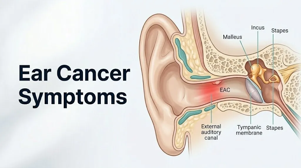
The external auditory canal (EAC) extends from the opening of the ear canal to the tympanic membrane (eardrum). Its outer third is cartilaginous and contains ceruminous glands (modified apocrine sweat glands) and sebaceous glands. Its inner two-thirds is bony, lined by thin stratified squamous epithelium. Primary EAC malignancy is most commonly squamous cell carcinoma. Ceruminous gland adenocarcinoma (arising from the ceruminous glands of the cartilaginous EAC) is a distinct and rarer entity.
The middle ear occupies the tympanic cavity within the temporal bone. Middle ear cancers are rare and most commonly squamous cell carcinoma. They arise from the mucosal lining of the tympanic cavity or extend from an EAC primary tumor.
| Cancer Site | Primary Cell Type | Most Affected Population | Characteristic Early Symptom |
|---|---|---|---|
| Pinna (auricle) | Basal cell or squamous cell carcinoma | Men over 60, outdoor workers | Visible non-healing lesion or ulcer |
| External auditory canal | Squamous cell carcinoma | Adults 50 to 70 | Chronic discharge mimicking otitis externa |
| Ceruminous glands (EAC) | Ceruminous gland adenocarcinoma | Adults 50 to 70 | Slowly enlarging canal mass |
| Middle ear | Squamous cell carcinoma | Adults 50 to 70 | Hearing loss, chronic otorrhea |
| Pinna/auricle | Melanoma | Adults 40 to 80, fair skin | Pigmented lesion changing color or shape |
| Middle ear (children) | Rhabdomyosarcoma | Children under 10 | Ear discharge, polypoid mass, hearing loss |
Children diagnosed with ear cancer almost exclusively have rhabdomyosarcoma, a soft tissue sarcoma of embryonal muscle origin, rather than squamous cell carcinoma. This distinct biology produces a different symptom timeline and a different treatment approach.
Early Stage Ear Cancer Symptoms
Early stage ear cancer symptoms are often subtle and easily attributed to benign conditions, which is precisely why the average delay from symptom onset to diagnosis in external auditory canal carcinoma is estimated at 6 to 12 months based on case series reviewed in the Journal of Laryngology and Otology.
Think of early EAC cancer like a slow leak in a pipe buried in the wall: the damage is happening, but the visible signs, a slight stain on the surface, are easily attributed to something else. By the time the stain is impossible to ignore, the underlying problem has been progressing unchecked for months.
For pinna cancer, the earliest stage presents as a visible change on the ear’s skin surface. A basal cell carcinoma of the pinna in its earliest stage may appear as a small, pearly or translucent papule with a rolled border and central depression, or a flat, skin-colored plaque with a subtle waxy surface. A squamous cell carcinoma may begin as a rough, scaly patch or a firm, pink-red nodule on sun-damaged skin, often on the helix (the outer curved rim) or the antihelix of the ear.
For EAC cancer, the earliest detectable sign is typically a small area of granulation tissue or a slightly thickened area of the canal skin that bleeds on contact with an otoscope speculum during routine ear examination. This is frequently mistaken for inflamed otitis externa skin.
Early stage ear cancer symptoms by site:
- Pinna/BCC: small pearly papule, translucent nodule, or flat scar-like plaque, often on the helix or preauricular skin, not resolving after 4 to 6 weeks
- Pinna/SCC: rough, scaly, red-pink patch or firm nodule that may crust or bleed when touched; arises on chronically sun-damaged skin
- EAC/SCC early: unilateral ear canal irritation, mild discharge, or contact bleeding during ear cleaning that does not resolve with standard otitis externa treatment
- Middle ear early: mild progressive unilateral conductive hearing loss with or without intermittent ear fullness, frequently misdiagnosed as serous otitis media (fluid in the middle ear)
Women diagnosed with pinna cancer tend to have later-stage disease at presentation compared to men, a pattern attributed to longer hair covering the pinna in many women, reducing the visibility of early lesions during routine self-inspection.
Outer Ear (Pinna) Cancer Symptoms
Outer ear cancer symptoms are primarily visible skin changes on the sun-exposed surface of the pinna, and because the pinna is external and accessible to direct inspection, these are potentially the most detectable of all ear cancer presentations.
The anterior (front) surface of the pinna receives direct sun exposure in most outdoor activities. The posterior (back) surface and the conchal bowl (the deeper cup-shaped central depression leading to the ear canal opening) receive reflected and ambient UV radiation. Basal cell carcinoma most frequently develops on the helix, the antihelix, and the periauricular skin. Squamous cell carcinoma preferentially involves the helix and the superior conchal bowl, where UV damage accumulates most.
The ABCDE framework used for melanoma assessment (Asymmetry, Border irregularity, Color variation, Diameter greater than 6 mm, Evolution or change over time) applies to any suspicious pinna lesion. However, BCC and SCC on the pinna do not always follow classic melanoma ABCDE criteria, because they may be skin-colored or pink rather than pigmented.
Warning signs on the outer ear that require evaluation by a board-certified dermatologist within 2 to 4 weeks:
- Any sore, ulcer, or wound on the pinna that has not healed after 4 weeks
- A firm, raised nodule on the helix or antihelix that was not previously present
- A scaly, crusty, or rough patch on the ear that bleeds when scratched or rubbed
- A pearly or translucent bump that gradually enlarges over weeks
- A pigmented lesion on the ear with any asymmetry, irregular border, or color variation
- Any pinna lesion that recurs after apparent healing
Older adults with fair skin, a history of significant sun exposure (including those who worked outdoors for decades), or a personal or family history of non-melanoma skin cancer have a substantially higher baseline risk for pinna SCC or BCC. For these individuals, annual full-skin examination by a board-certified dermatologist that specifically includes the pinna and periauricular skin is clinically appropriate.
Key Takeaway: A non-healing sore, ulcer, or growing nodule on the pinna (outer ear) that has not resolved after 4 weeks is the single most important early warning sign of outer ear cancer and requires evaluation by a board-certified dermatologist without further delay.
External Auditory Canal Cancer Symptoms
External auditory canal cancer produces a symptom pattern that closely resembles chronic otitis externa (swimmer’s ear) in its early stages, which is the primary reason EAC carcinoma is so frequently diagnosed late.
The EAC in an adult measures approximately 2.5 centimeters in length, curving slightly as it proceeds from the cartilaginous outer segment to the bony inner segment adjacent to the tympanic membrane. A tumor arising from the squamous epithelium of the bony inner EAC initially produces localized thickening and granulation tissue that is visible only with otoscopic examination and is indistinguishable visually from inflamed skin or reactive tissue in chronic infection.
The key clinical red flag for EAC cancer in the absence of visible lesion is persistence and treatment failure. Otitis externa from bacterial infection (Staphylococcus aureus, Pseudomonas aeruginosa) or fungal origin responds to appropriate topical antimicrobial treatment within 1 to 2 weeks. EAC cancer does not respond to any antimicrobial treatment, because the underlying problem is not infection but malignant tissue proliferation. A patient who has completed two or more courses of topical or oral antibiotics for ear canal symptoms without improvement has not been adequately evaluated for malignancy.
Observable symptoms of EAC carcinoma:
- Unilateral ear canal discharge persisting or recurring despite multiple antibiotic treatment courses
- Ear canal pain that is disproportionately severe relative to the degree of visible inflammation
- A visible mass, polyp, or granulation tissue in the ear canal that does not resolve with treatment
- Contact bleeding from the ear canal with minimal trauma (ear cleaning, otoscope insertion)
- A sensation that the ear canal is blocked or narrowed that is not explained by wax impaction
According to the Cleveland Clinic, EAC squamous cell carcinoma has a notably better prognosis when detected while confined to the EAC (Pittsburgh stage T1 or T2) compared to cases with middle ear, mastoid, or infratemporal fossa extension.
Immunocompromised individuals (those with HIV, post-organ transplant recipients, or those on long-term immunosuppressive therapy) develop EAC malignancies more frequently and at a younger age than immunocompetent adults, and their tumors may grow more rapidly.
Middle Ear and Temporal Bone Cancer Symptoms
Middle ear and temporal bone cancer symptoms are the most clinically serious of all ear cancer presentations, because by the time symptoms from this anatomical location are apparent, the tumor has typically grown within a closed bony space and often already involves critical structures.
The temporal bone is one of the densest bones in the human body. It houses the middle ear (tympanic cavity), the mastoid air cells, the cochlea, the semicircular canals, the facial nerve canal (fallopian canal), and portions of the internal carotid artery and the jugular bulb. When a cancer develops within or extends into the temporal bone, it is growing in a space surrounded on all sides by structures whose dysfunction produces the symptoms.
Primary middle ear cancer is most commonly squamous cell carcinoma arising from the mucosal lining of the tympanic cavity. It more frequently represents extension of EAC carcinoma through the tympanic membrane into the middle ear.
Symptoms of middle ear and temporal bone carcinoma:
- Progressive, unilateral conductive hearing loss (from involvement of the ossicular chain or tympanic membrane)
- Persistent, often blood-tinged discharge from the ear canal
- Deep, constant otalgia (ear pain) that is frequently described as boring or pressure-like, reflecting periosteal invasion of the bony temporal bone
- A polypoid or fleshy mass visible behind the tympanic membrane or emerging through a tympanic membrane perforation
- Gradual onset of facial weakness on the same side as the tumor (CN VII involvement, typically occurring in moderately advanced disease)
- Hearing loss that progresses to include sensorineural components as cochlear or CN VIII involvement occurs
The distinction between conductive and sensorineural hearing loss in middle ear cancer is clinically important. Conductive loss (from ossicular chain disruption) improves with amplification and has a characteristic low-frequency pattern on pure tone audiometry. Sensorineural loss (from cochlear or CN VIII invasion) does not improve with simple amplification and carries a different prognostic significance, indicating deeper temporal bone invasion.
Key Takeaway: Middle ear cancer symptoms, particularly unilateral hearing loss combined with persistent ear pain and discharge that does not respond to standard treatment, require urgent evaluation by a board-certified otolaryngologist because the temporal bone’s confined anatomy accelerates spread to critical neural and vascular structures.
Hearing Loss as a Symptom of Ear Cancer
Hearing loss in ear cancer is produced by three distinct physiological mechanisms that reflect the depth of tumor involvement, and distinguishing between them provides important information about the extent of disease.
Conductive hearing loss occurs when a tumor physically obstructs or disrupts the mechanical sound transmission pathway from the tympanic membrane through the ossicular chain (malleus, incus, stapes) to the oval window of the cochlea. A tumor mass in the EAC blocking the canal produces conductive loss by preventing sound waves from reaching the tympanic membrane at all. A middle ear tumor involving the ossicles disrupts the amplification mechanism of the ossicular lever system, which normally amplifies sound pressure by approximately 22 decibels through the combined effects of the tympanic membrane-to-stapes footplate area ratio and the ossicular lever arm.
Sensorineural hearing loss from ear cancer indicates a more advanced situation: the tumor has reached the cochlea itself or has compressed or invaded the cochlear division of the vestibulocochlear nerve (cranial nerve VIII). Sensorineural loss from cochlear damage reflects destruction of the outer hair cells within the organ of Corti, which are the primary sensory transducers that convert mechanical vibration into electrochemical nerve signals. Unlike conductive loss, sensorineural loss cannot be corrected by mechanically clearing the obstruction.
Mixed hearing loss (combined conductive and sensorineural components) occurs in ear cancer when the tumor has reached the middle ear and has begun encroaching on the cochlea or CN VIII. This pattern on audiometric testing in a patient with unilateral ear symptoms and no prior history of hearing problems is a clinical red flag requiring urgent specialist evaluation.
The American Academy of Otolaryngology-Head and Neck Surgery identifies new onset unilateral sensorineural hearing loss as a symptom that always warrants audiological evaluation followed by MRI if no obvious cause is found, because a small number of cases reflect an underlying retrocochlear mass or, less commonly, malignant temporal bone invasion.
Any new, unexplained unilateral hearing loss in an adult over 50 that is accompanied by ear pain or ear canal discharge should prompt evaluation by a board-certified otolaryngologist within 1 to 2 weeks, not a “watch and wait” approach.
Ear Pain (Otalgia) and Discharge in Ear Cancer
Ear pain in ear cancer is produced by two separate physiological mechanisms that produce distinct qualities of pain, and the character of the pain provides clinical information about tumor behavior.
The first mechanism is primary otalgia: direct stimulation of pain afferent fibers (primarily C-fibers carrying substance P) within the periosteum of the bony EAC, the tympanic membrane, the middle ear mucosa, or the periosteum of the temporal bone. This pain is typically described as constant, deep, boring, or pressure-like, and it is located specifically within or immediately behind the ear. It does not respond to standard analgesics in a satisfying or complete way, which is a clinical observation that attentive clinicians note in distinguishing malignancy-related pain from infection-related pain.
The second mechanism is referred otalgia: pain perceived in the ear that originates from a primary source elsewhere in the head and neck. The ear receives sensory innervation from multiple cranial nerves (V3/trigeminal, VII/facial, IX/glossopharyngeal, X/vagus, and C2-C3 cervical spinal nerves), creating a complex referred pain network. A tumor growing in the parotid gland, nasopharynx, tonsil, base of tongue, or larynx can produce pain perceived in the ear through these shared nerve pathways. Ear pain without a visible ear abnormality that persists for more than 4 weeks should prompt evaluation of all these referred pain sources, not just the ear itself.
Otorrhea (ear discharge) in ear cancer results from tumor ulceration and neovascularization. Rapidly dividing tumor tissue outgrows its blood supply, undergoes central necrosis, and produces a discharge that is characteristically blood-tinged or frankly bloody, and may have a foul odor from secondary bacterial colonization of the necrotic tissue. This is physiologically distinct from infectious otorrhea, which responds to antibiotics; tumor-related otorrhea does not.
Men over 60 with a history of chronic ear disease have a higher threshold for seeking evaluation for new ear pain because they may attribute it to their known ear history. Clinicians specifically note that a change in the character or severity of ear pain in a patient with chronic ear disease warrants re-evaluation, not reassurance based on the prior diagnosis.
Facial Nerve Palsy and Neurological Symptoms of Ear Cancer
Facial nerve palsy from ear cancer is one of the most alarming and diagnostically specific advanced symptoms, occurring when a tumor has invaded the fallopian canal, the bony tunnel within the petrous temporal bone through which the facial nerve (cranial nerve VII) travels from the brainstem to the face.
The facial nerve travels approximately 30 to 35 millimeters through the petrous temporal bone in close proximity to the middle ear cavity and the mastoid air cells before exiting the skull at the stylomastoid foramen and spreading into its five terminal branches (temporal, zygomatic, buccal, marginal mandibular, and cervical) that supply all muscles of facial expression. The proximity of this nerve pathway to the middle ear space means that a middle ear or EAC tumor growing toward the temporal bone will eventually encounter the fallopian canal.
When the facial nerve is compressed or invaded within the canal, the resulting palsy is a lower motor neuron (LMN) facial palsy: it affects both the upper face (forehead muscles, orbicularis oculi) and the lower face (mouth, cheek) on the same side as the tumor. This distinguishes it from a central (upper motor neuron) facial palsy from a stroke, which typically spares the upper forehead.
Tumor-induced facial nerve palsy from ear cancer differs from Bell’s palsy (idiopathic facial nerve palsy) in several clinically important ways:
- Ear cancer facial palsy is typically gradual in onset (over weeks), while Bell’s palsy is usually acute (over hours to days)
- Ear cancer facial palsy is accompanied by other ear cancer symptoms (discharge, hearing loss, otalgia)
- Ear cancer facial palsy does not resolve spontaneously
- Ear cancer facial palsy may be accompanied by other cranial nerve deficits if the tumor has spread to the jugular foramen (affecting CN IX, X, XI)
According to research reviewed in JAMA Otolaryngology-Head and Neck Surgery, the presence of facial nerve palsy in a patient with EAC or middle ear carcinoma generally indicates T3 or T4 disease by the Pittsburgh staging system, which is associated with substantially reduced 5-year disease-specific survival compared to earlier-stage disease.
Any new onset of facial weakness on one side, regardless of associated symptoms, requires evaluation by an emergency medicine physician or a board-certified neurologist the same day to exclude stroke before pursuing ear cancer-specific workup.
Key Takeaway: Gradually progressive facial weakness on the same side as chronic ear symptoms is a red flag for advanced ear or temporal bone malignancy and requires urgent evaluation by a board-certified otolaryngologist within days, not weeks.
Tinnitus and Dizziness in Ear Cancer
Tinnitus and dizziness in ear cancer are produced by tumor involvement of the auditory and vestibular neural pathways and represent a worsening of the local disease process rather than incidental findings.
Tinnitus (a perceived sound in the ear without an external acoustic source) in ear cancer is produced by two overlapping mechanisms. The first is the mechanical effect of a tumor mass within the EAC or middle ear creating an altered acoustic environment: the tumor changes the resonance characteristics of the canal and middle ear space, producing a low-frequency hum or fullness that the auditory cortex interprets as sound. The second mechanism involves direct or indirect compression of the cochlear division of CN VIII, which disrupts the organized tonotopic (frequency-mapped) firing pattern of cochlear hair cells and auditory nerve fibers. Spontaneous, disordered firing from compromised cochlear neurons produces the phantom sound perception of tinnitus.
Dizziness and balance disturbance from ear cancer indicate that the tumor has reached the vestibular apparatus of the inner ear or has affected the vestibular division of CN VIII. The semicircular canals (superior, posterior, and lateral) detect rotational head movement through endolymph fluid dynamics and the cupula of each ampulla. The utricle and saccule detect linear acceleration and gravity via otolith crystals (otoconia) on the macula. When tumor invasion disrupts the fluid dynamics of the membranous labyrinth or interrupts the afferent vestibular nerve signals traveling through CN VIII, the brain receives asymmetric or absent vestibular input from the affected side, producing the sensation of dizziness, instability, or spinning.
Tinnitus in ear cancer is most commonly unilateral (affecting only the ear on the side of the tumor). Bilateral tinnitus, or tinnitus that worsens and improves cyclically, is more consistent with other causes such as Meniere’s disease or medication effects.
Older adults frequently attribute new tinnitus to age-related hearing loss (presbycusis) without seeking evaluation. Tinnitus that is new, unilateral, and accompanied by any other ear symptom (pain, discharge, hearing change) requires evaluation by a board-certified otolaryngologist rather than being attributed to age without clinical assessment.
Ear Cancer Symptoms in Children
Ear cancer in children is dominated by rhabdomyosarcoma, an embryonal soft tissue sarcoma arising from primitive mesenchymal cells that retain the capacity to differentiate toward skeletal muscle, and its symptom pattern differs substantially from adult squamous cell carcinoma.
Rhabdomyosarcoma accounts for approximately 35 to 40% of all soft tissue sarcomas in children, with the head and neck region being the most common primary site, according to data from the National Cancer Institute. Within the head and neck, the middle ear and temporal bone are recognized sites of primary rhabdomyosarcoma in children under 10 years of age, though still rare in absolute terms.
The presenting symptoms of middle ear rhabdomyosarcoma in children are deceptively similar to common childhood ear conditions: chronic otorrhea, ear pain, and a polypoid mass visible in the ear canal or emerging through a tympanic membrane perforation. The mass in rhabdomyosarcoma is often described as a grape-like (botryoid) or fleshy, pale polypoid tissue protruding into the canal. This is frequently misidentified as a polyp from chronic otitis media, delaying diagnosis.
What distinguishes rhabdomyosarcoma from benign childhood ear conditions is the additional appearance of:
- Facial nerve palsy in a child with ear symptoms (a combination that should never be attributed to benign otitis)
- Rapid growth of a visible ear canal mass over days to weeks
- Symptoms that do not respond to antibiotic treatment for presumed otitis media or externa
- Presence of a firm, growing mass adjacent to the ear or in the parotid region
Any child with persistent ear symptoms, visible ear canal mass, or any degree of facial weakness should be evaluated urgently by a board-certified pediatric otolaryngologist, and biopsy of visible abnormal tissue should be performed without delay. A board-certified pediatric oncologist should be involved immediately if rhabdomyosarcoma is confirmed.
Rare Ear Cancer Types: Melanoma and Ceruminous Gland Tumors
Melanoma of the pinna and ceruminous gland adenocarcinoma are two rare but clinically important ear cancers with symptom profiles distinct from squamous cell carcinoma, and their recognition requires specific awareness of their presentation patterns.
Melanoma of the pinna arises from melanocytes, the pigment-producing cells located in the basal layer of the epidermis. The pinna is a sun-exposed site that can develop primary cutaneous melanoma. Early pinna melanoma presents as a pigmented lesion showing one or more ABCDE criteria: Asymmetry between the two halves of the lesion, Border irregularity with notching or indistinct margins, Color variation within the lesion (multiple shades of brown, black, red, or blue), Diameter greater than 6 millimeters, and Evolution (any change in size, shape, color, or surface texture over weeks to months).
Amelanotic melanoma (a melanoma variant that lacks pigment) can appear as a pink or skin-colored nodule and is diagnostically challenging because it does not trigger the same visual recognition response as a pigmented lesion. Any new, unexplained pinna nodule that grows over 4 to 6 weeks in a person with a prior melanoma history warrants evaluation by a board-certified dermatologist with low threshold for biopsy.
Ceruminous gland adenocarcinoma arises from the modified apocrine ceruminous glands of the outer cartilaginous EAC. Its symptoms include a slowly enlarging, smooth or lobulated mass in the outer ear canal, mild otalgia, and intermittent discharge. Unlike EAC squamous cell carcinoma, ceruminous adenocarcinoma may grow slowly over months to years before producing significant symptoms, and its growth pattern does not follow the aggressive local invasion pattern of SCC. It is diagnosed definitively by biopsy and immunohistochemical staining (positive for CK7, CAM5.2, and S100 markers).
According to the Mayo Clinic, both ceruminous gland adenocarcinoma and melanoma of the ear require evaluation and treatment at a tertiary cancer center with head and neck surgical oncology expertise because of the specialized resection margins and reconstructive planning required.
Ear Cancer vs Ear Infection: How to Tell the Difference
Ear cancer and ear infection share overlapping symptoms that make early clinical distinction genuinely difficult, and the critical differentiator in most cases is treatment response rather than symptom character alone.
Think of it this way: an ear infection is a fire that responds to the appropriate extinguisher. Ear cancer is a fire that looks the same but gets worse no matter what you throw at it. The symptom pattern of chronic discharge, ear pain, and hearing loss appears in both conditions. The clinical test of whether the symptoms resolve with appropriate treatment separates the two.
| Feature | Ear Infection (Otitis) | Ear Cancer |
|---|---|---|
| Response to antibiotics | Typically improves within 1 to 2 weeks | Does not respond; symptoms persist or worsen |
| Discharge character | May be purulent, yellow, or white; culture-positive | Blood-tinged or frank bleeding; does not culture as typical infection |
| Pain quality | Typically aching or throbbing; proportionate to inflammation | Deep, boring, constant; disproportionate to visible signs |
| Duration without treatment response | Resolves or changes over days to weeks | Persists and progresses over weeks to months |
| Visible canal tissue | Erythema (redness), swelling; no mass | Granulation tissue, polyp, or mass that bleeds on contact |
| Facial weakness | Absent (in standard otitis) | May be present in advanced EAC or middle ear carcinoma |
| Tympanic membrane | May show perforation from pressure or trauma | Perforation with fleshy or granular tissue behind it |
| Response to ear drops | Often symptom improvement | No improvement; may irritate friable tumor tissue |
The cholesteatoma is another condition that mimics ear cancer closely: it is a benign but locally destructive growth of stratified squamous epithelium within the middle ear that produces chronic discharge, hearing loss, and in advanced cases facial weakness from bone erosion. A cholesteatoma is not malignant, but it requires surgical removal and can be distinguished from cancer definitively only by biopsy or surgical exploration.
Adults who have received two complete courses of appropriate antibiotic therapy for ear symptoms without response should be referred to a board-certified otolaryngologist for direct canal inspection and biopsy of any abnormal tissue. Self-attribution of persistent ear symptoms to recurrent infection without specialist evaluation is the most consistently identified risk factor for late-stage ear cancer diagnosis.
Risk Factors That Increase Ear Cancer Likelihood
Ear cancer risk is shaped by a combination of carcinogen exposure, anatomical location, chronic inflammatory conditions, and immune function, and several of these factors are substantially modifiable.
The most consistent and well-documented risk factors, supported by clinical epidemiological data reviewed by the National Cancer Institute:
- Cumulative ultraviolet radiation exposure to the pinna: the primary risk factor for squamous cell carcinoma and basal cell carcinoma of the auricle; UV-B radiation induces pyrimidine dimer formation in keratinocyte DNA, producing C-to-T mutations in the p53 tumor suppressor gene that allow unregulated cell proliferation; cumulative lifetime exposure explains why risk increases steeply with age
- Chronic otitis externa: long-standing ear canal inflammation creates a chronic inflammatory microenvironment with elevated prostaglandins, interleukin-1, and tumor necrosis factor-alpha that promotes keratinocyte hyperproliferation and may facilitate malignant transformation over years to decades
- Male biological sex: men have higher incidence of both pinna and EAC carcinoma than women, attributed to higher cumulative UV exposure (more outdoor occupation and recreation without hat use), higher rates of chronic ear disease, and possibly hormonal differences in keratinocyte proliferation regulation
- Age over 60: the highest incidence of ear canal and pinna squamous cell carcinoma occurs in adults over 60, reflecting decades of cumulative exposure
- Prior radiation therapy to the head and neck region: radiation-induced secondary malignancies in the temporal bone and ear region have been documented in patients treated for parotid tumors, nasopharyngeal carcinoma, or lymphoma
- Human papillomavirus (HPV) infection: research published in JAMA Otolaryngology-Head and Neck Surgery has found an association between high-risk HPV (particularly HPV-16) and a subset of EAC squamous cell carcinomas, with HPV E6 and E7 oncoproteins disrupting p53 and Rb tumor suppressor pathways respectively
- Immunosuppression: HIV-positive individuals and organ transplant recipients receiving calcineurin inhibitor or mycophenolate mofetil immunosuppression have substantially elevated rates of cutaneous squamous cell carcinoma at all sites including the ear
Fair skin type (Fitzpatrick skin types I and II), blonde or red hair, and a personal or family history of non-melanoma skin cancer represent additional individual risk modifiers for pinna skin cancer.
Key Takeaway: Chronic otitis externa that has persisted for years and UV radiation exposure are the two most clinically actionable risk factors for ear cancer, and both warrant higher vigilance for early symptoms and lower threshold for otolaryngology referral.
How Ear Cancer Is Diagnosed
Ear cancer diagnosis follows a systematic clinical pathway that moves from direct visual inspection to tissue biopsy and then to imaging for staging, with each step informing the next.
The diagnostic process step by step:
- Clinical history and symptom assessment: the physician documents symptom duration, prior ear disease, treatment response history, UV exposure history, tobacco use, immune status, and any prior head and neck radiation
- Physical examination: complete head and neck examination including otoscopy (visual inspection of the ear canal and tympanic membrane) or otomicroscopy (magnified examination under an operating microscope in the clinic); inspection of the pinna for surface lesions; palpation of parotid and cervical lymph node chains for enlarged or firm nodes
- Biopsy of suspicious tissue: punch biopsy for pinna skin lesions; incisional biopsy or cup forceps biopsy of EAC or middle ear tissue under otomicroscopic guidance; fine needle aspiration (FNA) of suspicious neck lymph nodes; pathological examination with hematoxylin and eosin staining and immunohistochemistry to confirm cell type and grade
- Audiological assessment (pure tone audiogram and tympanometry): documents the type (conductive vs. sensorineural vs. mixed) and degree of hearing loss; provides baseline data and helps characterize the extent of middle ear involvement
- High-resolution computed tomography (CT) of the temporal bone: the primary imaging modality for assessing bony involvement; demonstrates canal wall erosion, mastoid involvement, and tympanic bulla extension with excellent bone detail; ordered immediately when EAC or middle ear malignancy is confirmed
- MRI of the temporal bone with gadolinium contrast: superior to CT for evaluating soft tissue extension, perineural invasion, dural involvement, and intracranial extension; complements CT for complete staging
- PET-CT (positron emission tomography combined with CT): evaluates for regional lymph node metastasis and distant metastasis in cases of confirmed high-grade or advanced-stage carcinoma
- Pittsburgh staging classification for EAC carcinoma: T1 (confined to EAC, no bony erosion), T2 (limited bony erosion or full-thickness bony EAC involvement), T3 (full-thickness bony EAC erosion with limited soft tissue extension or facial nerve palsy or middle ear/mastoid involvement), T4 (extensive soft tissue, skull base, or dural involvement)
Emergency Symptoms: When to Call 911 or Go to the ER
Certain symptoms associated with ear cancer and its complications require immediate emergency evaluation. Do not wait to see if these resolve on their own.
Call 911 or go to the nearest emergency room immediately if you experience:
- Sudden onset of complete facial paralysis on one side of the face (acute facial nerve palsy), because this requires same-day neurological and otolaryngological assessment to exclude stroke, which is time-critical, and to initiate urgent cancer evaluation if stroke is excluded
- Sudden, profound hearing loss in one ear developing over hours (sudden sensorineural hearing loss), because this has a time-sensitive treatment window and requires same-day evaluation by a board-certified otolaryngologist or emergency medicine physician
- Severe headache developing alongside ear pain, stiff neck, or fever (suggesting possible meningeal involvement from temporal bone tumor extension, which is a neurosurgical emergency)
- Altered consciousness, confusion, or seizure in a person with known ear cancer or persistent ear symptoms (suggesting intracranial extension with cerebral involvement)
- Rapidly enlarging, pulsatile neck mass with new onset hoarseness or difficulty swallowing (suggesting vascular or skull base involvement requiring urgent imaging)
- Profuse, uncontrolled bleeding from the ear canal
- Facial drooping, arm weakness, speech difficulty, or vision changes alongside any ear symptom (classic stroke symptoms requiring immediate 911 activation regardless of any ear-related context)
These presentations can indicate intracranial tumor extension, carotid artery involvement, meningeal infiltration, or concurrent stroke, and they require emergency medical assessment, not an urgent care appointment or a scheduled specialist visit.
When Ear Cancer Symptoms Require Urgent Evaluation
Urgent evaluation for ear cancer does not always mean an emergency room visit, but it does mean acting within days rather than waiting weeks for a routine appointment.
The urgency threshold framework for ear cancer symptoms:
| Symptom or Finding | Appropriate Action | Provider and Timeframe |
|---|---|---|
| Non-healing pinna sore or growth, 4 or more weeks | Urgent evaluation | Board-certified dermatologist within 1 to 2 weeks |
| Ear canal discharge not responding to 2 antibiotic courses | Urgent evaluation | Board-certified otolaryngologist within 1 week |
| Unilateral hearing loss with ear pain or discharge | Urgent evaluation | Board-certified otolaryngologist within 1 week |
| Ear canal mass, polyp, or tissue that bleeds on contact | Urgent evaluation | Board-certified otolaryngologist within days |
| Any degree of new ipsilateral facial weakness | Emergency or urgent | Emergency medicine physician same day; exclude stroke first |
| Sudden profound hearing loss in one ear | Emergency or urgent | Board-certified otolaryngologist or ER same day |
| Child with ear discharge plus facial weakness or canal mass | Emergency | Pediatric otolaryngologist and pediatric oncologist within 24 hours |
| Confirmed ear cancer with new headache or neurological signs | Emergency | Call 911 or go to emergency room immediately |
The guiding clinical principle is that any combination of two or more ear cancer symptoms (pain plus discharge, hearing loss plus facial weakness, canal mass plus bleeding) in the same person at the same time substantially raises the probability that the cause is not routine infection and requires specialist evaluation rather than another trial of antibiotics.
A board-certified head and neck surgical oncologist at a tertiary cancer center should be the primary managing specialist for confirmed ear cancer, because the surgical, radiation, and reconstruction planning required for temporal bone resection exceeds the scope of general otolaryngology practice.
People over 60 with prior chronic ear disease should specifically request that any new or changing ear symptom be evaluated with direct canal inspection and, if abnormal tissue is visible, biopsy, rather than being managed empirically with further antibiotic prescriptions without visualization.
Frequently Asked Questions About Ear Cancer Symptoms
What are the first signs of ear cancer?
The first signs of ear cancer depend on the location: for outer ear (pinna) cancer, the earliest sign is a non-healing sore, ulcer, or growing nodule on the ear’s surface that persists beyond 4 weeks; for ear canal cancer, the earliest detectable change is typically chronic discharge or ear pain that does not respond to antibiotic treatment.
Middle ear cancer in its earliest stage may produce only mild, progressive unilateral hearing loss or a feeling of ear fullness, which is frequently attributed to fluid behind the eardrum or wax impaction.
Any ear symptom that fails to resolve after appropriate treatment over 3 to 4 weeks warrants evaluation by a board-certified otolaryngologist to exclude malignancy.
Can ear cancer be mistaken for an ear infection?
Ear cancer is frequently mistaken for a chronic or recurrent ear infection, particularly external auditory canal squamous cell carcinoma, which produces discharge, ear pain, and canal inflammation indistinguishable from otitis externa at initial presentation.
The critical distinguishing factor is treatment response: ear infections caused by bacteria or fungi respond to appropriate topical or oral antimicrobial therapy within 1 to 2 weeks, while ear cancer does not respond to any antimicrobial treatment.
Any patient who has completed two or more complete courses of appropriate antibiotic therapy for ear symptoms without improvement should be referred to a board-certified otolaryngologist for direct canal inspection and biopsy of any abnormal tissue.
What does ear cancer look like on the outer ear?
Basal cell carcinoma of the pinna appears as a small, pearly or translucent papule with a rolled border, or a flat, scar-like plaque, most commonly on the helix or antihelix of the ear.
Squamous cell carcinoma of the pinna presents as a rough, scaly, red-pink patch or firm nodule on chronically sun-damaged skin that may crust, bleed when touched, or develop a central ulcer.
Melanoma of the pinna appears as a pigmented lesion with asymmetry, irregular borders, color variation, or growth over time; any pinna lesion with these features requires evaluation by a board-certified dermatologist with biopsy.
Is ear pain always a sign of ear cancer?
Ear pain alone is not a reliable sign of ear cancer; it is far more commonly caused by otitis externa, otitis media, temporomandibular joint dysfunction, or referred pain from the teeth, throat, or neck.
Ear pain becomes clinically concerning for cancer when it is persistent, severe, unresponsive to standard antibiotic treatment, accompanied by ear canal discharge or hearing loss, or present alongside a visible ear canal mass or facial weakness.
Ear pain that has persisted for more than 4 weeks without a clear diagnosis or treatment response requires evaluation by a board-certified otolaryngologist, not repeated empirical antibiotic courses.
How is ear cancer diagnosed?
Ear cancer is diagnosed through clinical examination by a board-certified otolaryngologist followed by biopsy of any suspicious tissue in the ear canal, tympanic membrane area, or pinna, with pathological examination confirming the cancer cell type.
Staging requires high-resolution CT of the temporal bone to assess bony invasion and MRI with gadolinium contrast to evaluate soft tissue, perineural, and potential intracranial extension.
PET-CT is added for confirmed high-grade or advanced carcinoma to evaluate for regional lymph node involvement or distant metastasis before treatment planning begins.
When should I see a doctor for ear cancer symptoms?
See a board-certified dermatologist within 1 to 2 weeks for any non-healing sore, ulcer, or growing lesion on the outer ear that has persisted for 4 or more weeks without healing.
See a board-certified otolaryngologist within 1 week for persistent ear canal discharge or ear pain that has not improved after completing at least two antibiotic treatment courses, for any unilateral hearing loss accompanied by ear pain or discharge, or for a visible mass or polyp in the ear canal.
Go to the nearest emergency room immediately or call 911 for sudden facial weakness on one side, sudden profound hearing loss, severe headache with ear pain, or any neurological symptoms alongside ear symptoms.
Closing
Ear cancer’s most dangerous characteristic is that its early symptoms are easy to misattribute. A sore on the outer ear that does not heal, discharge that does not clear with antibiotics, or unilateral hearing loss that gets slowly worse are not dramatic enough individually to trigger urgency, which is exactly why the condition is so frequently diagnosed late.
The single most practical clinical principle from this article: any ear symptom that has not responded to appropriate treatment within 3 to 4 weeks requires direct visual inspection of the ear canal by a board-certified otolaryngologist and biopsy of any abnormal tissue, not another round of the same treatment that has already failed.
If you are reading this because you or someone you care for has a pinna lesion that has been present for more than a month, or an ear problem that keeps coming back despite antibiotic treatment, make the appointment with an otolaryngologist now. A biopsy provides a definitive answer; continued watchful waiting does not.