Cat Ear Infection Symptoms: Vet Guide for 2026
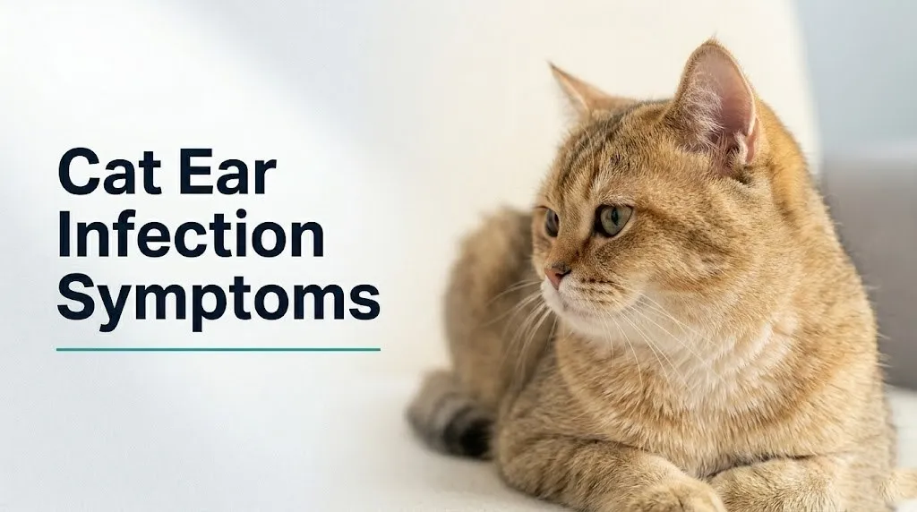
Cat ear infection symptoms range from subtle behavioral changes like increased irritability and reduced appetite to unmistakable signs like dark discharge, strong odor, and head shaking that no owner can miss. Catching them early, before infection spreads from the outer ear canal into the middle or inner ear, is the difference between a straightforward treatment course and a neurological emergency.
The American Association of Feline Practitioners recognizes ear disease as one of the most common reasons cats present for veterinary evaluation, with otitis externa (infection confined to the external ear canal) accounting for a substantial proportion of feline dermatological and ear complaints seen in general practice. What most cat owners do not realize is that cats are stoic animals that actively suppress outward signs of pain, which means the symptoms visible to you may represent a much more advanced infection than they initially appear.
This article covers every observable symptom of feline ear infection in plain language, explains the specific biological reason each symptom occurs, ranks them by severity, identifies which signs are veterinary emergencies, and tells you exactly what your veterinarian will do to diagnose and treat the condition.
Cat Ear Infection Symptoms: What Every Owner Needs to Know
Cat ear infection symptoms include head shaking, ear scratching, dark or colored discharge, ear odor, pain on ear handling, and, in severe cases, head tilt, loss of balance, and involuntary eye movements.
The full spectrum of cat ear infection symptoms varies considerably depending on which portion of the ear is affected and how advanced the infection has become. A cat with an early, localized infection of the external ear canal may show only mild occasional head shaking or a subtle reluctance to have their ears touched. A cat with an infection that has extended into the middle or inner ear may show dramatic neurological symptoms that look frightening and nothing like what most people associate with an “ear infection.”
Understanding the full symptom range matters because cats are physiologically and behaviorally wired to conceal discomfort. In their evolutionary history as both predator and prey, showing weakness is a liability. Clinical observation consistently shows that by the time a cat owner notices visible ear symptoms, the infection has often been present for days to weeks.
Complete list of cat ear infection symptoms by organ area affected:
- Repeated head shaking (most consistent early sign of ear canal irritation)
- Scratching or pawing at one or both ears, sometimes causing skin trauma around the ear base
- Dark brown, black, yellow, or reddish discharge visible at the ear canal opening
- Strong, unpleasant odor from one or both ears
- Redness or swelling visible at the ear canal opening
- Pain or flinching when the ear flap or base of the ear is touched
- Holding one ear lower than the other (early sign of discomfort)
- Behavioral changes: increased irritability, hiding, reduced grooming, reduced appetite
- Head tilt toward the affected side (middle or inner ear involvement)
- Stumbling, falling, or loss of balance (inner ear involvement)
- Rapid, involuntary eye movements (nystagmus), indicating inner ear or neurological involvement
- Third eyelid (nictitating membrane) partially covering one eye (Horner’s syndrome)
- Facial asymmetry or drooling from one side of the mouth (facial nerve involvement)
Kittens under 6 months of age with ear symptoms are more likely to have ear mites than bacterial or yeast infection, though the two can coexist, and veterinary diagnosis is the only reliable way to determine which organism is responsible.
Feline Ear Anatomy and Why Cats Get Ear Infections
The feline external ear canal is L-shaped, with a vertical portion descending from the ear opening and a horizontal portion turning toward the eardrum, and this architecture is a primary reason cats are vulnerable to ear infection development.
Think of the cat’s ear canal like a water trap under a kitchen sink: the bend in the pipe serves a purpose, but it also creates a space where material accumulates. In the cat’s case, cerumen (ear wax), shed epithelial cells, debris, and moisture collect in the deepest, horizontal portion of the canal where gravity cannot drain them and where the cat cannot self-clean. This warm, enclosed, debris-rich environment is ideal for bacterial and yeast overgrowth.
The canal lining consists of stratified squamous epithelium, which in a healthy ear performs a continuous self-cleaning function: cells migrate outward from the tympanic membrane (eardrum) toward the canal opening, carrying debris with them. When infection or inflammation develops, this epithelial migration is disrupted. Debris accumulates, inflammation thickens the canal walls, and the narrowed, inflamed canal retains moisture more effectively, worsening the cycle.
The ear canal also contains ceruminous glands (modified apocrine sweat glands) and sebaceous glands that produce the lipid and protein components of cerumen. When inflamed, these glands overproduce, generating the excess waxy or moist discharge that owners observe.
Unlike human ear canals, which are relatively straight and drain more effectively, the feline horizontal canal cannot be examined with a simple visual inspection. Owners who look into their cat’s ear from above see only the vertical canal opening. The infection source is typically deeper, in the horizontal canal near the tympanic membrane.
The Merck Veterinary Manual describes the total length of the feline external ear canal as approximately 5 to 8 centimeters in an adult cat, with the horizontal portion accounting for the majority of that length in breeds with standard ear confirmation.
Quick Tip:
- Never insert cotton swabs into a cat’s ear canal. Swabs compress debris deeper into the horizontal canal, worsening conditions for bacterial overgrowth.
- You can gently inspect the visible opening of the ear canal (the entrance to the vertical canal) for discharge or redness.
- Senior cats over 10 years may have reduced ceruminous gland function that alters cerumen composition, changing infection risk patterns compared to younger adults.
Types of Cat Ear Infections: Otitis Externa, Media, and Interna
Feline ear infections are classified by location into three types: otitis externa (outer ear canal), otitis media (middle ear), and otitis interna (inner ear), each producing a different and progressively more serious symptom profile.
Otitis externa is the most common form, confined to the external ear canal from the ear opening to the tympanic membrane. Its symptoms are the classic visible signs: discharge, odor, scratching, and redness. Most cats with otitis externa remain systemically well, eating and moving normally, though they are experiencing real discomfort in the ear.
Otitis media develops when infection extends past the tympanic membrane into the tympanic bulla, the bony middle ear cavity that houses the three tiny ossicles (malleus, incus, and stapes) responsible for transmitting sound vibrations. This extension can happen when a perforated eardrum allows organisms to migrate inward, or when infection ascends through the Eustachian tube from the nasopharynx. Otitis media produces all the symptoms of otitis externa, plus often increased pain and sometimes changes in hearing.
Otitis interna is the most serious form. Infection reaches the cochlea and semicircular canals of the inner ear, disrupting the two fundamental functions of this structure: hearing and vestibular (balance) signaling. The semicircular canals communicate spatial orientation data to the brain via the vestibulocochlear nerve (cranial nerve VIII). When this nerve is compromised by infection or inflammation, the cat loses normal balance signaling, producing the dramatic neurological symptoms described in later sections.
| Infection Type | Location | Key Symptoms | Urgency |
|---|---|---|---|
| Otitis externa | External ear canal | Discharge, odor, scratching, redness | Schedule vet within 24 to 48 hours |
| Otitis media | Middle ear (tympanic bulla) | All externa signs plus increased pain, possible hearing change | Vet appointment same day or next day |
| Otitis interna | Inner ear (cochlea, semicircular canals) | Head tilt, nystagmus, rolling, balance loss, neurological signs | Emergency veterinary evaluation immediately |
The American Association of Feline Practitioners emphasizes that otitis media and otitis interna are frequently underdiagnosed because owners and even some clinicians attribute early neurological signs to other conditions rather than recognizing them as ear-infection complications.
Early Signs of a Cat Ear Infection
The earliest signs of a cat ear infection are often behavioral rather than visible: a slight increase in head shaking, occasional pawing at one ear, and a subtle unwillingness to be touched near the ear.
These early behavioral signals are frequently dismissed as grooming behavior or personality quirks, especially in cats that are otherwise eating, drinking, and moving normally. That dismissal is understandable, because the visual signs of early otitis externa, mild redness at the canal entrance and a small amount of dark waxy material, can look similar to normal cerumen accumulation in a healthy ear.
The practical distinction is frequency and laterality. A healthy cat may briefly scratch an ear occasionally after grooming. A cat with an early ear infection tends to scratch the same ear repeatedly within a short time period, often with a focused intensity that differs from casual grooming. Scratching hard enough to produce redness or minor skin breaks at the base of the ear or on the inner ear flap is not normal grooming behavior.
According to the Cornell Feline Health Center, cats with early otitis may also show a subtle but detectable change in their response to sound on the affected side, a slight delay or reduced rotation of the affected ear toward sound sources, that attentive owners sometimes notice before other symptoms appear.
Early signs checklist for cat owners:
- Frequent head shaking, more than 3 to 4 times within a 30-minute period without an obvious trigger
- Repeated scratching at the same ear (unilateral focus is more concerning than symmetrical occasional scratching)
- Flinching or pulling away when you touch the base of the ear or the ear flap
- Small amount of dark brown or black waxy debris visible just inside the ear canal opening
- Subtle irritability when the head or ear area is approached or petted
Outdoor cats and cats in multi-cat households tend to show earlier ear mite-related symptoms than indoor single-cat household cats, because their exposure risk for Otodectes cynotis is higher.
Key Takeaway: The earliest reliable sign of a cat ear infection is repeated, focused scratching at one ear combined with resistance to ear handling, even when visible discharge or odor is not yet present.
Ear Discharge in Cats: What Color and Smell Mean
The color and texture of ear discharge in a cat with an ear infection directly reflects which organism is driving the infection, and distinguishing them helps your veterinarian choose the right diagnostic approach.
Normal feline cerumen is a small amount of light tan to pale golden waxy material with minimal odor. Anything darker, wetter, more abundant, or odorous than this baseline represents an abnormal change that warrants veterinary evaluation.
Dark brown to black, dry, crumbly discharge resembles used coffee grounds and is the hallmark of Otodectes cynotis (ear mite) infestation, though yeast overgrowth with dried exudate can look similar. The dark color comes from a combination of dried blood from mite feeding, mite feces, and cerumen oxidation. Under microscopy, ear mites and their eggs are definitively distinguishable from yeast cells.
Darker brown, waxy, moist discharge with a musty or yeasty smell indicates Malassezia pachydermatis (yeast) overgrowth. Malassezia is a lipid-dependent yeast normally present in small numbers in the healthy feline ear canal. When conditions favor its growth (warmth, moisture, excess cerumen), it proliferates rapidly, producing the characteristic rancid or musty odor from its lipid metabolism byproducts.
Yellow, greenish, or purulent (pus-like) discharge with a distinctly foul or fetid odor indicates bacterial infection, most commonly Staphylococcus pseudintermedius or, in chronic or resistant cases, Pseudomonas aeruginosa. Pseudomonas produces a particularly distinct sweet, grape-like or musty odor that experienced veterinary clinicians can sometimes identify on examination.
| Discharge Color and Texture | Likely Organism | Clinical Action |
|---|---|---|
| Light tan, minimal, waxy | Normal cerumen | Monitor; inspect for other symptoms |
| Dark brown to black, dry, crumbly | Ear mites (Otodectes cynotis) | Veterinary evaluation for diagnosis and treatment |
| Dark brown, moist, musty smell | Yeast (Malassezia pachydermatis) | Veterinary evaluation; ear cytology needed |
| Yellow, purulent, foul odor | Bacterial (Staphylococcus or Pseudomonas) | Veterinary evaluation; culture may be needed |
| Blood-tinged or bloody discharge | Trauma, severe infection, or polyp | Same-day veterinary evaluation |
Cats with bloody discharge or discharge mixed with visible blood should be evaluated the same day, as this can indicate severe infection, aural hematoma, or a polyp or mass within the ear canal.
Cat Shaking Head and Scratching at the Ear
Head shaking and ear scratching are the two most consistently observable cat ear infection symptoms because they are the cat’s direct behavioral response to pruritus (itching) and pain within the ear canal.
The physiological mechanism is straightforward. Inflamed ear canal tissue releases prostaglandin E2 and histamine from activated mast cells and the arachidonic acid inflammatory cascade, both of which are potent stimulators of the C-fiber nociceptors (itch-signaling nerve fibers) that densely innervate the ear canal skin. The cat responds to this intense, inescapable itch-pain signal the same way it would respond to any intense peripheral irritant: by attempting mechanical relief through shaking and scratching.
The problem with this response is that it compounds the injury. Repeated, forceful head shaking can rupture small blood vessels between the skin and cartilage of the ear flap, producing an aural hematoma (auricular hematoma), a pocket of blood that swells the ear flap dramatically. An aural hematoma is not a primary infection but a complication of the scratching behavior itself. It requires separate veterinary treatment, typically drainage and sometimes surgical management.
Repeated scratching with the hind claws produces excoriation (skin abrasion and breaking) of the skin behind the ear, along the ear flap margins, and at the base of the ear. These open skin areas are vulnerable to secondary bacterial skin infections, adding an additional infection site separate from the primary ear canal infection.
According to the Merck Veterinary Manual, intense, repetitive scratching focused on a single ear in a cat is one of the most reliable owner-observable indicators of unilateral otitis and should prompt veterinary evaluation within 24 hours, sooner if the cat appears to be in significant distress.
Bilateral (both ear) scratching in a kitten under 6 months strongly suggests ear mite infestation rather than primary bacterial or yeast infection, though the two conditions may coexist.
Cat Ear Infection Smell: What to Notice and Why
A noticeable odor from a cat’s ear is never normal and consistently indicates active infection, with the specific smell providing directional information about the causative organism.
Healthy cat ears should have minimal to no odor when you gently lift the ear flap and smell in the direction of the canal opening. This is a simple, owner-accessible check that takes seconds and can detect an early infection before visible discharge has fully developed.
The yeasty, musty, or rancid smell characteristic of Malassezia overgrowth is produced by the metabolic byproducts of yeast lipid metabolism. Malassezia organisms metabolize the lipid fractions of cerumen and sebum as their primary nutritional source, producing short-chain fatty acids, indoles, and volatile organic compounds as metabolic waste. These compounds are responsible for the distinctive odor.
Bacterial infections, particularly those involving Pseudomonas aeruginosa, produce a different and often more acrid or fetid odor profile. Pseudomonas generates pyocyanin and other pigmented metabolites that contribute to the distinctive smell of gram-negative bacterial ear infections. Staphylococcal infections tend to produce a sour or slightly cheesy odor from their protein-metabolizing activity.
A sweet, syrupy, or unusually strong odor from a cat’s ear that has not been present before should prompt veterinary evaluation within 24 hours. A fetid, putrid, or deeply foul odor, especially combined with thick or bloody discharge, warrants same-day evaluation.
Quick Tip:
- Perform a weekly “sniff check” by gently lifting your cat’s ear flap and smelling from an inch away from the canal opening. A change in odor from normal (minimal or neutral) to any recognizable unpleasant smell is a reliable early indicator of infection.
- Cats with confirmed diabetes mellitus may have altered cerumen composition and are at higher risk of recurrent Malassezia ear infections, making this routine check particularly valuable for diabetic cat owners.
- Never attempt to clean the ear canal with any substance (vinegar solutions, hydrogen peroxide, rubbing alcohol) without veterinary guidance, as these can damage the inflamed canal epithelium and worsen the infection environment.
Key Takeaway: A detectable odor from your cat’s ear, whether yeasty, musty, sour, or fetid, is an abnormal finding that always warrants veterinary evaluation, because healthy feline ears produce no noticeable smell.
Hidden and Atypical Cat Ear Infection Symptoms
Some cats with established ear infections show no obvious scratching or discharge at all, presenting instead with subtle behavioral changes that owners rarely connect to ear pain.
This presentation is particularly common in cats receiving systemic corticosteroids or prednisolone for other conditions (such as feline asthma, inflammatory bowel disease, or allergic skin disease). Corticosteroids suppress the prostaglandin E2 and histamine signaling that produces itching, which means the cat does not scratch, does not shake its head, and does not draw the owner’s attention to the ear. The infection progresses silently while the anti-inflammatory effect of the steroid masks the warning signs.
Behavioral changes that may represent hidden ear infection in cats include:
- Increased hiding or reduced desire to interact with household members
- Uncharacteristic aggression when touched near the head, neck, or ears
- Reduced grooming of the head and face (because touching that region causes discomfort)
- Changes in eating behavior: slower eating, dropping food, or preference for soft food (chewing movement transmits vibration to the temporomandibular joint and adjacent ear structures in middle ear disease)
- Reduced responsiveness to familiar sounds on one side (hearing loss from otitis media)
- Increased sleep or reduced activity without other obvious illness signs
Cats receiving long-term topical otic preparations (ear drops) for a previous infection may also develop a rebound presentation when treatment stops, appearing suddenly symptomatic even though the infection was never fully resolved.
According to the Journal of Feline Medicine and Surgery, veterinary clinicians specifically note that cats are more likely than dogs to have unilateral ear infections that remain clinically “quiet” for extended periods before owners notice behavioral changes, making a complete physical examination of the ear canal part of every routine wellness visit essential.
Otitis Interna in Cats: Neurological Symptoms
Otitis interna in cats produces neurological symptoms rather than the typical ear symptoms of discharge and scratching, because the infection has reached the inner ear structures that control balance and hearing.
The inner ear contains two systems within a fluid-filled membranous labyrinth housed in the bony petrous temporal bone: the cochlea (hearing) and the semicircular canals plus utricle and saccule (vestibular function). When infection or its associated inflammation reaches these structures, the fluid equilibrium and hair cell function that provide spatial orientation data to the brain are disrupted. The affected cat’s nervous system receives conflicting or absent vestibular signals, producing the clinical signs of vestibular dysfunction.
The specific neural pathway responsible is the vestibulocochlear nerve (cranial nerve VIII), which carries sensory information from both the cochlea (hearing) and the semicircular canals (balance) to the brainstem. Inflammatory edema, direct bacterial invasion, or toxic damage to this nerve or the structures it serves produces the characteristic signs of peripheral vestibular disease.
Neurological symptoms of otitis interna in cats:
- Persistent head tilt toward the affected side (the most consistent sign)
- Spontaneous nystagmus (rapid, involuntary, repetitive eye movements): horizontal, vertical, or rotary
- Falling or rolling toward the affected side
- Ataxia (loss of coordinated movement, appearing drunk or wobbly)
- Circling in tight circles toward the affected side
- Inability to stand or severe difficulty maintaining an upright position in severe cases
- Reduced or absent hearing response (cochlear involvement)
Central vestibular disease (brainstem involvement rather than peripheral ear disease) produces similar signs but with additional features: vertical nystagmus (rather than horizontal), ipsilateral or paradoxical head tilt, and signs of other cranial nerve deficits. Distinguishing peripheral from central vestibular disease requires veterinary neurological examination and often advanced imaging.
A board-certified veterinary neurologist should be involved in cases where the clinical signs suggest central rather than peripheral vestibular disease, because the treatment and prognosis differ substantially.
Cat Head Tilt and Loss of Balance from Ear Infection
A persistent head tilt in a cat toward one side, combined with loss of normal balance, is the most clinically recognizable sign that an ear infection has reached the middle or inner ear and requires immediate veterinary attention.
The head tilt produced by otitis media and otitis interna reflects asymmetric vestibular input to the brain. Under normal conditions, the paired semicircular canals in both inner ears send equal and opposite signals through CN VIII to the cerebellum and brainstem. The brain interprets this balanced input as a stable sense of position. When one inner ear is diseased or compromised, its signal is reduced or absent. The brain receives unequal input, interpreting the discrepancy as continuous tilting or movement. The cat tilts its head to “compensate” for the false signal, and this tilt is persistent as long as the vestibular dysfunction continues.
Nystagmus, the involuntary rapid eye movement that accompanies otitis interna, is produced by the same mechanism: the vestibulo-ocular reflex, which normally coordinates eye position with head movement, receives distorted input from the asymmetric vestibular signal and produces rhythmic, repetitive eye movements as it attempts to compensate for perceived continuous motion.
Think of this like a compass that has had a magnet placed next to it on one side. The needle no longer points accurately north; it is pulled consistently toward the interfering signal. A cat’s head tilt from inner ear disease is the neurological equivalent of that compass needle: the vestibular system is consistently pulled toward the side of the damaged ear.
The American Veterinary Medical Association notes that vestibular signs in cats may also be caused by feline idiopathic vestibular syndrome (also called “old cat vestibular disease”), a non-infectious condition that produces identical symptoms but resolves spontaneously in 72 hours to 3 weeks. Distinguishing idiopathic vestibular syndrome from infectious otitis interna requires veterinary examination and sometimes imaging. A cat owner cannot make this distinction without professional evaluation.
Young adult cats presenting with sudden head tilt after any period of ear symptoms should be seen the same day.
Key Takeaway: A cat with a persistent head tilt toward one side and any degree of balance loss has vestibular dysfunction that requires same-day veterinary evaluation, because distinguishing inner ear infection from other serious neurological causes requires professional examination and imaging, not home monitoring.
Cat Ear Infection vs Ear Mites: How to Tell the Difference
Ear mites and bacterial or yeast ear infections in cats produce overlapping symptoms, but several characteristics help distinguish them, and definitive differentiation requires veterinary ear cytology.
Both conditions produce head shaking, ear scratching, and discharge. The characteristics of the discharge, the pattern of affected animals in the household, and the cat’s age and lifestyle provide the most useful differentiating information at home, though these are directional clues rather than definitive diagnostic answers.
Otodectes cynotis (ear mites) are the most common cause of ear disease in kittens and young cats, particularly those from multi-cat environments, shelters, or with outdoor access. Ear mites are highly contagious between cats in the same household. If one cat in a household develops ear symptoms, other cats sharing the same environment will frequently show similar signs within days to weeks, because mites transfer readily through direct contact. Dogs in the household can also carry Otodectes, though they are less commonly symptomatic.
The discharge from ear mite infestation is classically dark brown to black, dry, granular, and described as resembling coffee grounds. It typically fills the visible canal opening. The discharge from yeast or bacterial infection is more likely to be moist, pasty, or liquid, and may be lighter brown, yellowish, or greenish depending on the organism.
| Feature | Ear Mites (Otodectes cynotis) | Yeast or Bacterial Infection |
|---|---|---|
| Age group most affected | Kittens, young cats, outdoor cats | Any age, most commonly adults |
| Discharge appearance | Dark brown/black, dry, coffee-ground-like | Moist, waxy, pasty, variable color |
| Spread to other pets | High; highly contagious | Not contagious between pets |
| Odor | Minimal to mild earthy | Often notable: musty, sour, or fetid |
| Bilateral presentation | Usually both ears affected | May be one or both ears |
| Definitive diagnosis | Microscopy: mites and eggs visible | Microscopy: yeast cells, bacteria, neutrophils |
Cats living exclusively indoors with no contact with other animals have a low probability of ear mite infestation but can still develop bacterial or yeast infections from environmental factors and altered immune function.
What Causes Cat Ear Infections
Cat ear infections are caused by overgrowth of organisms normally present in small numbers in the ear canal, triggered by changes in the ear environment that favor their proliferation over the host’s ability to suppress them.
The three most common causative categories, in order of clinical frequency according to the Merck Veterinary Manual, are: parasitic infestation (primarily Otodectes cynotis), yeast overgrowth (primarily Malassezia pachydermatis), and bacterial infection (Staphylococcus pseudintermedius most commonly in primary infections, Pseudomonas aeruginosa in chronic or treatment-refractory cases).
The ear canal environment that enables these organisms to overgrow is shaped by several factors. Moisture retention in the horizontal canal from water exposure, excessive ear cleaning, or anatomical narrowing provides the warm, humid conditions that both yeast and bacteria prefer. Excess cerumen production from inflamed ceruminous glands provides nutritional substrate. Disruption of the normal epithelial migration self-cleaning mechanism allows debris accumulation.
Secondary causes that are less common but clinically important:
- Nasopharyngeal polyps: benign inflammatory masses that originate from the Eustachian tube or middle ear mucosa and extend into the ear canal or nasopharynx; they are seen most often in young cats and cause mechanical obstruction of the canal rather than infectious inflammation
- Allergic skin disease (feline atopic dermatitis or food allergy): chronic inflammation of the ear canal skin alters the microenvironment and predisposes to secondary yeast or bacterial overgrowth
- Foreign bodies (grass seeds, plant material) lodged in the vertical canal
- Neoplasia (ear canal tumors): less common but occurring in older cats, producing unilateral discharge and sometimes bleeding
- Systemic immunosuppression from FIV, FeLV, or medication
Cats on long-term immunosuppressive medication for autoimmune or inflammatory conditions have a higher rate of recurrent or difficult-to-resolve ear infections, because the same immune suppression that controls their primary disease also reduces local ear canal defense against opportunistic organisms.
Breeds and Individual Cats at Highest Risk
Certain cat breeds have anatomical or physiological characteristics that place them at substantially higher risk for ear infections, and individual health factors including immune status and age further modify that risk.
Persian cats are at elevated risk due to their brachycephalic (flat-faced) skull conformation. The compressed craniofacial anatomy alters the geometry of the ear canal relative to the skull base, which may impair normal cerumen drainage and ventilation. Persians also tend to have naturally oilier skin with higher sebaceous gland activity, which provides additional nutritional substrate for Malassezia overgrowth in the ear canal.
Scottish Fold cats carry a mutation causing Osteochondrodysplasia, a systemic cartilage abnormality affecting all cartilaginous structures, including the auricular cartilage that forms the ear canal walls. The altered cartilage in Scottish Fold cats may affect ear canal compliance and drainage, and these cats often require more frequent veterinary monitoring for ear health across their lifetime.
Cornish Rex and Devon Rex cats have altered coat structure (reduced or absent guard hairs) that is accompanied by differences in ceruminous gland secretion. Their cerumen composition may create a more favorable environment for Malassezia colonization compared to standard-coated cats.
Individual risk factors beyond breed:
- FIV-positive cats: the feline immunodeficiency virus depletes CD4+ T lymphocytes progressively, reducing adaptive immune surveillance of the ear canal; these cats are at high risk for chronic, recurrent, and treatment-resistant ear infections
- FeLV-positive cats: feline leukemia virus causes bone marrow suppression and immune dysfunction; infected cats have impaired neutrophil and lymphocyte function that compromises their ability to clear ear canal infections
- Kittens under 6 months: maternal immunity provides some protection initially, but wanes by 6 to 8 weeks; ear mites are the primary risk in this age group because Otodectes transmits readily from queen to offspring
- Senior cats over 10 years: immune senescence reduces pathogen clearance efficiency; chronic otitis is more common in geriatric cats and more likely to involve atypical organisms or resistant strains
A board-certified veterinary dermatologist should be involved in managing ear infections in breeds with known structural predispositions, and in any cat with confirmed recurrent infection (more than two episodes per year despite completed treatment courses).
Emergency Signs: When to Take Your Cat to the Vet Immediately
Certain cat ear infection symptoms require immediate emergency veterinary evaluation. Do not wait for a routine appointment if your cat shows any of the following signs.
Take your cat to an emergency veterinary hospital or call your licensed veterinarian immediately if you observe:
- Persistent head tilt toward one side that appeared suddenly (within hours)
- Falling, rolling, or inability to stand and walk normally
- Rapid, involuntary, repetitive eye movements (nystagmus): eyes moving back and forth or rotating continuously
- Third eyelid (the pale tissue at the inner corner of the eye) protruding and covering part of one eye, especially when combined with a smaller pupil on that side (signs of Horner’s syndrome)
- Facial asymmetry: drooping of one side of the face, inability to blink one eye fully, saliva drooling from one side of the mouth (signs of facial nerve/cranial nerve VII involvement)
- Complete loss of balance or collapse when attempting to walk
- Circling continuously in one direction, particularly tight circles toward one side
- Sudden loss of hearing response on one or both sides
- Bloody or blood-mixed discharge from the ear canal
- Extreme pain when the head or ear area is touched, combined with any other neurological sign
- Rapid swelling of the ear flap (aural hematoma developing from vigorous scratching)
These presentations indicate that infection may have extended to the middle ear, inner ear, or adjacent neurological structures and require emergency veterinary assessment, not a scheduled appointment.
If you are uncertain whether your cat’s symptoms meet emergency criteria, call your nearest 24-hour emergency veterinary hospital or your regular veterinarian immediately for telephone guidance. Do not apply any substance, drops, or cleansers to the ear canal before the veterinary examination, as this can obscure diagnostic findings.
How Vets Diagnose Cat Ear Infections
Veterinarians diagnose cat ear infections through a systematic examination sequence that begins with visual assessment and progresses to microscopic analysis, with additional imaging or laboratory testing when middle or inner ear disease is suspected.
The diagnostic process at a veterinary visit:
- History and behavioral assessment: the veterinarian asks when symptoms began, whether the cat has had previous ear infections, what medications the cat is currently receiving, whether other pets in the household are affected, and whether the cat is indoor-only or has outdoor access
- General physical examination: body temperature, lymph node assessment, neurological screening including gait evaluation and cranial nerve testing, overall body condition and hydration assessment
- Otoscopic examination: a handheld otoscope or video-otoscope is inserted into the vertical canal to visualize the canal walls, the tympanic membrane (if visible), and the type and distribution of discharge; this examination can be uncomfortable for a cat with painful otitis, and sedation may be required for complete examination
- Ear swab collection: a sterile swab is introduced into the canal and rotated to collect a representative sample of discharge; the swab is rolled onto a glass slide
- Cytology (microscopic examination): the slide is stained (typically with Diff-Quik or similar rapid stain) and examined under 40x and 100x magnification to identify and quantify yeast cells (Malassezia), bacteria (rods vs. cocci, suggesting gram-negative vs. gram-positive species), mites and mite eggs (Otodectes), and inflammatory cells (neutrophils, macrophages)
- Culture and sensitivity testing: ordered when cytology shows heavy bacterial infection, when the infection has not responded to previous treatment, or when rod-shaped bacteria (suggesting Pseudomonas or other gram-negative species) are seen on cytology; identifies the specific organism and determines which antibiotics will be effective
- Imaging (skull radiographs or CT scan): performed when otitis media or interna is suspected, to visualize the tympanic bulla for fluid, thickening, or bony changes; CT imaging provides substantially more anatomical detail than plain radiographs for this region
The Journal of Feline Medicine and Surgery has reported that ear cytology is the single most clinically impactful diagnostic test for feline otitis, because it directly guides treatment selection by identifying the causative organism type before any treatment begins.
Cat Ear Infection Treatment and Home Monitoring
Cat ear infection treatment is determined by the causative organism and the anatomical location of infection, ranging from topical otic preparations for uncomplicated otitis externa to systemic antibiotic or antifungal therapy and surgical flushing for middle ear disease.
For uncomplicated otitis externa caused by yeast (Malassezia), veterinarians typically prescribe topical otic preparations containing an antifungal agent such as clotrimazole or miconazole, often combined with a corticosteroid to reduce canal inflammation and swelling. Treatment courses typically run 7 to 14 days. For bacterial otitis externa, the choice of antibiotic component in the topical otic preparation is guided by the cytology findings (gram-positive versus gram-negative bacteria) and, for resistant cases, by culture and sensitivity results.
Ear mite infestation is treated with acaricide agents: topical ear preparations containing pyrethrins or ivermectin, or systemic parasiticide agents applied topically to the skin (selamectin or moxidectin formulations). All cats in the household must be treated simultaneously, because untreated household contacts will reinfect treated cats.
Otitis media typically requires systemic antibiotic therapy in addition to topical ear treatment, because topical agents cannot reliably penetrate the tympanic bulla. Treatment courses for otitis media are substantially longer than for externa, often 4 to 8 weeks. In cases with tympanic bulla involvement confirmed by imaging, veterinary flushing of the bulla under anesthesia (myringotomy and bulla lavage) may be necessary.
Home monitoring during treatment:
- Apply all prescribed medications for the full prescribed duration, even if symptoms appear to resolve early; stopping treatment before the ear canal is fully cleared allows organisms to regrow and develop resistance
- Monitor for symptom improvement within 5 to 7 days of treatment initiation; if symptoms are not improving or are worsening, contact your veterinarian for reassessment
- Return for the scheduled follow-up appointment; the veterinarian will repeat ear cytology to confirm resolution rather than clinical improvement alone
- Watch for new onset of neurological signs (head tilt, balance problems) during treatment, which could indicate progression to inner ear involvement, requiring same-day contact with your veterinarian
The American Association of Feline Practitioners recommends that cats with more than two ear infection episodes within a 12-month period be evaluated for underlying primary causes such as allergic skin disease, hypothyroidism (rare in cats), anatomical abnormality, or systemic immune compromise, because treating each episode in isolation without addressing the predisposing factor results in ongoing recurrence.
Key Takeaway: A full course of veterinary-prescribed treatment with a follow-up cytology check, not just symptom observation, is the standard of care for cat ear infections, because clinical improvement does not reliably confirm organism clearance at the microscopic level.
Frequently Asked Questions About Cat Ear Infection Symptoms
What are the first signs of a cat ear infection?
The first signs of a cat ear infection are usually repeated head shaking, focused scratching at one ear, and mild reluctance to have the ear touched, often before visible discharge or odor develops.
Behavioral changes such as reduced interaction, increased hiding, or irritability when touched near the head may appear before any visible ear symptoms in cats that are suppressing pain.
Any of these early signs warrant a veterinary appointment within 24 to 48 hours, because early treatment prevents progression to middle or inner ear involvement.
Can a cat ear infection go away on its own?
Cat ear infections do not reliably resolve without treatment, and delaying care allows the infection to progress deeper into the ear canal, increasing the risk of middle or inner ear involvement.
The Merck Veterinary Manual consistently recommends veterinary evaluation for all suspected ear infections in cats, because the causative organism cannot be identified without cytology, and treatment without knowing the organism leads to treatment failure.
Attempting to manage a cat ear infection at home with over-the-counter products or home remedies can worsen the canal environment and obscure diagnostic findings at the veterinary examination.
What does cat ear infection discharge look like?
Cat ear infection discharge varies by causative organism: dark brown to black, dry, and crumbly resembles coffee grounds and suggests ear mites; moist, dark brown, and waxy with a musty odor suggests yeast overgrowth; yellow, green, or purulent with a foul odor indicates bacterial infection.
Any discharge that appears different from a small amount of light tan cerumen, or that is accompanied by odor, is abnormal and warrants veterinary evaluation.
Bloody or blood-mixed discharge requires same-day veterinary assessment.
How do I know if my cat’s ear infection has spread to the inner ear?
Signs that a cat ear infection has spread to the inner ear include persistent head tilt toward one side, loss of normal balance or stumbling, rapid involuntary eye movements (nystagmus), and difficulty walking in a straight line.
These neurological signs appear because the infection has compromised the semicircular canals and vestibulocochlear nerve (cranial nerve VIII) that control spatial orientation and balance.
A cat showing any of these signs needs same-day emergency veterinary evaluation, not a routine appointment.
Is a cat ear infection an emergency?
A cat ear infection is a veterinary emergency if the cat shows neurological signs including head tilt, rolling, nystagmus, facial asymmetry, or third eyelid protrusion, because these indicate middle or inner ear involvement.
Uncomplicated otitis externa (outer ear infection only) with discharge and scratching but no neurological signs requires a veterinary appointment within 24 to 48 hours rather than emergency-level urgency.
When in doubt about whether your cat’s symptoms are emergency-level, call an emergency veterinary hospital immediately for telephone triage guidance.
How long does a cat ear infection take to clear up with treatment?
Uncomplicated yeast or bacterial otitis externa typically shows clinical improvement within 5 to 7 days of prescribed topical treatment, with full resolution confirmed by follow-up ear cytology at 14 to 21 days.
Otitis media requires substantially longer treatment, often 4 to 8 weeks of systemic antibiotic therapy, with repeat imaging to confirm tympanic bulla resolution.
Ear mite treatment requires a full acaricide course of 3 to 4 weeks in most cases, with simultaneous treatment of all household cats to prevent reinfection.
Closing
Cat ear infection symptoms tell a clear story if you know what to look for. Head shaking and ear scratching are the starting point. Discharge color and smell give you directional information about the organism. Neurological signs mean the infection has moved deeper and the clock is running.
The most important thing a cat owner can take from this article is the urgency distinction: outer ear symptoms warrant a veterinary appointment within 24 to 48 hours; neurological symptoms (head tilt, rolling, nystagmus, facial asymmetry) require same-day emergency veterinary care. That distinction could determine whether your cat makes a full recovery or sustains permanent vestibular damage from a treatable infection.
If your cat is showing any of the symptoms described here, contact a licensed veterinarian. Bring notes on when the symptoms started, which ear is affected, what the discharge looks like, and any medications your cat is currently receiving. That information will allow your veterinarian to move directly to targeted diagnosis rather than starting from scratch.