White Coating on Tongue Causes: Full 2026 Clinical Guide
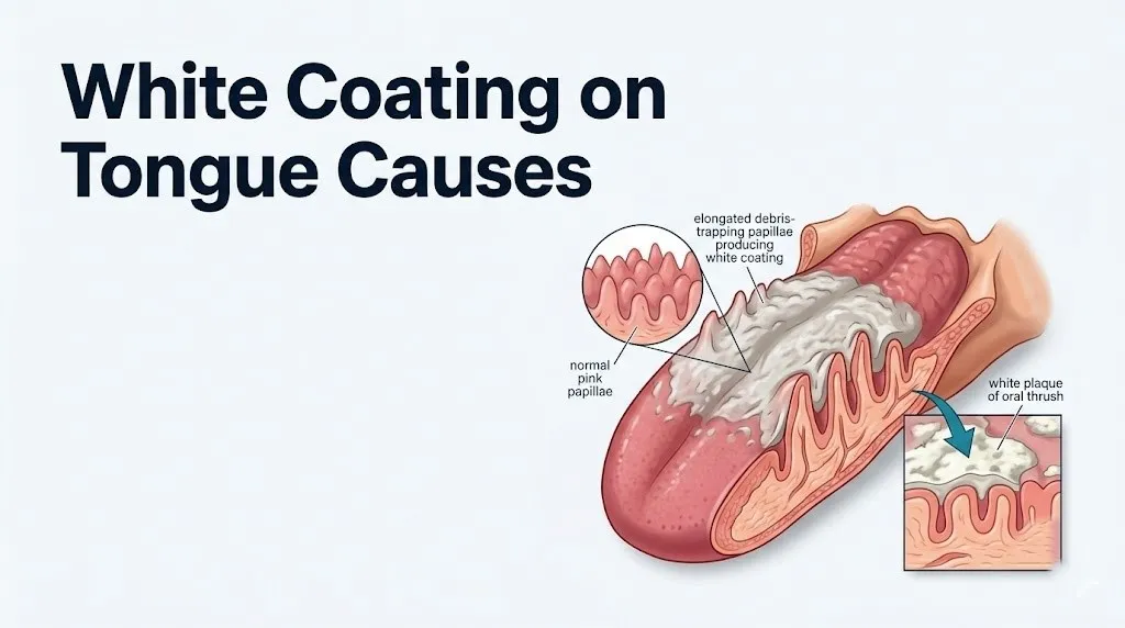
A white coating on tongue surfaces forms when filiform papillae (the hair-like projections covering the dorsal tongue surface) elongate and trap layers of dead epithelial cells, bacteria, and debris that saliva and tongue movement would normally clear, or when a specific infection, immune condition, or cell growth abnormality independently produces white patches on the oral mucosa. The specific cause ranges from entirely benign and self-resolving (dehydration, poor oral hygiene, recent antibiotic use) to conditions requiring urgent professional evaluation (leukoplakia, secondary syphilis, oral cancer precursor lesions).
According to the National Institute of Dental and Craniofacial Research (NIDCR), oral mucosal diseases collectively affect a substantial proportion of the adult population, and white patches on the tongue and oral mucosa are among the most frequently observed oral findings in clinical dental and medical practice. The clinical significance of those findings spans from completely inconsequential to a potential sign of oral cancer, which is precisely why the cause matters so much.
This article covers every established cause of white tongue, the specific cellular and physiological mechanism behind each, who is at higher risk for each cause, how to distinguish benign from concerning presentations, and when a white coating on the tongue requires professional evaluation by a named specialist.
White Coating on Tongue Causes: The Short Answer
The most frequent causes of a white coating on the tongue are dehydration, poor oral hygiene, mouth breathing, recent antibiotic use, and oral candidiasis (oral thrush), with less common but clinically important causes including leukoplakia, oral lichen planus, secondary syphilis, and smoking-related changes to oral mucosa.
The vast majority of white tongue presentations are benign and resolve with improved hydration, better oral hygiene, and treatment of any underlying infection. The minority that represent a genuine health concern are identifiable by specific clinical features: most importantly, whether the white coating wipes off cleanly (benign coating or oral thrush) or does not wipe off (leukoplakia, scarring, lichen planus, or neoplastic change).
| Cause | Wipeable? | Urgency | Who Is Most At Risk |
|---|---|---|---|
| Dehydration / dry mouth | Yes | Self-resolving | Anyone; mouth breathers, elderly |
| Poor oral hygiene | Yes | Self-resolving | Anyone with irregular tongue cleaning |
| Oral thrush (Candida) | Yes (reveals red base) | Treat promptly | Antibiotic users, immunocompromised, infants |
| Antibiotic-related dysbiosis | Yes | Self-resolving or treat | Anyone after broad-spectrum antibiotics |
| Leukoplakia | No | Urgent evaluation | Smokers, tobacco users, alcohol users, older adults |
| Geographic tongue | Partially (borders stable) | Monitor | Adults, stress-related flares |
| Oral lichen planus | No | Evaluate and monitor | Middle-aged adults, women slightly more |
| Secondary syphilis | Yes or no | Urgent testing | Sexually active adults |
| Hairy leukoplakia (EBV) | No | Evaluate | HIV-positive individuals |
| Nutritional deficiency (pale) | Not coating per se | Evaluate | Vegans, elderly, malabsorption conditions |
According to the Mayo Clinic, most cases of white tongue are benign and resolve without medical treatment. The clinical threshold that shifts a white tongue from “monitor and maintain hygiene” to “see a professional this week” is primarily whether the white patch persists beyond 2 weeks without improvement.
How the Tongue Produces a White Coating
A white coating forms on the tongue when the normal self-cleaning cycle of the dorsal tongue surface breaks down: specifically, when filiform papillae stop shedding their surface keratinocytes fast enough, and saliva stops washing away the accumulated debris effectively enough, to keep the tongue surface pink and visible.
Think of filiform papillae like the carpet fibers of a rug. When the carpet is used and vacuumed regularly, debris doesn’t accumulate visibly. When the carpet sits in a corner, unused, and the vacuum isn’t run, debris builds up between the fibers and the surface looks dull and grey. The tongue’s papillae work the same way: normal eating, drinking, and tongue movement against the palate are the daily “vacuuming” that keeps the surface clear.
Filiform papillae are the most numerous papillae on the tongue, covering most of the dorsal (top) surface in dense rows. Each papilla is a column of stratified squamous epithelium topped with a keratinized tip. Normally, the basal layer of the papilla continuously produces new keratinocytes that push the older surface cells outward, where they desquamate (shed). Saliva from the parotid gland (which opens near the upper second molar) and the submandibular and sublingual glands mechanically irrigates the tongue surface with each swallow, carrying shed cells and debris into the pharynx.
When salivary flow decreases, keratinocyte desquamation rate slows, or the tongue moves less than normal (during illness, fasting, or sleep), filiform papillae elongate and accumulate bacteria, dead cells, and food particles within their projections. The coating appears most prominently on the posterior third of the dorsal tongue because this region is farthest from the parotid duct opening and receives the least effective salivary irrigation.
Older adults experience this cycle more acutely than younger adults because salivary gland output declines with age and polypharmacy (the use of multiple medications) increases xerostomia risk dramatically. Individuals with reduced tongue mobility from neurological conditions, post-surgical swallowing difficulty, or mechanical ventilation show particularly pronounced coating due to the complete absence of the mechanical tongue-palate contact that normally clears papillae debris.
Dehydration and Dry Mouth as Causes of White Tongue
Dehydration and dry mouth (xerostomia) are the most prevalent causes of a white coating on the tongue across all age groups, operating through the same basic mechanism: reduced salivary flow diminishes the mechanical irrigation that normally clears dead epithelial cells, bacteria, and debris from the elongated filiform papillae.
Saliva is not merely a digestive fluid. It is the primary antimicrobial and mechanical defense mechanism of the oral cavity. Each of the three major salivary gland pairs (parotid, submandibular, sublingual) produces saliva with a specific composition: the parotid produces predominantly serous saliva rich in secretory immunoglobulin A (sIgA) and amylase, while the sublingual glands produce mucous saliva that lubricates mucosal surfaces and supports film-barrier protection. When salivary output falls, both the mechanical cleansing function and the antimicrobial protection of saliva are simultaneously reduced.
Dehydration reduces saliva production through two pathways: reduced plasma volume decreases the hydrostatic pressure available for salivary acinar cell secretion, and osmoreceptors in the hypothalamus trigger antidiuretic hormone (ADH) release, which simultaneously promotes water retention systemically and reduces fluid available for salivary secretion. The result is a dry, tacky oral environment where saliva is too viscous to flow effectively over the tongue surface.
Xerostomia specifically refers to the subjective sensation of dry mouth, which commonly results from medications rather than true dehydration. More than 400 medication classes are known to cause dry mouth as a side effect, including antihistamines, antidepressants (particularly tricyclics and SSRIs), antihypertensives, diuretics, and anticholinergic agents. According to the American Dental Association (ADA), older adults taking multiple medications are the population most severely affected by medication-induced xerostomia and the associated white tongue coating it produces.
Quick Tip:
- Drinking at least 8 cups of water daily supports adequate salivary flow for tongue surface clearance.
- Sleeping with your mouth open (from nasal congestion or habitual mouth breathing) dramatically reduces overnight salivary irrigation, explaining why white tongue is often most pronounced in the morning.
- Individuals with Sjögren’s syndrome (an autoimmune condition causing salivary and lacrimal gland destruction) experience severe chronic xerostomia and are at disproportionately elevated risk for both white coating and oral Candida colonization, and should be monitored by their rheumatologist and a board-certified dentist or oral medicine specialist for oral mucosal changes.
Key Takeaway: White coating on the tongue is most commonly caused by reduced saliva flow allowing filiform papillae to elongate and trap debris. The coating is heaviest on the posterior tongue where salivary irrigation is weakest.
Poor Oral Hygiene Causing White Tongue
Poor oral hygiene causes a white tongue coating when inadequate mechanical cleaning of the tongue surface allows bacteria, dead cells, and food particles to accumulate progressively within the filiform papillae over days to weeks, producing a thickening white-to-grey coating that worsens without intervention.
The tongue surface is the single largest reservoir of oral bacteria in the human mouth. A 2019 study published in the Journal of Oral Microbiology confirmed that the posterior dorsal tongue harbors the highest bacterial load of any oral surface, with the most coating-associated bacterial genera including Streptococcus, Prevotella, Fusobacterium, and Porphyromonas species dominating the papillary debris layer. These bacteria thrive in the low-oxygen, moist environment created within elongated papillary projections.
The coating from poor oral hygiene is almost always wipeable and lacks the specific clinical features of fungal infection (no erythematous base when wiped) or mucosal disease (no persistent underlying redness, ulceration, or induration). It also produces a notable clinical side effect: volatile sulfur compound (VSC) production from anaerobic bacteria within the papillae is the primary cause of halitosis (bad breath) associated with a white-coated tongue. When patients present complaining of persistent bad breath and notice a white tongue simultaneously, the two symptoms almost always share the same root cause.
Daily tongue scraping removes significantly more bacteria from the tongue surface than tooth brushing alone reaches. A 2022 Cochrane Review on tongue scraping interventions found that consistent mechanical tongue cleaning reduces VSC-based halitosis and tongue coating scores in short-term trials, though the evidence base for specific long-term oral health outcomes beyond halitosis remains limited.
Individuals who are hospitalized, bedridden, or receiving tube feeding are at high risk for severe hygiene-related tongue coating because they lack the mechanical self-cleaning that eating and drinking provide. Nursing home residents in particular are frequently observed to have pronounced white tongue coating from a combination of reduced mobility, polypharmacy-induced xerostomia, and inadequate daily oral hygiene care. Oral hygiene protocols for this population should include tongue cleaning as a standard component, according to American Dental Association guidelines for elderly patient oral care.
Oral Thrush as a Cause of White Coating on Tongue
Oral thrush (oral candidiasis, ICD-10: B37.0) is a fungal infection of the oral mucosa caused by the overgrowth of Candida albicans from its normal commensal yeast state into its pathogenic pseudohyphal and hyphal forms, producing creamy-white, wipeable plaques on the tongue and oral mucosa that reveal a red, inflamed base when removed.
Candida albicans is normally present in the oral cavity of approximately 40 to 60% of healthy adults as a harmless commensal organism, according to U.S. National Library of Medicine (NLM) MedlinePlus data. Its growth is continuously suppressed by salivary sIgA, epithelial antimicrobial peptides (defensins and histatins), and the competitive presence of normal oral bacteria that occupy the same ecological niche. When any of these suppression mechanisms fails, Candida transitions from its yeast morphotype to its more invasive pseudohyphal and hyphal forms through a morphological switch driven by temperature, pH, and CO2 changes in the oral environment.
The hyphal form is the clinically important one. Candida hyphae penetrate the superficial keratin layer of the oral epithelium, secreting aspartyl proteases that degrade local tissue proteins and allow deeper invasion. The white plaque that forms represents the accumulated Candida hyphae, desquamated epithelial cells, and inflammatory exudate sitting on top of the viable (and inflamed, hence red) underlying epithelium. This is why oral thrush plaques wipe off: the pathology is above the viable cell layer, not within it.
The wipeable white plaque with an erythematous (red) base when removed is the single most clinically reliable distinguishing feature of oral thrush compared to other white tongue causes. No other cause of white coating produces this specific combination.
Individuals using inhaled corticosteroids for asthma or COPD are at elevated oral candidiasis risk because aerosolized steroid particles deposit on the pharyngeal and laryngeal mucosa, producing localized immunosuppression in that region. This risk is substantially reduced by rinsing the mouth with water and spitting after each inhaled corticosteroid dose, which is a standard recommendation in the British National Formulary and by the American Academy of Allergy, Asthma, and Immunology.
Antibiotics and White Coated Tongue Causes
Antibiotic use causes a white tongue coating through a mechanism called oral microbiome dysbiosis: broad-spectrum antibiotics eliminate the diverse bacterial community that normally suppresses Candida albicans growth, creating a vacant ecological niche in the oral cavity that Candida rapidly colonizes and expands into.
The oral microbiome in a healthy adult contains approximately 700 bacterial species in a dynamic equilibrium. The streptococcal and other gram-positive species that dominate the tongue surface directly compete with Candida for epithelial adhesion sites and nutritional substrates. When a broad-spectrum antibiotic (such as amoxicillin-clavulanate, clindamycin, fluoroquinolones, or cephalosporins) is taken systemically, it reaches salivary concentrations sufficient to suppress this bacterial community throughout the oral cavity. With competing bacteria eliminated, Candida expands from its low-level commensal state to the clinically visible white coating of oral candidiasis within days of antibiotic initiation.
Antibiotic-associated oral candidiasis is self-limiting in otherwise healthy adults once the antibiotic course ends, as the normal oral bacterial community re-establishes. However, in individuals who are also immunosuppressed, diabetic, or elderly, the re-establishment of the normal oral microbiome may not occur automatically, and persistent oral thrush following antibiotic cessation warrants evaluation and treatment by a primary care physician.
Not all post-antibiotic white tongue is candidiasis. Some individuals develop a coated tongue after antibiotics from the combination of reduced eating during illness, disrupted tongue movement, and antibiotic effects on salivary gland bacterial populations, without frank fungal infection. This coating is wipeable and produces no erythematous base, distinguishing it from candidiasis.
People with Type 1 or Type 2 diabetes are at disproportionately elevated risk for antibiotic-triggered oral candidiasis because elevated blood glucose concentrations increase oral glucose levels, providing additional carbon substrate for Candida growth at the very time when bacterial competition has been removed by antibiotic therapy.
Key Takeaway: Antibiotics cause white tongue by eliminating the normal oral bacteria that compete with Candida for space in the mouth. In otherwise healthy adults this resolves when the antibiotic course ends. In diabetic or immunocompromised individuals, it warrants treatment.
Leukoplakia and White Patches on Tongue
Leukoplakia (ICD-10: K13.21) is a white patch on the oral mucosa or tongue surface that cannot be wiped off and cannot be clinically attributed to any other identifiable cause, representing a potentially premalignant condition that requires biopsy and histopathological evaluation to determine the degree of epithelial dysplasia.
The word leukoplakia means simply “white plaque” in Greek, but its clinical significance lies in what it represents at the cellular level. Unlike a white coating from debris accumulation or fungal infection, leukoplakia reflects an intrinsic change in the oral mucosal epithelium itself: hyperkeratosis (abnormal thickening of the keratinized surface layer) combined with varying degrees of epithelial dysplasia (cellular atypia and disordered maturation in the epithelial layers). The white appearance comes from the thickened keratin surface layer, which prevents the underlying blood vessels from showing the normal pink color through it.
According to data published in the Journal of Oral Pathology and Medicine, the malignant transformation rate of oral leukoplakia ranges from 1% to 17% depending on histopathological grade, location (lateral tongue and floor of mouth carry higher transformation risk than other sites), and the presence of tobacco and alcohol exposure. Leukoplakia on the lateral border of the tongue or floor of the mouth in a tobacco and alcohol user represents a clinical situation requiring urgent biopsy and evaluation by a board-certified oral and maxillofacial surgeon or oral medicine specialist.
The non-wipeable nature of leukoplakia is the single most important clinical distinguishing feature. Any white patch on the tongue or oral mucosa that cannot be removed by firm wiping with a gauze pad warrants professional evaluation within 2 weeks, per American Academy of Oral Medicine clinical guidelines.
Leukoplakia risk is dramatically higher in tobacco users (smokers and smokeless tobacco users), heavy alcohol users (with tobacco and alcohol showing synergistic carcinogenic effect on oral mucosa), and adults over 50. Males are affected approximately twice as frequently as females in most epidemiological surveys, though rates in females are increasing with changing tobacco use patterns.
Geographic Tongue and White Coating
Geographic tongue (benign migratory glossitis, ICD-10: K14.1) is an inflammatory condition of the tongue in which patches of filiform papillae undergo acute atrophy, producing smooth, red, map-like areas on the dorsal tongue surrounded by elevated white or yellowish-white irregular borders that migrate across the tongue surface over days to weeks.
The white component of geographic tongue is the raised, slightly keratinized border surrounding each area of papillary atrophy, rather than a generalized white coating. These white borders consist of a mixed inflammatory infiltrate beneath thickened epithelium at the margin of the atrophic area. Within the red zones, filiform papillae have been shed and the epithelium has thinned, revealing the underlying vascular lamina propria and giving the area its characteristic red appearance.
The mechanism of geographic tongue involves T-lymphocyte-mediated inflammation that targets filiform papillae for atrophy. The specific trigger remains incompletely characterized, but the condition is associated with psoriasis (sharing several immune pathway similarities), atopic dermatitis, and stress. It is not caused by infection, it is not contagious, and it carries no malignant transformation risk, distinguishing it categorically from leukoplakia.
According to the Mayo Clinic, geographic tongue affects approximately 1 to 3% of the general population and is typically more noticeable during periods of stress, hormonal change, or certain food exposures. The white borders of geographic tongue do shift position over days to weeks (hence “migratory”), which is a characteristic feature that distinguishes it from fixed white patches.
Geographic tongue can cause discomfort or sensitivity to spicy, acidic, or highly seasoned foods during flares. It is more frequently observed in women during hormonal fluctuations and in individuals with a personal or family history of psoriasis. No specific treatment reverses the condition, though avoiding trigger foods during symptomatic periods and using over-the-counter topical anesthetics (for context, not as a recommendation) may reduce discomfort.
Oral Lichen Planus and White Tongue
Oral lichen planus (ICD-10: L43.8) is a chronic autoimmune inflammatory condition of the oral mucosa produced by CD8+ cytotoxic T-lymphocyte-mediated attack on basal keratinocytes of the oral epithelium, producing characteristic white lace-like patterns (Wickham’s striae), white plaques, or erosive ulcerations on the tongue, buccal mucosa, and gingiva.
The white appearance of oral lichen planus results from the thickened, hyperkeratotic epithelial surface produced in response to the subepithelial inflammatory infiltrate. The classic reticular pattern (interlacing white lines called Wickham’s striae) on the buccal mucosa is pathognomonic enough for an experienced clinician to recognize clinically, but tongue presentation can be more variable: white plaques on the dorsal tongue, white erosions on the lateral tongue, or diffuse whitening of the tongue dorsum without the classic striae pattern.
Unlike geographic tongue, oral lichen planus does not migrate and does not resolve spontaneously without treatment. It is classified as a potentially malignant disorder by the World Health Organization, with an estimated malignant transformation rate of approximately 1% per year across large cohort studies, making long-term monitoring by a board-certified oral medicine specialist an essential component of management.
Research published in the British Dental Journal has confirmed that the erosive variant of oral lichen planus (where erosions and ulcerations accompany the white striations) produces considerably more discomfort than the reticular variant and carries a somewhat higher malignant transformation risk, particularly when lesions are located on the lateral tongue.
Oral lichen planus affects women somewhat more frequently than men and most commonly presents in adults over 40, with peak incidence in the fifth and sixth decades of life. It is also associated with hepatitis C virus infection (the mechanism is thought to involve molecular mimicry between hepatitis C viral antigens and basal keratinocyte surface proteins, triggering T-cell cross-reactivity). Any adult presenting with oral lichen planus who has not had hepatitis C serology should have it tested through their primary care physician.
Key Takeaway: Oral lichen planus white patches do not wipe off, do not migrate, and carry a roughly 1% per year malignant transformation risk, making ongoing monitoring by an oral medicine specialist essential rather than optional.
Syphilis Causing White Tongue Patches
Secondary syphilis (Treponema pallidum infection, ICD-10: A51.39) can cause mucous patches on the tongue and oral mucosa: painless, slightly raised, grayish-white lesions that can closely mimic oral thrush or leukoplakia and are highly infectious through oral contact.
The mucous patches of secondary syphilis appear during the disseminated phase of Treponema pallidum infection, typically 2 to 10 weeks after the primary chancre (which may have been painless and unnoticed, particularly on internal mucosal surfaces). These oral lesions represent direct spirochetal invasion of the oral mucosa with associated inflammatory response, producing the characteristic white-grey, slightly eroded surface. Unlike oral thrush, the underlying mucosa when the patch is wiped is not consistently erythematous. Unlike leukoplakia, the patches resolve with appropriate antibiotic treatment.
The clinical importance of recognizing syphilis as a white tongue cause cannot be overstated given current epidemiological trends. The Centers for Disease Control and Prevention reported that syphilis cases in the United States reached their highest level since 1952 in 2022, with primary and secondary syphilis cases increasing by 10.3% from 2021 to 2022 alone. The upward trend continued in 2023 and 2024 surveillance data. Any sexually active adult presenting with a white tongue patch that does not fit the pattern of simple oral thrush (no antibiotic use, no clear immunosuppression) and who has not been recently tested for syphilis should have rapid plasma reagin (RPR) or Venereal Disease Research Laboratory (VDRL) serology ordered by their primary care physician or through a sexual health clinic.
Concurrent HIV infection dramatically increases the risk of both syphilis acquisition and atypical syphilis presentations, including extensive oral mucous patches. Adults presenting with oral syphilis findings should be offered HIV testing concurrently.
White Tongue Causes in Specific Populations
White tongue causes vary substantially by population group, and understanding which causes are most likely in a specific individual’s demographic and medical context is the most practical tool for determining urgency.
| Population | Most Likely White Tongue Cause | Why This Population Is at Higher Risk |
|---|---|---|
| Healthy adults, morning presentation | Dehydration/dry mouth from sleep, mouth breathing | Overnight reduced salivary flow, no eating/drinking |
| Adults after antibiotic course | Oral candidiasis, microbiome dysbiosis | Bacterial competition with Candida eliminated |
| Women on oral contraceptives | Oral candidiasis | Progesterone upregulates oral epithelial glycogen, Candida substrate |
| Pregnant women | Oral candidiasis | Elevated progesterone, immune modulation of pregnancy |
| Adults with poorly controlled diabetes | Oral candidiasis | Elevated oral glucose concentrations support Candida growth |
| HIV-positive adults | Oral candidiasis, oral hairy leukoplakia | CD4+ T-cell depletion removes mucosal Candida defense; EBV reactivation |
| Older adults on multiple medications | Xerostomia-related coating | 400+ medication classes cause dry mouth as a side effect |
| Adults with Sjögren’s syndrome | Xerostomia and Candida | Autoimmune destruction of salivary glands |
| Tobacco and alcohol users, over 50 | Leukoplakia | Synergistic carcinogenic mucosal irritant effect |
| Sexually active adults, new partner | Secondary syphilis | Highest-incidence STI in decades per CDC |
| Users of inhaled corticosteroids | Oral candidiasis | Localized pharyngeal and oropharyngeal immunosuppression |
According to the Cleveland Clinic, the white tongue presentations that most frequently turn out to require medical treatment are oral candidiasis (requiring antifungal therapy in symptomatic cases), leukoplakia (requiring biopsy and surveillance), and secondary syphilis (requiring antibiotic treatment and partner notification). All three of these can appear superficially similar to a simple benign coated tongue, making the wipeable-versus-non-wipeable assessment the most practically important clinical starting point for any adult with a persistent white tongue.
Smoking and Alcohol as Causes of White Tongue
Tobacco smoking and heavy alcohol consumption cause white tongue through overlapping but distinct mechanisms: tobacco produces direct thermal and chemical mucosal irritation that drives keratinocyte hyperproliferation, while alcohol acts as a solvent that increases mucosal permeability and enhances the penetration of carcinogenic tobacco compounds into the oral epithelium, creating a synergistic risk for mucosal dysplasia that exceeds the risk of either substance alone.
Tobacco smoke contains more than 70 confirmed carcinogens, including polycyclic aromatic hydrocarbons, nitrosamines, and formaldehyde, that directly bind to DNA in oral keratinocytes, causing oxidative DNA damage and upregulating the EGFR/Ras/MAPK signaling pathway that drives keratinocyte proliferation. Chronic low-level mucosal irritation from heat and chemical exposure produces a progressive hyperkeratotic response in the epithelium, manifesting as a white, thickened, non-wipeable patch that is the defining feature of leukoplakia.
Smokeless tobacco products (chewing tobacco, dip, snus) produce similar oral mucosal changes specifically at the site of tobacco placement, typically the lower lip and buccal mucosa, producing what is clinically termed smokeless tobacco-associated keratosis, which carries its own malignant transformation risk distinct from smoking-associated leukoplakia.
Alcohol consumption alone is a significantly weaker independent risk factor for oral leukoplakia than tobacco, but in combination with tobacco use, the malignant transformation risk is approximately 15 times higher than in non-tobacco, non-alcohol users, according to data published in the Oral Surgery, Oral Medicine, Oral Pathology, Oral Radiology (OOOO) journal. Any adult who uses both tobacco and alcohol and notices a white patch on the tongue, lateral tongue border, or floor of the mouth that does not wipe off should be evaluated by a board-certified dentist or oral medicine specialist within 2 weeks, without waiting for additional symptoms to develop.
White Tongue in Babies and Neonates
White tongue in babies and neonates most commonly represents neonatal oral candidiasis (oral thrush in newborns), acquired when the infant passes through the birth canal of a mother with vaginal Candida colonization, with colonization rates in healthy neonatal nurseries estimated at 5 to 10% of newborn admissions.
Neonatal oral thrush presents as white patches on the tongue, inner cheeks, and soft palate in the first weeks of life. It is wipeable but reveals a red, irritated base on removal. Infants with oral thrush may feed with apparent discomfort or nipple reluctance because the oral surfaces are tender. The maternal breast nipple may simultaneously develop candidal infection (Candida mastitis), producing breast pain during and after nursing, and both mother and infant should be evaluated and treated concurrently when neonatal oral thrush is confirmed.
White tongue in a breastfed infant can also represent milk residue from formula or breast milk pooling on the tongue surface. Milk residue is distinguishable from thrush by location (milk residue coats the tongue in the feeding position, typically the anterior tongue; thrush plaques appear on the palate, cheeks, and posterior tongue as well) and by ease of removal (milk residue wipes off completely with no red base; thrush plaques leave an inflamed red base when wiped).
Any white patch on an infant’s tongue that persists for more than a few days, is accompanied by feeding difficulty, or that the parents are uncertain about should be evaluated by the child’s pediatrician rather than managed at home, because oral thrush in infants can spread to the maternal nipple and create a cycle of re-infection that interrupts breastfeeding if both partners in the nursing dyad are not treated.
Key Takeaway: In babies, white tongue is most likely either harmless milk residue (wipes off completely, no red base) or oral thrush (leaves red, inflamed base when wiped). A pediatrician evaluation resolves the distinction quickly and guides treatment for both the infant and the nursing mother if applicable.
Pale Tongue Causes and Anemia
A pale tongue (rather than a coating or white patches specifically) is a distinct clinical presentation caused by conditions that reduce hemoglobin concentration in the lingual mucosal blood vessels, primarily iron deficiency anemia, vitamin B12 deficiency, and folate deficiency, producing a tongue that appears washed-out, light pink, or nearly white across its entire surface rather than the normal deep pink of a well-vascularized oral mucosa.
The tongue’s normal color reflects the rich vascular supply of the lamina propria shining through a thin epithelial surface layer. When circulating hemoglobin levels fall (below approximately 8 to 10 g/dL in significant anemia), the reduced oxygen-carrying pigment in the mucosal capillaries produces the characteristic pale appearance. This is the same physiological principle that makes pale conjunctivae and pale nail beds reliable clinical signs of anemia.
Iron deficiency anemia specifically produces atrophic glossitis: a smooth, beefy-red or pale, depapillated tongue where filiform and fungiform papillae are partially or fully atrophied. The tongue surface feels smooth and sometimes burning (glossodynia). This presentation looks different from a coating or white patches: the tongue is uniformly pale or reddened with a smooth rather than textured surface.
Vitamin B12 deficiency produces a similar atrophic glossitis through impaired DNA synthesis in the rapidly dividing basal keratinocytes of the tongue epithelium (the same mechanism that produces macrocytic megaloblastic anemia in other rapidly dividing cell populations). The tongue in B12 deficiency can appear bright red and inflamed (“beefy red” glossitis) or pale and smooth, depending on the stage and severity.
A person with a persistently pale tongue without obvious cause should have a complete blood count (CBC), serum ferritin, vitamin B12 level, and folate level ordered by their primary care physician to determine whether a nutritional deficiency or hematological condition is responsible.
When White Tongue Is a Warning Sign
A white tongue becomes a warning sign requiring professional evaluation when specific clinical features are present that distinguish it from benign self-resolving causes: most importantly, persistence beyond 2 weeks, inability to wipe the coating off, pain or burning at the site, associated difficulty swallowing, or the presence of additional symptoms suggesting systemic illness.
Here is the urgency framework for white tongue presentations:
| Presentation | Clinical Significance | Action Required |
|---|---|---|
| White coating that wipes off, resolves with hydration and hygiene | Benign debris accumulation | Self-care; monitor |
| White coating after antibiotics, wipeable, no red base | Microbiome dysbiosis or Candida | Monitor; if persisting after antibiotic course ends, see primary care physician |
| White wipeable plaque revealing red base | Likely oral candidiasis | Evaluate by primary care physician or dentist within 1 to 2 weeks |
| White patch that does not wipe off, persisting more than 2 weeks | Leukoplakia or lichen planus; biopsy indicated | Board-certified oral medicine specialist or oral and maxillofacial surgeon within 2 weeks |
| White patches in sexually active adult, no clear cause | Secondary syphilis possible | RPR or VDRL serology through primary care physician or sexual health clinic this week |
| White patches in HIV-positive individual | Oral hairy leukoplakia or candidiasis | Evaluation by HIV specialist or infectious disease physician |
| Pale, smooth tongue, not a coating | Iron or B12 deficiency anemia | CBC, ferritin, B12 level through primary care physician |
| Any white tongue with fever, swollen neck, or difficulty swallowing | Possible deep space infection or systemic illness | Same-day emergency evaluation |
The American Academy of Oral Medicine specifies that any white oral mucosal lesion persisting for more than 2 to 3 weeks without an identifiable cause should be biopsied to exclude dysplasia and malignancy, regardless of the patient’s perceived risk level. Oral squamous cell carcinoma can occur in non-smokers and non-drinkers, though at substantially lower rates than in tobacco and alcohol users.
Emergency Symptoms: When to Call 911 or Go to the ER
Certain presentations associated with white tongue and oral infection require immediate emergency evaluation. Do not wait to see if these resolve on their own.
Call 911 or go to the nearest emergency room immediately if you experience:
- Swelling of the floor of the mouth, neck, or throat combined with white tongue or oral infection: This can indicate Ludwig’s angina (a rapidly spreading deep space neck infection) or deep space neck abscess, both of which can cause airway obstruction within hours. The airway can close rapidly in these conditions.
- Difficulty breathing or swallowing combined with any oral swelling or oral infection: Impending airway compromise is a life-threatening emergency requiring immediate intervention.
- Severe trismus (inability to open the mouth) combined with fever and neck swelling: This combination in the context of oral infection indicates spread to the masticator or parapharyngeal spaces and requires emergency surgical drainage evaluation.
- Sudden onset of white patches covering the entire oral cavity in a severely immunocompromised individual combined with systemic symptoms (high fever, difficulty breathing): Extensive oral and esophageal candidiasis in a severely immunocompromised person can be part of a systemic fungal infection requiring emergency antifungal therapy.
These presentations can indicate life-threatening deep space oral or neck infection, airway compromise, or systemic fungal infection and require emergency medical assessment, not an urgent care appointment or a dentist callback.
How to Tell Which Cause of White Tongue You Have
The single most clinically reliable first step in determining the cause of a white tongue is the wipe test: using a clean piece of gauze or a tissue to firmly wipe the white area and observing whether it removes cleanly, leaves a red inflamed base, or does not wipe off at all. This one observation narrows the differential dramatically before any professional evaluation is needed.
To assess your white tongue systematically before a medical or dental appointment:
- Use a clean piece of gauze and firmly wipe the white area on your tongue. Note: does it come off completely (no residue, no redness underneath), come off to reveal a red or irritated surface, or not come off at all?
- Consider your recent history: have you taken antibiotics in the last 2 to 4 weeks? Have you been ill or eaten less than usual? Are you currently dehydrated, sleeping with your mouth open, or using a medication that causes dry mouth?
- Note the location: is the white coating across the whole dorsal tongue (most consistent with debris, dehydration, or candidiasis)? Or is it a localized patch on the lateral tongue border, floor of the mouth, or buccal mucosa (more consistent with leukoplakia or lichen planus)?
- Note how long it has been present: less than 2 weeks, more than 2 weeks, or has it been present for months?
- Photograph the tongue in good natural lighting with a measurement reference (a ruler or coin) for scale and bring the photograph to your appointment.
- List your current medications, including all inhalers, to share with the evaluating provider.
If the white coating comes off completely with no redness underneath and your history is consistent with dehydration, poor oral hygiene, or recent antibiotic use, improved hydration and tongue cleaning for 5 to 7 days is a reasonable first step. If the coating reveals a red base when wiped, evaluation by a primary care physician or dentist within 1 to 2 weeks is appropriate. If it does not wipe off at all, a board-certified oral medicine specialist or dentist should evaluate it within 2 weeks regardless of other factors.
Frequently Asked Questions About White Coating on Tongue Causes
Is a white coating on the tongue normal?
A transient white coating on the tongue, particularly visible in the morning, is very frequent and in most cases reflects benign debris accumulation from reduced overnight salivary flow and less tongue movement during sleep.
The coating is normal in the sense of being a physiologically explainable response to reduced saliva production during sleep, and it typically resolves after drinking water, eating, and brushing and cleaning the tongue.
A white coating that persists throughout the day, does not respond to oral hygiene improvement over 5 to 7 days, or cannot be wiped off is not a normal finding and warrants professional evaluation.
What does it mean if a white patch on my tongue won’t wipe off?
A white patch that cannot be wiped off is not a coating from surface debris or fungal infection and instead reflects a change within the oral mucosal epithelium itself, most commonly leukoplakia or oral lichen planus.
Leukoplakia represents abnormal keratinocyte proliferation (hyperkeratosis) and possibly epithelial dysplasia, with a malignant transformation rate of 1 to 17% depending on histopathological grade, making biopsy and evaluation by a board-certified oral medicine specialist or oral and maxillofacial surgeon necessary within 2 weeks.
Any non-wipeable white patch that has been present for more than 2 weeks should not be monitored at home without professional evaluation, per American Academy of Oral Medicine clinical guidelines.
Can antibiotics cause a white tongue?
Yes. Antibiotics cause white tongue by eliminating the oral bacterial community that normally competes with Candida albicans for adhesion sites and nutritional substrate in the oral cavity, allowing Candida to expand into its pathogenic hyphal form and produce oral thrush.
This antibiotic-associated oral candidiasis produces a creamy-white, wipeable coating that reveals a red, inflamed base when removed, distinguishing it from a simple debris coating that leaves no redness underneath.
In otherwise healthy adults the condition typically resolves after the antibiotic course ends, but in people with diabetes, HIV, or other immunosuppressive conditions, antifungal treatment directed by a primary care physician is often needed.
How do I get rid of the white coating on my tongue?
If the white tongue is from dehydration, poor oral hygiene, or recent antibiotic use, increased water intake, twice-daily tongue cleaning (scraping or brushing the tongue surface), and improved overall oral hygiene typically resolve the coating within 3 to 5 days.
If the cause is oral candidiasis (wipeable coating with red base on removal), antifungal therapy prescribed by a primary care physician or dentist is required, as mechanical cleaning alone will not eliminate the Candida infection.
White patches that do not respond to oral hygiene improvement after 1 week, or that cannot be wiped off, require professional evaluation to determine the specific cause and appropriate management.
What does white tongue look like with oral thrush vs. leukoplakia?
Oral thrush appears as creamy-white or cottage-cheese-textured plaques that wipe off with gauze to reveal a red, inflamed, sometimes bleeding mucosal surface underneath, and can occur on the tongue, inner cheeks, soft palate, and throat.
Leukoplakia appears as a flat or slightly raised white or grey-white patch that does not wipe off, has no consistent texture (it can be smooth, wrinkled, or velvety), and is located most often on the lateral border of the tongue, floor of the mouth, or buccal mucosa.
The wipe test is the most reliable home distinction: if it wipes off with a red base, oral thrush is the likely cause; if it stays put, evaluation for leukoplakia by a board-certified oral medicine specialist is warranted within 2 weeks.
When should I see a doctor for a white tongue?
See a primary care physician or dentist if a white tongue coating persists for more than 2 weeks without improvement from hydration and oral hygiene, if any white area cannot be wiped off, or if the coating is accompanied by pain, a burning sensation, difficulty swallowing, or systemic symptoms such as fever.
White tongue in an individual with HIV, poorly controlled diabetes, or recent chemotherapy always warrants prompt evaluation regardless of duration, because the risk of a clinically important cause is substantially higher in these groups than in the general population.
Any white tongue with swelling of the neck, floor of the mouth, or throat, or any difficulty breathing or swallowing, requires immediate emergency evaluation at the nearest emergency room rather than a scheduled appointment.
The Clinical Line That Matters
The most practical clinical knowledge from everything in this article is one binary question: does the white coating wipe off? That single observation separates the vast majority of benign, self-resolving presentations from the minority that require professional evaluation, biopsy, or urgent testing.
If your white tongue wipes off cleanly with no red base, improved hydration and consistent tongue cleaning for one week is a reasonable starting point. If it wipes off to a red, inflamed surface, a primary care physician or dentist should evaluate for oral candidiasis within 1 to 2 weeks. If it does not wipe off, a board-certified oral medicine specialist or dentist should see you within 2 weeks for a clinical examination and potential biopsy.
The 2-week rule is not arbitrary. It reflects the clinical standard that any oral mucosal change persistent beyond 2 weeks without a clear benign explanation requires histopathological assessment to exclude dysplasia and malignancy. Acting on that timeline is what gives you the best chance of catching any concerning cause early.