Eye Allergy Symptoms: Your 2026 Clinical Diagnosis Guide
Eye allergy symptoms are produced when airborne allergens trigger an IgE-mediated immune response in the conjunctiva, the transparent membrane covering the white of the eye and lining the inner eyelids, releasing histamine and other inflammatory chemicals that cause intense itching, redness, tearing, and swelling. The severity of symptoms ranges from mild seasonal annoyance to vision-threatening corneal inflammation, depending on the type of eye allergy involved.
Eye allergies, clinically called allergic conjunctivitis, affect a substantial portion of the global population. According to the American College of Allergy, Asthma and Immunology (ACAAI), allergic eye disease affects up to 40 percent of the population in the United States, making it one of the most prevalent conditions managed in both primary care and ophthalmology practice. The condition is closely linked to other atopic diseases: approximately 80 percent of people with allergic rhinitis have concurrent allergic conjunctivitis.
This article covers every recognized symptom of eye allergies with the specific physiological mechanism behind each one. It distinguishes the five clinical types of allergic conjunctivitis, explains how symptoms progress from early to late-phase reactions, and identifies the precise thresholds that require ophthalmological evaluation rather than self-management.
Eye Allergy Symptoms: What They Are and Why They Occur
Eye allergy symptoms are the clinical result of mast cell degranulation in the conjunctival substantia propria, triggered by allergen exposure in a previously sensitized individual, releasing a cascade of inflammatory mediators that act on the conjunctival vasculature, epithelium, and sensory nerve endings.
The conjunctiva is the body’s most immunologically active external mucosal surface. It contains approximately 6,000 to 10,000 mast cells per square millimeter, each armed with IgE antibodies on their surface, poised to respond to recognized allergens. This exceptionally high mast cell density is one reason the eyes react so dramatically and quickly to allergens.
The primary eye allergy symptoms, ranked by clinical frequency:
- Intense bilateral itching of the eyes (pruritus)
- Redness of the conjunctiva and surrounding white of the eye
- Excessive tearing or watery discharge (epiphora)
- Swollen, puffy eyelids
- Burning or stinging sensation
- Gritty or foreign body sensation
- Clear, mucoid discharge
- Sensitivity to light (photophobia) in more severe types
- Blurred vision (from discharge or severe conjunctival swelling)
- Eyelid skin thickening and darkening in chronic cases (allergic shiners)
People with comorbid atopic conditions (asthma, atopic dermatitis, allergic rhinitis) experience more intense conjunctival symptoms because systemic Th2 immune activation elevates IgE levels throughout the body, including in conjunctival mast cells, lowering the threshold at which allergen exposure triggers degranulation.
What Is Eye Allergy and How Does the Conjunctiva React to Allergens?
Eye allergy, clinically termed allergic conjunctivitis (ICD-10: H10.1), is an inflammatory condition of the conjunctiva mediated by IgE-dependent mast cell activation in response to environmental allergens including pollen, dust mite proteins, pet dander, and mold spores.
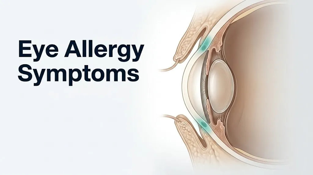
The conjunctiva consists of two regions: the bulbar conjunctiva, which covers the visible white scleral surface, and the palpebral conjunctiva, which lines the inner surface of the upper and lower eyelids. Both regions contain mast cells, goblet cells (which produce mucin for the tear film), and blood vessels. When the conjunctival surface is exposed to allergens, these allergens penetrate the epithelial layer and interact with IgE antibodies already bound to mast cell surface receptors.
Think of each conjunctival mast cell like a booby-trapped balloon. In a sensitized person, the balloon is already stretched thin, filled with histamine and other inflammatory chemicals, just waiting for the right trigger. When an allergen molecule bridges two nearby IgE antibodies on the cell surface (a process called cross-linking), it is like striking the balloon. The cell ruptures, releasing its contents in seconds. That is why eye allergy symptoms can begin within minutes of allergen exposure.
The bulbar conjunctival vessels dilate rapidly in response to histamine, producing the visible redness. Goblet cells hypersecrete mucin in response to the inflammatory stimulus, producing the gummy discharge. The tarsal conjunctiva (over the tarsal plate of the eyelid) can develop follicles and papillae in more chronic or severe presentations.
According to the National Eye Institute, allergic conjunctivitis is the most frequent allergic disease of the eye and one of the leading causes of recurrent eye discomfort in adults and children with atopic disease.
The IgE-Mast Cell Cascade: The Mechanism Behind Eye Allergy Symptoms
The IgE-mast cell cascade produces eye allergy symptoms through two sequential inflammatory phases: an immediate phase within minutes of allergen exposure driven by preformed mediators, and a late phase 4 to 8 hours later driven by newly synthesized inflammatory chemicals.
In the immediate phase, allergen cross-links IgE antibodies bound to high-affinity FcεRI receptors on conjunctival mast cell surfaces. This cross-linking event triggers rapid degranulation: preformed granule contents are released within seconds to minutes. Histamine is the dominant immediate mediator. It binds H1 receptors on corneal C-fibers (unmyelinated sensory nerves at the corneal surface and limbus) to produce the characteristic intense itch. Histamine also acts on H1 and H2 receptors on conjunctival blood vessel walls, causing vasodilation and increased vascular permeability, producing redness and swelling. Prostaglandin D2 and thromboxane, synthesized rapidly via the COX-2 pathway, amplify vasodilation and recruit additional inflammatory cells.
The late phase begins 4 to 8 hours after initial exposure and is driven by newly synthesized mediators. Cysteinyl leukotrienes (LTC4, LTD4, LTE4) produced via the 5-lipoxygenase pathway attract eosinophils and sustain conjunctival inflammation. Platelet-activating factor (PAF) further increases vascular permeability and activates eosinophils. Cytokines IL-4, IL-5, and IL-13 maintain Th2 dominance and promote eosinophil survival.
This two-phase mechanism explains why many patients notice their eye symptoms improve in the afternoon, then return with renewed severity in the evening without additional allergen exposure. They have experienced the gap between the immediate and late phases.
Research published in the Ocular Surface journal has documented the biochemical composition of these two phases in human conjunctival lavage fluid, confirming that histamine predominates in the first hour and leukotrienes become the primary mediators after 4 hours.
Key Takeaway: Eye allergy symptoms occur in two waves: intense itch and redness within minutes (from histamine), then a return of inflammation 4 to 8 hours later (from leukotrienes), which is why symptoms often feel like they reset in the late afternoon even without new allergen exposure.
Eye Itching as an Eye Allergy Symptom
Eye itching, clinically called ocular pruritus, is the most pathognomonic symptom of allergic conjunctivitis, present in virtually all cases and distinguishing it from most other forms of conjunctivitis, which produce pain or burning rather than itch as the primary complaint.
The itch sensation originates at corneal C-fibers and conjunctival sensory nerve endings in the limbal area (the junction between the cornea and sclera). Histamine, released from mast cells during degranulation, directly activates H1 receptors on these C-fibers, generating itch signals that travel via the ophthalmic branch of the trigeminal nerve (cranial nerve V1) to the trigeminal sensory nucleus in the brainstem. The itch is intense, bilateral, and worsened by rubbing.
Eye rubbing is one of the most clinically consequential behaviors in allergic conjunctivitis. Rubbing the eyes mechanically degranulates additional mast cells directly, amplifying histamine release and intensifying symptoms. Research published in JAMA Ophthalmology has found an association between chronic, forceful eye rubbing in patients with vernal keratoconjunctivitis and the development of keratoconus (a progressive thinning and steepening of the cornea). This association, while requiring further study, is consistent with the mechanical stress hypothesis of keratoconus in atopic patients.
Children who rub their eyes persistently, particularly those with the more severe forms of allergic conjunctivitis, should be evaluated by a board-certified ophthalmologist to assess whether the rubbing is producing corneal changes, and caregivers should be informed that eye rubbing can worsen rather than relieve the itch.
Quick Tip:
- Cold compresses applied to closed eyelids constrict conjunctival blood vessels and temporarily suppress mast cell degranulation by cooling the tissue.
- Rubbing always makes the itching worse within minutes. It feels like relief but releases more histamine immediately.
- Patients with atopic dermatitis who also rub their eyes forcefully need a lower threshold for ophthalmology referral due to the documented keratoconus association.
Eye Redness and Watering in Eye Allergies
Eye redness in allergic conjunctivitis is produced by vasodilation of the conjunctival blood vessels driven by histamine acting on H1 and H2 receptors on vascular smooth muscle cells and endothelial cells, causing the vessels to widen and become visible through the transparent conjunctiva.
Normal conjunctival blood vessels are narrow enough that they are largely invisible against the white sclera. When histamine and prostaglandin D2 bind to vascular receptors after mast cell degranulation, the vessels dilate to many times their resting diameter. The white of the eye turns pink to red as these dilated vessels become visible. Unlike the localized redness of a subconjunctival hemorrhage (which produces a bright, sharply bordered red patch), allergic conjunctivitis redness is diffuse, extends over the entire bulbar conjunctival surface, and is bilateral in over 90 percent of cases.
Watering, called epiphora, occurs through two concurrent mechanisms. First, the increased vascular permeability from histamine allows plasma fluid to leak across conjunctival vessel walls into the conjunctival space, adding fluid volume to the conjunctival surface. Second, the lacrimal gland is stimulated by reflex tearing triggered by the itch sensation and the irritation of the ocular surface. The combination produces the characteristic profuse, clear, watery discharge of allergic conjunctivitis.
The redness and watering of allergic conjunctivitis is reliably bilateral and is associated with itch, which distinguishes it from bacterial conjunctivitis (which is often unilateral initially and produces purulent, not watery, discharge). Bacterial conjunctivitis does not produce the dominant itch of allergic disease.
According to the American Academy of Ophthalmology (AAO), the combination of bilateral ocular itch and clear watery discharge in a patient with known allergic rhinitis has a positive predictive value sufficient for a clinical diagnosis of allergic conjunctivitis without further testing.
Swollen Eyelids as an Eye Allergy Symptom
Swollen eyelids in allergic conjunctivitis, clinically called periorbital edema or chemosis when involving the conjunctival tissue itself, result from increased vascular permeability in the eyelid dermis and palpebral conjunctiva, allowing plasma proteins and fluid to accumulate in the surrounding tissue.
Chemosis specifically refers to swelling of the bulbar conjunctiva itself, producing a gel-like protrusion of the transparent conjunctival membrane over the eyelid margin or around the cornea. In severe acute allergic reactions, chemosis can be dramatic enough to prevent the eyelid from fully closing, which exposes the cornea to drying and secondary damage.
The eyelid skin is among the thinnest and most loosely organized skin in the body, with minimal subcutaneous connective tissue to constrain swelling. When histamine-driven vascular permeability increases in the eyelid’s rich vascular network, fluid accumulates rapidly in this loose tissue, producing the characteristic puffy, swollen appearance within minutes of allergen exposure.
The AAAAI (American Academy of Allergy, Asthma and Immunology) notes that periorbital edema from allergic reactions can occasionally be severe enough to completely shut the eye, particularly in children and in individuals with acute high-dose allergen exposure such as a bee sting near the eye.
Pregnant women with pre-existing allergic conjunctivitis often experience more pronounced eyelid swelling during pregnancy because progesterone increases baseline vascular permeability throughout the body and the Th2 immune bias of pregnancy amplifies the allergic response. An obstetric provider should be informed if a pregnant woman’s ocular allergy symptoms deteriorate substantially, as systemic antihistamines have varying safety profiles across pregnancy trimesters.
Key Takeaway: Swollen eyelids in allergic conjunctivitis result from histamine-driven vascular permeability in the eyelid’s extremely thin, loosely organized skin. Chemosis (swelling of the conjunctiva itself) in a severe acute reaction may require same-day evaluation by an ophthalmologist to assess corneal exposure risk.
Discharge and Mucus as Eye Allergy Symptoms
Discharge from the eye in allergic conjunctivitis is characteristically clear to slightly white and stringy or mucoid, produced by excessive secretion from conjunctival goblet cells stimulated by the inflammatory mediators released during mast cell degranulation, and is distinct from the yellow-green purulent discharge of bacterial infection.
The conjunctival goblet cells are specialized mucin-secreting cells embedded in the conjunctival epithelium. In non-allergic conditions, they secrete controlled amounts of mucin (primarily MUC5AC) that forms the innermost, mucous layer of the tear film, helping the tear film adhere uniformly to the corneal surface. During allergic inflammation, cytokines including IL-13 and IL-4 directly stimulate goblet cell hypersecretion. The mucin volume increases dramatically, creating visible stringy, ropy, or web-like clear discharge.
The mucoid discharge of allergic conjunctivitis often causes the eyelids to feel sticky, particularly upon waking. This happens because the discharge accumulates overnight and dries at the lash margin. Patients commonly describe crusting at the inner corner of the eye (the medial canthus) that is easily removed with a warm washcloth. The discharge is not purulent, meaning it contains no pus, and gentle washing removes it completely.
Knowing the discharge character helps distinguish eye allergies from other conditions. Purulent (yellow-green, thick) discharge indicates bacterial infection. Watery, profuse discharge with no mucoid component is more characteristic of viral conjunctivitis. The stringy, clear mucoid discharge that forms threads or ropes when the eye is rubbed is specific to allergic and autoimmune conjunctivitis and is not produced by either bacterial or viral infections.
Any discharge accompanied by reduced vision, eye pain, or light sensitivity should prompt evaluation by an optometrist or ophthalmologist the same day, as these additional findings suggest the condition has moved beyond simple allergic conjunctivitis.
Early and Late-Phase Eye Allergy Symptom Patterns
The early and late phases of the allergic conjunctival response produce distinctly different symptom patterns separated by a period of partial or apparent improvement, and understanding this two-phase structure explains why many patients experience symptom relapse in the late afternoon or evening without additional allergen exposure.
The immediate phase begins within seconds to 5 minutes of allergen contact and peaks by 15 to 30 minutes. It is dominated by histamine-driven symptoms: intense itch, rapid onset redness, profuse tearing, and burning. These symptoms often feel explosive in onset. Most patients are accustomed to this phase because it is the one that brings them to reach for antihistamine eye drops.
The late phase begins approximately 4 to 8 hours after the initial allergen exposure and is driven primarily by leukotrienes, PAF, and recruited eosinophils and basophils. Symptom quality in the late phase shifts: the acute itch often gives way to a more sustained ache, burning, and a heavy, uncomfortable feeling in the eyes. Eyelid swelling and chemosis may actually be worse in the late phase than in the immediate phase, because eosinophil-driven cytokines sustain vascular permeability after histamine’s initial effect has waned.
| Phase | Timing | Dominant Mediators | Primary Symptoms |
|---|---|---|---|
| Immediate | 0 to 30 minutes | Histamine, tryptase, PGD2 | Intense itch, redness, tearing, burning |
| Late | 4 to 8 hours | Leukotrienes, PAF, IL-5, eosinophils | Sustained itch, ache, eyelid swelling, mucoid discharge |
| Chronic (repeated exposure) | Days to weeks | Th2 cytokines, eosinophils, TGF-beta | Conjunctival thickening, papillae formation, persistent redness |
Topical antihistamine eye drops primarily address the immediate phase. Mast cell stabilizing drops (cromolyn sodium, lodoxamide) require pre-treatment before allergen exposure to be effective, as they prevent degranulation rather than blocking already-released histamine. For late-phase symptoms, leukotriene-modifying treatments may be beneficial, but this requires evaluation by a board-certified allergist/immunologist or ophthalmologist.
Seasonal vs. Perennial Eye Allergy Symptoms
Seasonal allergic conjunctivitis (SAC) produces acute, episodic eye allergy symptoms that correlate with specific pollen seasons, while perennial allergic conjunctivitis (PAC) produces lower-grade but persistent year-round symptoms from continuously present indoor allergens such as dust mite proteins, pet dander, and mold.
SAC is the most prevalent form of allergic conjunctivitis, typically driven by tree pollen in spring, grass pollen in early summer, and ragweed pollen in late summer and early fall, depending on geographic location. Symptoms appear suddenly with the relevant pollen season and resolve when that season ends. The symptom intensity in SAC can be severe during peak pollen days, with intense bilateral itch, redness, tearing, and eyelid swelling appearing within minutes of outdoor exposure.
PAC symptoms tend to be milder in any single episode but more persistent throughout the year. Dust mite allergens (primarily the glycoprotein Der p 1 from Dermatophagoides pteronyssinus) and cat allergen (Fel d 1) are the most frequent PAC triggers. Symptoms often worsen in the morning after sleeping (increased dust mite exposure from bedding) and in environments with poor air filtration. Many PAC patients describe their eye symptoms as a chronic low-grade irritation rather than acute attacks.
People who work outdoors (agricultural workers, landscapers, construction workers) have disproportionately high exposure to SAC triggers and often experience more severe seasonal symptoms than office-based workers, according to clinical observational data reviewed by the ACAAI. These individuals should specifically mention occupational pollen exposure to their allergist, as it changes the risk-benefit calculation for immunotherapy consideration.
Key Takeaway: SAC produces intense acute attacks during specific pollen seasons, while PAC causes milder, year-round symptoms from indoor allergens. The distinction matters because their triggers, avoidance strategies, and management approaches differ significantly.
Vernal and Atopic Keratoconjunctivitis: Severe Eye Allergy Symptoms
Vernal keratoconjunctivitis (VKC) and atopic keratoconjunctivitis (AKC) are severe, chronic forms of allergic eye disease that produce corneal involvement alongside the conjunctival symptoms of simpler allergic conjunctivitis, and they carry a risk of permanent vision loss from corneal scarring if not managed aggressively.
VKC predominantly affects boys between ages 5 and 25 in warm, dry climates, with a male-to-female ratio of approximately 3:1. The hallmark finding of VKC is the formation of cobblestone papillae: giant, flat-topped papillae visible on the upper tarsal conjunctiva that resemble the surface of a cobblestone street under slit-lamp examination. These papillae form because eosinophil-derived major basic protein (MBP) and eosinophil cationic protein (ECP) stimulate fibroblasts in the conjunctival substantia propria via TGF-beta, producing subepithelial fibrosis. The papillae then mechanically abrade the corneal surface with every blink.
This mechanical abrasion can produce corneal shield ulcers: horizontal, shield-shaped erosions in the upper corneal epithelium that represent one of the most serious complications of allergic eye disease. Shield ulcers can permanently scar the cornea if untreated, directly impairing vision.
AKC affects adults with severe atopic dermatitis, typically ages 30 to 50. Unlike VKC’s predominantly upper tarsal papillae, AKC involves the entire conjunctival surface and frequently causes subepithelial fibrosis and fornix foreshortening (scarring of the conjunctival pocket where the eyelid meets the globe). AKC carries the highest risk of vision loss among all allergic conjunctivitis types.
Any child with persistent photophobia, mucoid discharge, and corneal discomfort should be evaluated by a pediatric ophthalmologist within one week, as VKC can produce amblyopia (reduced visual development in the affected eye) if corneal scarring develops during the critical period of visual maturation.
Giant Papillary Conjunctivitis Symptoms in Contact Lens Wearers
Giant papillary conjunctivitis (GPC/ICD-10: H10.4) produces symptoms of itching, lens intolerance, mucoid discharge, and a foreign body sensation under the upper eyelid in contact lens wearers, caused by a combination of mechanical irritation from the lens and an immune hypersensitivity reaction to lens deposits.
GPC is mechanistically distinct from the IgE-mediated allergic conjunctivitis of SAC and PAC. Its pathogenesis involves two components: mechanical trauma to the upper tarsal conjunctiva from the contact lens edge with every blink, and immune sensitization to protein deposits (primarily lysozyme, albumin, and lipids) that accumulate on the lens surface. This sensitization involves both IgE-mediated and non-IgE-mediated immune pathways, making GPC a partially allergic, partially mechanical condition.
The defining symptom of GPC is lens intolerance: patients who previously wore their lenses comfortably for 12 to 16 hours per day find they can tolerate lenses for only 2 to 4 hours before the itching, foreign body sensation, and blurred vision become intolerable. The large papillae on the upper tarsal conjunctiva create surface irregularity that the contact lens edge contacts with every blink, producing a cyclical irritation-inflammation pattern.
According to research published in Contact Lens and Anterior Eye, GPC affects approximately 1 to 5 percent of soft contact lens wearers and a higher proportion of hard (rigid gas-permeable) lens wearers, with the risk increasing with lens wearing duration and the use of old or dirty lenses.
Contact lens wearers who develop new eye allergy symptoms should temporarily discontinue lens wear and switch to spectacles while awaiting evaluation. They should bring their lenses, the lens case, and the care solution to their optometrist or ophthalmologist appointment, as the type and condition of the lens material provides diagnostic information.
Eye Allergy Symptoms in Children: What to Watch For
Eye allergy symptoms in children follow the same IgE-mediated mechanism as in adults, but children are at higher risk for the more severe forms (VKC and AKC) that can permanently damage vision, and the symptom expression in younger children differs because they often cannot accurately describe itch, leading to persistent eye rubbing as the primary observable sign.
Children who rub their eyes persistently, particularly in seasonal patterns or in response to outdoor exposure, should not have this attributed to fatigue or habit without evaluating for allergic conjunctivitis. Eye rubbing in a child without any described symptom is often the child’s behavioral response to intense ocular pruritus they lack the language to describe.
The American Academy of Pediatrics (AAP) recommends that pediatricians screen children with known atopic disease (asthma, atopic dermatitis, allergic rhinitis) for concurrent allergic conjunctivitis, as these conditions frequently coexist. The classic facial signs of chronic allergic eye disease in children include allergic shiners (darkened, bluish discoloration beneath the lower eyelids from chronic venous stasis from persistent eyelid swelling and rubbing) and Dennie-Morgan lines (extra skin folds at the lower eyelid margin associated with chronic periorbital edema).
Any child with photophobia (covering eyes in normal indoor lighting), a large amount of stringy discharge, or pain in addition to itching should be evaluated by a pediatric ophthalmologist or a board-certified ophthalmologist the same week, not managed empirically with OTC drops. These additional signs indicate VKC or AKC rather than simple seasonal allergic conjunctivitis, and these conditions require prescription management and monitoring for corneal complications.
Eye Allergy Symptoms vs. Pink Eye and Other Eye Conditions
Eye allergy symptoms are distinguished from bacterial conjunctivitis (pink eye) by the presence of intense bilateral itch (specific to allergy), the character of the discharge (clear and mucoid vs. yellow-green purulent), and the absence of fever or other infectious systemic symptoms.
Pink eye from a bacterial infection (most commonly Staphylococcus aureus, Streptococcus pneumoniae, or Haemophilus influenzae in children) typically begins in one eye, produces a thick, yellow-green discharge that crusts the eyelashes on waking, and may be associated with a recent upper respiratory infection. It does not produce the intense itch that is the hallmark of allergic conjunctivitis.
| Feature | Allergic Conjunctivitis | Bacterial Conjunctivitis | Viral Conjunctivitis | Dry Eye Disease |
|---|---|---|---|---|
| Primary symptom | Intense bilateral itch | Burning, discharge | Gritty, burning | Burning, gritty |
| Discharge | Clear, mucoid, stringy | Yellow-green, purulent | Watery | Minimal or none |
| Onset | Often bilateral immediately | Often unilateral, then bilateral | Often starts one eye | Both eyes chronically |
| Redness pattern | Diffuse, bilateral | Diffuse, often unilateral first | Diffuse, periorbital | Mild diffuse |
| Fever possible | No | Sometimes in children | Sometimes | No |
| Pollen/allergen history | Yes | No | No | No |
| Response to antihistamine eye drops | Yes | No | No | No |
| Corneal involvement | Rarely (SAC); yes in VKC/AKC | Sometimes | Sometimes | Yes |
Viral conjunctivitis (most commonly from adenovirus) produces a watery discharge, a gritty sensation, and visible follicles (small bumps) on the lower tarsal conjunctiva that differ from the papillae of allergic conjunctivitis. It is highly contagious and frequently occurs alongside pharyngitis and upper respiratory symptoms.
Differentiation between these conditions is not always possible by self-assessment. Any eye condition that does not clearly respond to antihistamine drops within 48 to 72 hours of use warrants evaluation by an optometrist or primary care physician.
Key Takeaway: Intense bilateral itch is the single most reliable symptom distinguishing allergic conjunctivitis from bacterial pink eye, viral conjunctivitis, and dry eye disease. If there is no itch, the condition is unlikely to be an eye allergy.
Blurry Vision and Eye Pain as Eye Allergy Symptoms
Blurry vision in allergic conjunctivitis is usually transient, caused by the mucoid discharge or chemosis distorting the tear film and ocular surface, and clears with blinking or gentle eye irrigation. Persistent blurry vision or any eye pain in the context of allergic eye symptoms is a red flag requiring same-day evaluation.
Standard seasonal allergic conjunctivitis does not damage the cornea and does not cause persistent blurring. When blurring occurs, it is momentary: the stringy discharge temporarily obscures vision until it is cleared by the tear film or a blink. If blinking clears the vision completely and it returns immediately, this pattern is consistent with discharge-related transient blurring and is not alarming on its own.
Eye pain is not a feature of simple allergic conjunctivitis. The conjunctiva is insensitive to pain in a way the cornea is not. If a patient with known allergic conjunctivitis develops new eye pain (not just itch or discomfort, but a sharp, aching, or persistent pain), this indicates corneal involvement rather than pure conjunctival disease. This is the clinical threshold that separates SAC (manageable with antihistamine drops) from VKC with corneal shield ulcers (which requires urgent ophthalmological management).
Photophobia (light sensitivity significant enough to cause squinting or discomfort in normal indoor lighting) in the context of an allergic eye presentation is another corneal sign. The cornea is densely innervated with pain-sensitive C-fibers. When the corneal epithelium is damaged or inflamed (from shield ulcers in VKC or from secondary infection), light stimulates these C-fibers, producing photophobia.
Any patient reporting eye allergy symptoms plus eye pain, photophobia, or vision that does not clear completely with blinking must be evaluated by a board-certified ophthalmologist the same day to rule out corneal involvement.
When Eye Allergy Symptoms Require Urgent Evaluation
Eye allergy symptoms require urgent evaluation when they include reduced visual acuity, eye pain, photophobia, or failure to improve with appropriate OTC antihistamine treatment within 48 to 72 hours, as these features indicate the possibility of corneal involvement, secondary infection, or a more severe allergic conjunctivitis type.
Certain presentations of eye-related symptoms in the context of a known or suspected allergen exposure require emergency evaluation rather than routine scheduling.
Go to the emergency room or call an ophthalmologist for same-day urgent evaluation if you experience:
- Any reduction in visual acuity (objects not appearing sharp after blinking and clearing discharge)
- Sharp or aching eye pain rather than the itch or discomfort typical of allergic conjunctivitis
- Photophobia: pain or significant discomfort in normal indoor lighting
- White or cloudy spot visible on the colored part of the eye (possible corneal ulcer or infection)
- Severe chemosis (conjunctival swelling protruding beyond the eyelid margin)
- Eye symptoms developing after chemical or substance exposure to the eye (possible chemical burn)
- Eye symptoms combined with facial rash, hives, throat tightening, or shortness of breath (possible systemic anaphylaxis)
Call 911 immediately if eye symptoms occur alongside throat swelling, difficulty breathing, widespread hives, or any signs of anaphylaxis following allergen exposure. Anaphylaxis is a life-threatening systemic allergic emergency.
These presentations require emergency medical assessment. Corneal ulceration, chemical burns, and anaphylaxis cannot wait for a scheduled appointment.
How Eye Allergy Symptoms Are Diagnosed and Evaluated
Eye allergy symptoms are diagnosed primarily through clinical history and physical examination, with targeted laboratory and allergy testing added when the diagnosis is unclear, the condition is severe, or management decisions require identification of the specific causative allergen.
A board-certified ophthalmologist or optometrist evaluates eye allergy symptoms by examining the eyelid skin (for periorbital edema and Dennie-Morgan lines), the conjunctival surface under slit-lamp biomicroscopy (assessing for papillae, follicles, chemosis, and discharge character), the corneal surface with fluorescein staining (identifying any epithelial defects, including shield ulcers), and tear film stability with tear break-up time (TBUT) testing.
A board-certified allergist/immunologist evaluates the underlying allergic sensitization to identify specific triggers:
- Skin prick testing (SPT): the standard for identifying specific IgE sensitization to pollens, dust mites, pet dander, mold, and food allergens; results available in 15 to 20 minutes in the clinic
- Serum-specific IgE testing (ImmunoCAP/RAST): blood test identifying specific IgE levels to named allergens; useful when skin testing is not possible (severe eczema, recent antihistamine use)
- Conjunctival provocation test: placing a small amount of allergen directly on the conjunctival surface and observing whether the characteristic allergic response is produced; used primarily in research settings
To prepare for your ophthalmology or allergy appointment:
- Note the timing of symptoms: do they follow a seasonal pattern, appear after specific indoor exposures, or occur with contact lens wear?
- List all current eye drops, oral antihistamines, and contact lens care products being used.
- Record whether symptoms are bilateral or unilateral.
- Note whether itch, pain, or burning is the primary symptom.
- Bring your contact lenses and case if you wear them.
- Tell the provider about any other atopic conditions (asthma, eczema, allergic rhinitis) and any family history of atopic disease.
Frequently Asked Questions About Eye Allergy Symptoms
What are the most common symptoms of eye allergies?
The most common eye allergy symptoms are intense bilateral itching, redness, excessive tearing, and eyelid swelling, produced by histamine released from conjunctival mast cells during IgE-mediated mast cell degranulation.
According to the American College of Allergy, Asthma and Immunology, itching is present in virtually all cases and is the most reliable feature distinguishing allergic conjunctivitis from bacterial or viral pink eye.
The clear, mucoid, stringy discharge that forms threads when the eye is rubbed is another characteristic finding that is specific to allergic and autoimmune conjunctivitis.
How do I tell the difference between eye allergies and pink eye?
Eye allergies produce intense bilateral itch, clear watery or mucoid discharge, and a correlation with allergen exposure, while bacterial pink eye produces burning discomfort, purulent yellow-green discharge, and often starts in one eye before spreading to the other.
Viral conjunctivitis causes watery discharge and a gritty sensation without the dominant itch of allergic disease, and is often associated with a recent upper respiratory infection.
If antihistamine eye drops provide no relief within 24 to 48 hours, the diagnosis of allergic conjunctivitis should be reconsidered and the condition evaluated by an optometrist or primary care physician.
Can eye allergies cause blurry vision?
Transient blurry vision in eye allergies is caused by mucoid discharge and conjunctival swelling distorting the tear film, and it clears completely with blinking in simple allergic conjunctivitis.
Persistent blurry vision that does not clear with blinking is not a feature of seasonal allergic conjunctivitis and requires evaluation by a board-certified ophthalmologist the same day to rule out corneal involvement or secondary infection.
Children with severe eye allergy types (vernal keratoconjunctivitis) can develop corneal shield ulcers that produce persistent vision blurring requiring urgent ophthalmological management.
Why are my eye allergy symptoms worse at night?
Eye allergy symptoms often worsen at night because the late-phase allergic reaction, driven by leukotrienes and eosinophils rather than histamine, peaks approximately 4 to 8 hours after initial daytime allergen exposure.
Increased indoor allergen exposure at night (from dust mites in bedding) worsens perennial allergic conjunctivitis symptoms specifically during and after sleep.
Removing and washing bedding covers regularly, keeping pets out of the bedroom, and using an air purifier with a HEPA filter can reduce nighttime perennial allergen load while awaiting formal allergy evaluation.
When should I see a doctor for eye allergy symptoms?
Eye allergy symptoms lasting more than two weeks or not responding to OTC antihistamine eye drops within 48 to 72 hours warrant evaluation by an optometrist or primary care physician.
Any eye allergy symptom accompanied by eye pain, photophobia, or reduced visual acuity requires same-day evaluation by a board-certified ophthalmologist to exclude corneal complications.
Children with persistent eye rubbing, mucoid discharge, and any light sensitivity should be evaluated by a pediatric ophthalmologist within one week, not managed empirically with over-the-counter drops.
What do swollen eyelids from allergies feel like?
Swollen eyelids from eye allergies feel puffy, heavy, and mildly tender, similar to the feeling after crying, and appear most prominent after waking or after acute allergen exposure.
The swelling is caused by histamine-driven vascular permeability in the extremely thin, loosely structured eyelid skin, allowing plasma fluid to accumulate rapidly in the periorbital tissue.
Swelling that involves the eyeball surface itself (chemosis, where the transparent conjunctiva protrudes beyond the lid margin like a water blister) represents more severe inflammation and warrants evaluation by an ophthalmologist the same day if it prevents normal eyelid closure.
The most clinically actionable fact from this entire guide is the itch rule: intense, bilateral itch is the defining symptom of allergic conjunctivitis, and its presence or absence reliably separates eye allergies from bacterial pink eye, viral conjunctivitis, and dry eye disease. If your eyes itch intensely and symmetrically, allergic conjunctivitis is the most likely explanation. If they burn, ache, or produce thick yellow discharge without itch, the picture points elsewhere.
For mild seasonal symptoms in an adult with no corneal involvement, OTC antihistamine-mast cell stabilizer combination drops are a reasonable first step while you arrange evaluation. For any child with persistent symptoms, any adult with symptoms that don’t respond in 72 hours, or any patient with eye pain or vision change, the next contact should be a board-certified ophthalmologist.
You now have the mechanistic framework to describe your eye symptoms precisely, identify which type of allergic conjunctivitis your pattern resembles, and ask the right questions at your evaluation.