Left Flank Pain Causes: 16 Reasons and When to Act
Left flank pain causes include kidney stones, kidney infection, splenic pathology, left-sided diverticulitis, musculoskeletal strain, shingles, and aortic disease, with the specific cause determined by the character of the pain, accompanying symptoms, and which left-sided organ is involved. Because several of these causes are medical emergencies and others are entirely self-limiting, knowing how to read the specific features of your pain is the most clinically useful skill you can bring to this situation.
According to the National Institute of Diabetes and Digestive and Kidney Diseases (NIDDK), kidney stones affect approximately 11% of men and 6% of women in the United States at some point in their lives, making renal causes collectively the most frequent explanation for acute flank pain. But the left flank specifically sits over a set of anatomical structures that the right flank does not share, including the spleen, the tail of the pancreas, and the splenic flexure of the colon, meaning left flank pain has a distinct differential diagnosis that a general “flank pain” article cannot adequately address.
This article covers every major left flank pain cause with the physiological mechanism behind each one, distinguishes left-specific from right-specific causes, addresses how the pain pattern and associated symptoms help narrow the diagnosis, and gives you the exact clinical threshold at which left flank pain requires a 911 call rather than a doctor’s appointment.
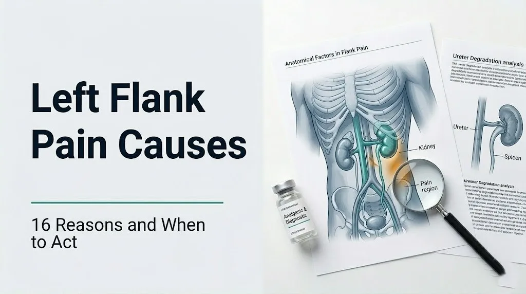
Left Flank Pain Causes: The Anatomy That Explains Everything
The left flank pain causes that matter most are determined by which specific organs and structures occupy the left flank anatomical region, and understanding this geography is the foundation for interpreting any flank pain symptom pattern.
The left flank refers to the region of the body between the lower edge of the left rib cage (approximately the 12th rib) and the left iliac crest (top of the left hip bone), lateral to the lumbar spine. This area includes both the posterior flank (the lateral back surface) and the lateral abdomen. The structures that lie within or immediately adjacent to this region and can produce pain when diseased or injured include:
- Left kidney: Positioned retroperitoneally (behind the peritoneum) at the T12-L2 vertebral level
- Left ureter: Running from the left renal pelvis downward through the retroperitoneal space to the bladder
- Left adrenal gland: Sitting superior (above) the left kidney
- Spleen: Positioned intraperitoneal at the left 9th to 11th ribs, covered by the splenic capsule
- Splenic flexure of the descending colon: The leftward bend of the colon as it transitions from the transverse colon to the descending colon, sitting in the left upper flank
- Tail of the pancreas: Extending leftward from the body of the pancreas toward the splenic hilum
- Abdominal aorta: Descending along the left lateral surface of the lumbar vertebral column
- Erector spinae and quadratus lumborum muscles: Running along the posterior flank bilaterally
- Intercostal nerves T10-T12: Running along the lower rib margins into the flank
Right-sided structures absent from the left include the liver, gallbladder, and appendix. Left-sided structures absent from the right include the spleen, splenic flexure, and pancreatic tail. This anatomical asymmetry is precisely why left flank pain has specific causes that right flank pain does not, and vice versa.
What Is the Left Flank and What Organs Are There?
The left flank is the lateral region of the body between the 12th rib and the left iliac crest, containing the left kidney, left ureter, spleen, splenic flexure of the colon, pancreatic tail, left adrenal gland, and major vascular structures including the abdominal aorta and left renal artery and vein.
The retroperitoneal space is the anatomical compartment behind the peritoneal cavity lining the abdominal cavity. The left kidney, left ureter, and left adrenal gland all reside in this retroperitoneal space. Pain from retroperitoneal structures tends to be felt in the posterior flank, between the spine and the lateral abdominal wall, rather than in the anterior abdomen.
The spleen sits in the intraperitoneal space, covered by the peritoneum. It rests against the left 9th to 11th ribs and is connected to the stomach by the gastrosplenic ligament and to the left kidney by the splenorenal ligament. Its rich innervation by the left phrenic nerve (C3-C5) explains why splenic pathology can produce not only left upper quadrant and flank pain but also referred left shoulder pain via the phrenic nerve pathway, a phenomenon also seen with diaphragmatic irritation.
Understanding that the spleen, the descending colon’s splenic flexure, and the pancreatic tail are left-specific is the essential starting point for any evaluation of left flank pain. A right-sided diagnosis like gallbladder disease or appendicitis cannot produce left flank pain through its primary mechanism, though referred pain patterns can occasionally cross midline at lower intensities.
Key left flank anatomical structures and their pain referral patterns:
| Structure | Position | Pain Location | Pain Character |
|---|---|---|---|
| Left kidney | Retroperitoneal, T12-L2 | Posterior flank, CVA | Dull, constant aching |
| Left ureter | Retroperitoneal, T12-pelvis | Flank to groin, inner thigh | Colicky, wave-like |
| Spleen | Intraperitoneal, 9th-11th ribs | Left upper flank, shoulder | Sharp, constant |
| Splenic flexure | Intraperitoneal, left upper abdomen | Left flank, lateral abdomen | Cramping, gas-like |
| Pancreatic tail | Retroperitoneal, left upper abdomen | Left flank, back, epigastrium | Deep, boring, constant |
| Aorta | Retroperitoneal, anterior lumbar spine | Left flank, lower back, abdomen | Severe, tearing |
Kidney Stone as a Cause of Left Flank Pain
A kidney stone (nephrolithiasis) is the single most frequent cause of acute, severe left flank pain, producing its characteristic colicky, wave-like pain pattern through a specific mechanism involving ureteral smooth muscle contractions against an obstructing stone.
When a stone forms in the left kidney and migrates from the renal pelvis into the left ureter, it lodges most commonly at one of three anatomically narrow points: the left ureteropelvic junction (UPJ), the point where the ureter crosses the iliac vessels at the pelvic brim, or the ureterovesical junction (UVJ) where the ureter enters the bladder. At any of these locations, the obstruction prevents urine flow and triggers vigorous ureteral peristaltic contractions that attempt to push the stone forward.
Think of the ureter as a narrow garden hose with a pebble stuck in it. The hose doesn’t just sit still. It contracts rhythmically and forcefully to dislodge the obstruction. Each contraction produces a wave of intense cramping pain. Between contractions, the pain partially subsides, only to return with the next wave. This wave-like pattern is what clinicians mean by “colicky” pain.
The pain is referred to the left flank because the ureteral sensory afferents travel through the T10-L2 spinal nerve segments, the same segments that receive sensory input from the flank skin. When the ureter signals pain, the brain interprets it as coming from the flank skin served by those same segments.
According to the American Urological Association, the gold standard diagnostic test for a suspected kidney stone is a non-contrast CT urogram (computed tomography of the urinary tract without intravenous contrast), which detects stones of virtually all compositions with high sensitivity and specificity.
Kidney stones are approximately twice as prevalent in men as in women, though the female-to-male ratio has been narrowing over recent decades. Women on topiramate (an antiepileptic medication that increases urine pH and reduces citrate excretion) have elevated stone risk regardless of sex-based baseline rates.
Key Takeaway: Left flank pain that comes in waves of severe cramping separated by partial relief, radiating toward the groin or inner thigh, without fever, is the textbook presentation of a left ureteral stone and warrants urgent evaluation with a non-contrast CT urogram.
Kidney Infection (Pyelonephritis) Causing Left Flank Pain
Pyelonephritis, the bacterial infection of the left renal parenchyma (kidney tissue), causes left flank pain through a mechanism of renal capsule distension from inflammatory edema and congestion within the kidney, producing a constant, dull aching pain that differs distinctly from the colicky pattern of a stone.
The renal capsule is a tough, fibrous outer covering of the kidney with no capacity to expand. When the kidney becomes infected, inflammatory cells and fluid accumulate within the renal parenchyma. This swelling pushes outward against the inelastic capsule, activating stretch receptors in the capsule’s innervation network and producing the constant, pressure-like pain felt at the costovertebral angle (CVA), the anatomical junction of the 12th rib and the lateral border of the erector spinae muscles. CVA tenderness on percussion (gently tapping with a fist over this area) is the clinical sign that most specifically suggests pyelonephritis rather than a lower urinary tract infection.
Pyelonephritis almost always begins as a lower urinary tract infection (UTI) that ascends via the left ureter to reach the left kidney. The causative organism is Escherichia coli in approximately 80% of community-acquired cases, according to a clinical review published in JAMA. The infection triggers a full inflammatory response in the renal parenchyma, producing systemic symptoms alongside the local flank pain.
Clinical features that distinguish pyelonephritis from kidney stone:
- Fever typically above 38°C (100.4°F), often with shaking chills
- Constant rather than colicky pain
- Dysuria (burning on urination) and urinary frequency
- Nausea and vomiting
- Urine dipstick positive for leukocyte esterase, nitrites, and blood
Women experience pyelonephritis approximately 5 to 10 times more frequently than men, primarily because a shorter urethra (approximately 4 cm in women vs. 20 cm in men) allows ascending bacterial colonization to reach the bladder and subsequently the kidney more easily. Pregnant women with pyelonephritis face a higher risk of preterm labor and require hospital-level intravenous antibiotic treatment.
Spleen-Related Causes of Left Flank Pain
The spleen is a left-specific anatomical structure, and when it becomes enlarged, infarcted, infected, or ruptured, it produces left flank and left upper quadrant pain through direct capsular distension or peritoneal irritation.
Several distinct splenic conditions cause left flank pain. Splenic infarction occurs when the blood supply to a segment of the spleen is interrupted, most commonly by a blood clot (thrombus or embolus) traveling through the splenic artery (a branch of the celiac trunk). Infarction causes ischemic necrosis of splenic tissue, which triggers an intense inflammatory response in the splenic capsule. The pain is typically sudden in onset, located in the left upper flank and left upper quadrant, and may radiate to the left shoulder via the phrenic nerve pathway.
Splenomegaly (enlargement of the spleen) from any cause, including infectious mononucleosis, portal hypertension from cirrhosis, hematological malignancies such as lymphoma, or hemolytic anemias, can produce a dull, persistent left flank aching as the enlarged organ stretches its capsule. According to the Mayo Clinic, the spleen in splenomegaly can expand from its normal weight of approximately 150 grams to over 1,000 grams, placing progressive stretch on its capsular innervation.
Splenic abscess is a less common but potentially life-threatening condition in which bacterial infection within the splenic parenchyma forms a pus-filled cavity. It produces left flank pain with systemic infection signs: fever, elevated white blood cell count, and malaise. Immunocompromised individuals, including those with HIV, organ transplant recipients, and those on chronic immunosuppressive therapy, face disproportionately higher risk.
Splenic rupture, either from trauma or, in rare cases, from spontaneous rupture in an enlarged spleen (most classically after infectious mononucleosis), is a surgical emergency. Call 911 immediately if left flank or left upper abdominal pain follows any trauma, particularly in a patient with known splenomegaly.
Left-Sided Diverticulitis and Colon Causes of Left Flank Pain
The splenic flexure of the descending colon and the sigmoid colon are left-sided colonic structures whose inflammation or distension produces left flank pain through visceral afferent pathways that refer pain to the lateral left abdomen and left flank.
Diverticulitis occurs when small outpouchings of the colonic wall (diverticula) become inflamed or infected. Diverticula form most commonly in the sigmoid colon, which is located in the left lower abdomen and pelvis. When sigmoid diverticula become inflamed, the pain is typically felt in the left lower flank and left lower quadrant, often described as a constant, aching pain that does not wax and wane in the way ureteral colic does.
According to the American College of Gastroenterology, diverticulitis affects approximately 1 in 200,000 younger adults annually, but the incidence rises substantially with age: it affects approximately 10% of people over 40 and up to 50 to 60% of people over 70 years of age. The mechanism involves perforation or microperforation of a diverticulum, triggering pericolonic inflammation and sometimes frank abscess or perforation of the colonic wall.
Splenic flexure syndrome refers to gas trapping at the splenic flexure of the colon, producing left upper quadrant and left flank pain that is cramping and often temporarily relieved by passing gas or having a bowel movement. The pain occurs because distension of the colonic wall at the flexure activates visceral stretch receptors innervated by the left intercostal nerves. While not an emergency, persistent splenic flexure syndrome warrants evaluation by a board-certified gastroenterologist to exclude inflammatory bowel disease or a colonic mass.
Older adults on low-fiber diets are at elevated risk for diverticular disease, while individuals with inflammatory bowel disease (Crohn’s disease or ulcerative colitis) are at elevated risk for left colonic inflammation producing flank symptoms.
Key Takeaway: Left flank pain that is constant rather than colicky, worsens with eating, is accompanied by fever or change in bowel habits in an adult over 50, and is located in the lower left flank strongly suggests left-sided diverticulitis and warrants same-day evaluation by a primary care physician with CT imaging of the abdomen and pelvis with contrast.
Pancreatic Tail and Left Adrenal Causes of Left Flank Pain
The tail of the pancreas and the left adrenal gland are two anatomically left-specific retroperitoneal structures that can produce left flank pain when inflamed, infected, or occupied by a tumor.
The tail of the pancreas extends leftward from the body of the pancreas, approaching the hilum of the spleen. Because the pancreatic tail is retroperitoneal in position, inflammation of the pancreatic tail (as occurs in pancreatitis when the tail is preferentially involved) produces deep, boring pain in the left upper abdomen and left flank that often radiates through to the back. This is clinically distinct from the typical epigastric pancreatitis presentation because the tail’s position against the left flank produces a more lateral, flank-dominant pain pattern in some patients.
According to a review published in the New England Journal of Medicine, acute pancreatitis produces retroperitoneal inflammation that extends into the perirenal fat and retroperitoneal fascial planes, which is why left flank pain is a recognized component of pancreatitis when the tail is involved.
The left adrenal gland (suprarenal gland) sits on the superior pole of the left kidney. Adrenal pathology, including adrenal hemorrhage, adrenal tumor (adrenal adenoma, pheochromocytoma, or adrenal carcinoma), or adrenal abscess, can produce left flank pain through capsular distension of the adrenal gland and surrounding retroperitoneal fat. Pheochromocytoma, a catecholamine-secreting adrenal tumor, may produce hypertensive crises with associated back and flank pain as a presenting feature.
Patients with newly diagnosed hypertension who develop left flank pain should be evaluated by a primary care physician for adrenal pathology, including measurement of plasma metanephrines, given the association between pheochromocytoma and secondary hypertension.
Musculoskeletal Causes of Left Flank Pain
Musculoskeletal strain is the most frequently self-diagnosable and self-limiting cause of left flank pain, produced by injury or overuse of the erector spinae muscle group, the quadratus lumborum, or the intercostal muscles and their associated nerves.
The erector spinae group (composed of the iliocostalis, longissimus, and spinalis muscles) runs bilaterally along the length of the spine from the sacrum to the skull, with its most superficial fibers easily palpable in the paraspinal area. The quadratus lumborum runs from the 12th rib and lumbar transverse processes to the iliac crest, lying deep to the erector spinae. Either of these muscles, when acutely strained by lifting, twisting, or prolonged postural stress, produces localized somatic pain in the posterior flank that is worsened by movement and improved with rest.
The distinguishing features of musculoskeletal flank pain are critically important for differentiation from visceral causes. Musculoskeletal pain typically:
- Worsens with specific movements (rotation, lateral flexion, forward bending)
- Is reproduced by direct pressure on the tender muscle
- Does not accompany fever, dysuria, hematuria, nausea, or systemic symptoms
- Has a clear precipitating activity (lifting heavy objects, sudden twisting motion)
- Improves within 48 to 72 hours with rest and anti-inflammatory measures
Athletes and manual laborers have the highest baseline risk of musculoskeletal flank pain from erector spinae and quadratus lumborum overuse. Older adults with osteoporosis face a different musculoskeletal risk: a vertebral compression fracture of the T12 or L1 vertebra can produce sudden severe posterior flank pain that mimics visceral pathology. A history of osteoporosis and sudden pain onset after minimal force (coughing, standing from a chair) in a postmenopausal woman warrants urgent imaging to exclude vertebral fracture.
L1-L2 nerve root compression from disc herniation at the T12-L1 or L1-L2 intervertebral level produces radicular pain in the flank and groin distribution, mimicking ureteral colic. The distinction requires imaging: MRI of the lumbar spine versus CT urogram depending on which diagnosis is suspected.
Shingles (Herpes Zoster) as a Cause of Left Flank Pain
Shingles (herpes zoster) is a clinically underrecognized cause of left flank pain because the burning, unilateral dermatomal pain it produces can precede the characteristic skin rash by 1 to 5 days, a period during which it is indistinguishable from renal or musculoskeletal causes on history alone.
Shingles results from reactivation of varicella-zoster virus (VZV), which remains dormant in the dorsal root ganglia of sensory nerve roots after childhood chickenpox infection. Reactivation, triggered by immune suppression, stress, aging, or immunosenescence, causes VZV to replicate within the ganglion and travel down the sensory nerve axons to the skin of the corresponding dermatome. The T10, T11, and T12 dermatomal distributions, which cover the lateral trunk and flank, are among the most frequently affected locations.
The prodromal pain of shingles is burning, stabbing, or electric in character. It is strictly unilateral and does not cross the midline, following the course of the intercostal nerve from the posterior paravertebral area around the flank to the anterior chest or abdominal wall. The skin over the affected dermatome may be exquisitely sensitive to light touch (allodynia), even before the rash appears.
According to the Centers for Disease Control and Prevention, approximately 1 in 3 people in the United States will develop shingles in their lifetime, with risk rising sharply after age 50. Adults aged 60 and older account for the majority of clinical cases.
If a patient presents with burning, unilateral left flank pain without fever, without hematuria, and without a clear mechanical trigger, a shingles prodrome should be considered. A careful skin examination looking for early erythema or even a single vesicle along the T10-T12 distribution can clinch the diagnosis before the full rash erupts.
Key Takeaway: Burning, strictly unilateral left flank pain in an adult over 50 that follows a dermatomal stripe from the back around the flank toward the front, without hematuria or fever, should prompt examination of the skin in that distribution for early shingles vesicles before renal or musculoskeletal causes are assumed.
Aortic and Vascular Causes of Left Flank Pain
Vascular causes of left flank pain include abdominal aortic aneurysm, renal artery stenosis, renal vein thrombosis, and retroperitoneal hematoma, and one of these, a ruptured abdominal aortic aneurysm, is among the most immediately life-threatening conditions that can present as flank pain.
The abdominal aorta descends along the left lateral surface of the lumbar vertebral column, bifurcating into the iliac arteries at the L4 level. An abdominal aortic aneurysm (AAA) is defined as a focal dilation of the aorta to a diameter greater than 3 centimeters. When an AAA expands rapidly or begins to leak blood into the retroperitoneal space, it produces sudden severe left flank and back pain, often described as tearing or ripping in quality. Blood accumulating in the retroperitoneal space causes intense pain by distending the retroperitoneal fascial compartment. A ruptured AAA is fatal without emergency surgical repair.
Left renal artery stenosis, a narrowing of the left renal artery (most commonly from atherosclerosis or fibromuscular dysplasia), reduces blood flow to the left kidney, triggering a chronic ischemic process that can produce intermittent left flank discomfort and, more clinically, causes renovascular hypertension through renin-angiotensin-aldosterone system (RAAS) activation by the ischemic kidney.
Renal vein thrombosis, a clot in the left renal vein, causes sudden severe left flank pain from venous congestion and renal capsule distension. It occurs in the setting of nephrotic syndrome, hypercoagulable states, renal cell carcinoma with vascular invasion, or severe dehydration. Hematuria is a frequent accompanying sign.
Retroperitoneal hematoma develops when bleeding occurs into the retroperitoneal space, most commonly in patients taking anticoagulant medications (warfarin, apixaban, rivaroxaban, heparin) who experience either spontaneous vessel wall bleeding or minor trauma that would be inconsequential without anticoagulation. Sudden severe flank pain in any patient on anticoagulant therapy is a medical emergency.
Older men (aged 65 to 75) who have ever smoked have the highest AAA risk, and the U.S. Preventive Services Task Force (USPSTF) recommends one-time abdominal ultrasound screening for AAA in this population.
Renal Cell Carcinoma and Left Flank Pain
Renal cell carcinoma (RCC) is a malignancy of the renal tubular epithelial cells that, when arising in the left kidney, produces left flank pain through two mechanisms: mass effect with renal capsule distension as the tumor grows, and internal hemorrhage within the tumor producing sudden acute pain superimposed on a chronic dull ache.
The classic clinical triad of RCC is flank pain, hematuria (blood in the urine), and a palpable abdominal mass. According to the National Cancer Institute, this complete triad is present in fewer than 10% of RCC patients at diagnosis, because most tumors are now detected incidentally on imaging before they reach sufficient size to produce all three findings. Flank pain as an isolated presenting symptom is possible but typically indicates a larger tumor or one with capsular involvement.
RCC grows within the renal parenchyma. As it enlarges, it pushes outward against the inelastic renal capsule, producing the same constant, dull, capsular-distension pain that pyelonephritis produces, but without fever, without urinary symptoms, and without the acute onset pattern. The pain is typically chronic, weeks to months in duration, and gradually worsening.
Hematuria accompanying left flank pain without fever, dysuria, or a clear stone passage history, particularly in an adult over 50, warrants urgent evaluation by a board-certified urologist including CT abdomen and pelvis with and without intravenous contrast, and a urology consultation for cystoscopy and upper tract evaluation.
Men aged 50 to 70 have the highest incidence of renal cell carcinoma, with RCC approximately twice as prevalent in men as in women. Smoking, obesity, hypertension, and a family history of RCC all independently increase risk, according to data from the National Cancer Institute.
Right Flank Pain Causes: How They Differ from the Left
Right flank pain causes differ from left flank causes specifically because the right flank contains the liver, gallbladder, right kidney and ureter, appendix (in its retrocecal position), and right adrenal gland, none of which are present on the left side.
The most clinically important right-specific cause of right flank pain is acute cholecystitis (gallbladder inflammation), which produces right upper flank and right upper quadrant pain from gallbladder wall distension and the stretching of the common bile duct and hepatic capsule. This is an entirely right-sided condition with no left flank equivalent.
Appendicitis, particularly when the appendix is in a retrocecal position (pointing backward behind the cecum rather than downward), produces right flank pain rather than the classic right lower quadrant McBurney’s point pain. This anatomical variant makes retrocecal appendicitis one of the most diagnostically challenging presentations of right flank pain.
The right kidney and right ureter produce stone and infection symptoms identical in character to left-sided renal causes but lateralized to the right flank. The right kidney sits slightly lower than the left in most individuals because the liver occupies space above it, pushing the right kidney inferiorly.
| Cause | Left Flank | Right Flank | Explanation |
|---|---|---|---|
| Kidney stone | Yes | Yes | Bilateral anatomical equivalence |
| Pyelonephritis | Yes | Yes | Bilateral anatomical equivalence |
| Gallbladder/liver | No | Yes | Right-sided organs only |
| Appendix (retrocecal) | No | Yes | Right-sided structure |
| Spleen pathology | Yes | No | Left-sided organ only |
| Diverticulitis (sigmoid) | Yes | No | Left-sided colon |
| Pancreatic tail | Yes | No | Extends leftward |
For a person experiencing right flank pain, the priority differential includes right kidney stone, right pyelonephritis, cholecystitis, retrocecal appendicitis, and right musculoskeletal causes, evaluated by a primary care physician with right-specific imaging ordered based on the dominant clinical picture.
Key Takeaway: The specific organ anatomy of the left vs. right flank determines which causes are possible on each side, and causes that are left-specific (spleen, sigmoid colon, pancreatic tail) literally cannot be the source of right flank pain through their primary mechanisms.
Left Flank Pain in Women: Specific Causes and Differences
Left flank pain in women has a broader differential diagnosis than in men because the female reproductive organs, including the left ovary and fallopian tube, lie in close anatomical proximity to the left lower flank and can produce referred pain to this region from several distinct gynecological conditions.
A left ovarian cyst is one of the most frequently overlooked causes of left lower flank pain in women of reproductive age. Ovarian cysts produce pain through two primary mechanisms: capsular distension from fluid accumulation within the cyst, producing a dull, constant aching pain, or acute torsion (ovarian torsion), in which the ovary rotates on its ligamentous attachments, cutting off its blood supply and producing sudden, severe, unilateral lower abdominal and flank pain that requires emergency surgical intervention.
Ectopic pregnancy, in which a fertilized embryo implants in the left fallopian tube rather than the uterine cavity, can produce left lower flank pain as the tube distends with the growing pregnancy. If the fallopian tube ruptures, catastrophic intraperitoneal hemorrhage follows. The pain becomes sudden and severe, and the patient may become hemodynamically unstable from blood loss. Any woman of reproductive age with left lower flank pain and a missed period must be evaluated for ectopic pregnancy immediately, regardless of contraceptive use.
Endometriosis affecting the left uterosacral ligament or left ovary can produce cyclical left flank and pelvic pain that correlates with the menstrual cycle, worsening in the days before and during menstruation due to prostaglandin-mediated smooth muscle contractions and inflammatory tissue response.
Women with interstitial cystitis (bladder pain syndrome) often describe their pain as extending into the flank region bilaterally, though the mechanism is primarily bladder wall inflammation rather than true flank pathology.
Pregnant women who develop left flank pain should be evaluated urgently for pyelonephritis, ureteral obstruction from uterine compression of the left ureter (which crosses the iliac vessels and can be compressed by the gravid uterus), and placental abruption if late in pregnancy. Imaging is limited to renal ultrasound as first-line to avoid fetal radiation exposure.
Left Flank Pain in Men: Specific Risk Factors
Left flank pain in men shares most causes with women but carries a higher prior probability for abdominal aortic aneurysm, uric acid kidney stones, and certain musculoskeletal patterns from occupational exposures, while lacking the gynecological causes that expand the female differential.
Men have approximately twice the baseline rate of kidney stones compared to women, with higher rates of uric acid stones specifically, because men have higher serum uric acid levels from greater purine turnover and, historically, higher dietary purine intake. Uric acid stones are radiolucent, meaning they are not visible on plain radiographs and may appear less clearly on non-contrast CT than calcium-containing stones. In a man over 50 with left flank pain, blood and urine uric acid levels and a CT urogram provide the most complete diagnostic evaluation.
The risk of abdominal aortic aneurysm is approximately 6 to 10 times higher in men than in women, particularly in men over 65 who have a smoking history. The USPSTF recommends one-time AAA screening ultrasound for men aged 65 to 75 who have ever smoked. Any sudden severe left flank or back pain in a man of this demographic should be treated as a possible AAA until proven otherwise.
Testicular pathology can refer pain to the left flank through the shared T10-L2 spinal nerve pathway. The left testicular vein drains directly into the left renal vein (unlike the right testicular vein, which drains into the inferior vena cava), which is why left-sided varicocele (dilation of the testicular veins) occurs more frequently on the left. A new left flank pain with a feeling of heaviness in the left scrotum warrants evaluation for varicocele or, in young men, left testicular torsion.
Men who perform heavy manual labor are at elevated risk for quadratus lumborum and erector spinae strain, particularly with repetitive asymmetric lifting or twisting that preferentially loads the left paraspinal muscles.
How to Diagnose the Cause of Left Flank Pain
Diagnosing the cause of left flank pain follows a sequential clinical evaluation process that begins with history and physical examination and progresses to targeted laboratory and imaging studies based on the dominant symptom pattern.
The diagnostic sequence a clinician follows:
- History characterization: Is the pain colicky or constant? Did it start suddenly or gradually? Is it accompanied by fever, hematuria, dysuria, nausea, or bowel changes? Does it radiate toward the groin (ureteral) or shoulder (splenic)? What is the patient’s age, sex, medication list, and relevant history?
- Physical examination: CVA tenderness on percussion (pyelonephritis), abdominal tenderness with guarding (peritonitis), palpable pulsatile abdominal mass (AAA), left lower quadrant tenderness (diverticulitis), skin examination for early vesicles (shingles).
- Urinalysis with microscopy: Detects hematuria (kidney stone or renal tumor), pyuria and bacteriuria (pyelonephritis), proteinuria (renal disease). This is the fastest and most informative initial test for most flank pain presentations.
- Blood tests: Complete blood count (CBC, elevated WBC suggests infection), comprehensive metabolic panel (serum creatinine and BUN for renal function), C-reactive protein and ESR (inflammation), lipase (pancreatitis), uric acid (stone risk assessment).
- Imaging selection per the American College of Radiology Appropriateness Criteria:
| Clinical Scenario | Preferred Imaging | Rationale |
|---|---|---|
| Suspected kidney stone | Non-contrast CT urogram | Highest sensitivity for all stone types |
| Suspected pyelonephritis | Renal ultrasound or CT with contrast | Excludes abscess, obstruction |
| Suspected AAA | CT abdomen with contrast | Defines aneurysm size and leak |
| Suspected diverticulitis | CT abdomen/pelvis with contrast | Defines inflammation, abscess |
| Pregnant patient | Renal ultrasound first | Avoids fetal radiation |
| Suspected renal mass | CT with and without contrast | Characterizes lesion vascularity |
- Specialist referral based on findings: Board-certified urologist for confirmed stone or renal mass; board-certified gastroenterologist for diverticulitis or pancreatic pathology; board-certified vascular surgeon for AAA; primary care physician for initial shingles evaluation.
Women of reproductive age should have a urine or serum pregnancy test before CT imaging, both to exclude ectopic pregnancy and to modify imaging approach if pregnant.
When to Go to the ER for Left Flank Pain
The threshold for emergency room evaluation of left flank pain depends on the specific features of the pain and accompanying symptoms, and several presentations require 911 or immediate emergency evaluation without any delay for a scheduled appointment or urgent care visit.
Immediate emergency evaluation (call 911 or go to the ER without delay):
- Sudden, severe left flank or back pain in a man over 60, especially with a pulsatile abdominal mass, low blood pressure, or lightheadedness (possible ruptured abdominal aortic aneurysm)
- Left flank pain with high fever (above 39°C), shaking chills, confusion, rapid heart rate, or low blood pressure (possible urosepsis from complicated pyelonephritis or obstructed infected kidney)
- Left lower flank pain in a woman of reproductive age with a missed period, lightheadedness, or shoulder pain (possible ruptured ectopic pregnancy)
- Left upper flank pain after trauma, particularly in a patient with known splenomegaly (possible splenic rupture)
- Severe left flank pain in a patient on anticoagulant medication (possible retroperitoneal hematoma)
Urgent evaluation within 24 hours:
- Left flank pain with blood in the urine (hematuria), without fever
- Left flank pain with fever below 39°C but persistent over 24 hours
- Severe colicky pain that is not improving after 2 to 3 hours
Schedule appointment with primary care physician within 48 to 72 hours:
- Mild to moderate left flank pain without systemic symptoms, possibly musculoskeletal in origin, not improving after 48 hours of rest and anti-inflammatory measures
Emergency Symptoms: When to Call 911 or Go to the ER
Certain left flank pain presentations require immediate emergency evaluation. Do not wait to see if these resolve on their own.
Call 911 or go to the nearest emergency room immediately if you experience:
- Sudden, severe left flank or back pain with a pulsatile sensation in the abdomen, dizziness, or low blood pressure (possible ruptured abdominal aortic aneurysm, which is fatal without immediate surgical intervention)
- Left flank pain with high fever, shaking chills, confusion, or rapid heart rate (possible urosepsis from an obstructed, infected kidney, which can progress to septic shock within hours)
- Left lower flank pain in a woman of reproductive age with a missed period and lightheadedness or shoulder pain (possible ruptured ectopic pregnancy with intraperitoneal hemorrhage)
- Severe left flank pain with new inability to urinate (possible complete ureteral obstruction requiring emergency decompression)
- Left flank pain after trauma with rapid-onset abdominal rigidity or hard, board-like abdomen (possible intraperitoneal hemorrhage from splenic or renal injury)
- Left flank pain in any patient on warfarin, apixaban, rivaroxaban, or heparin with sudden severe pain onset (possible retroperitoneal hematoma)
These presentations can indicate ruptured aortic aneurysm, urosepsis, ruptured ectopic pregnancy, or life-threatening hemorrhage, and they require emergency medical assessment with immediate diagnostic imaging and possible surgical intervention, not an urgent care appointment or a phone consultation.
Frequently Asked Questions About Left Flank Pain Causes
What organ is causing pain in my left flank?
Left flank pain can arise from the left kidney, left ureter, spleen, splenic flexure of the colon, tail of the pancreas, left adrenal gland, abdominal aorta, or the erector spinae and quadratus lumborum muscles.
The specific organ responsible is suggested by the pain character: colicky wave-like pain points to the ureter; constant dull aching with CVA tenderness points to the kidney; sudden sharp pain with left shoulder referral points to the spleen.
Only a clinical evaluation with urinalysis, blood tests, and targeted imaging can determine the specific organ causing pain in a given individual.
How do I know if my left flank pain is a kidney stone or something else?
Kidney stone pain is classically colicky: it comes in waves of severe cramping separated by intervals of partial relief, radiating from the left flank toward the groin or inner thigh, often without fever.
Kidney infection (pyelonephritis) produces constant rather than colicky pain, always accompanied by fever, and often by dysuria and urinary frequency.
Musculoskeletal pain worsens with specific movements, is reproduced by pressing on the tender area, and has no accompanying fever, hematuria, or urinary symptoms.
Can a kidney infection cause left flank pain?
A left kidney infection (pyelonephritis) causes left flank pain by producing inflammatory swelling within the renal parenchyma that presses against the inelastic renal capsule.
This capsular distension produces constant, dull left flank pain concentrated at the costovertebral angle, always accompanied by fever above 38°C, often with shaking chills and urinary symptoms.
Pyelonephritis is confirmed by urinalysis showing pyuria and bacteriuria plus urine culture, and it requires antibiotic treatment prescribed by a primary care physician or emergency medicine physician.
What does left flank pain from the spleen feel like?
Left flank pain from splenic pathology (infarction, abscess, or rupture) is typically sharp, sudden in onset, and located in the left upper flank and left upper quadrant, often radiating to the left shoulder.
The left shoulder referral occurs because the spleen’s capsule is innervated by the left phrenic nerve (C3-C5), and diaphragmatic irritation from splenic inflammation or bleeding refers pain to the shoulder via the same nerve pathway.
Splenic causes of left flank pain require urgent imaging and are not self-limiting conditions.
When is left flank pain a medical emergency?
Left flank pain is a medical emergency when it is sudden and severe in an older adult with a possible pulsatile abdominal mass (possible ruptured aortic aneurysm), when it is accompanied by high fever and systemic signs of infection (possible urosepsis), or when it occurs in a woman of reproductive age with a missed period and lightheadedness (possible ruptured ectopic pregnancy).
Any of these presentations warrants calling 911 or going to the nearest emergency room without delay.
An urgent care clinic or scheduled physician appointment is not the appropriate destination for any of these presentations.
How is the cause of left flank pain diagnosed?
Diagnosing left flank pain begins with a clinical history and physical examination, followed by urinalysis with microscopy as the fastest informative initial test for most renal and urinary causes.
The American College of Radiology Appropriateness Criteria recommends non-contrast CT urogram for suspected kidney stone, CT abdomen with contrast for suspected diverticulitis or aortic pathology, and renal ultrasound as first-line imaging in pregnant patients.
Blood tests including CBC, comprehensive metabolic panel, lipase, and CRP help identify infectious, inflammatory, renal function, and pancreatic causes.
Reading Your Left Flank Pain Correctly
The most clinically useful thing to take from this article is the pain character distinction: colicky, wave-like pain that radiates toward the groin points to the ureter; constant, dull, tender-to-percussion pain at the costovertebral angle points to the kidney parenchyma; sudden sharp pain with left shoulder radiation points to the spleen; constant aching with no systemic symptoms and worsening with movement points to muscle.
If any of the emergency presentations described above apply to your situation, stop reading and call 911 or go to the nearest emergency room. A ruptured aortic aneurysm, urosepsis, and a ruptured ectopic pregnancy are all conditions in which time from symptom to treatment directly determines survival.
For flank pain that does not meet emergency criteria, contact your primary care physician with a clear description of the pain character, location, radiation pattern, and associated symptoms. That specific clinical information, not a general complaint of “side pain,” is what allows a physician to order the right test the first time and avoid a diagnostic delay.