Acoustic Neuroma Symptoms: Full 2026 Guide to Every Sign
Acoustic neuroma symptoms arise when a benign, slow-growing tumor on the vestibular branch of cranial nerve VIII compresses the hearing and balance nerve fibers within the internal auditory canal, producing a characteristic pattern of unilateral hearing loss, tinnitus, and imbalance that progressively involves additional cranial nerves as the tumor grows toward the brainstem. Recognizing the specific clinical features that distinguish acoustic neuroma from other, far more common inner ear conditions is the difference between timely diagnosis and years of misattributed symptoms.
According to the National Institute on Deafness and Other Communication Disorders (NIDCD), acoustic neuroma (clinically termed vestibular schwannoma) accounts for approximately 6 to 10% of all brain tumors and affects approximately 1 per 100,000 persons annually in the United States. The American Academy of Otolaryngology-Head and Neck Surgery (AAO-HNS) notes that the median time from symptom onset to diagnosis exceeds 24 months in most published series, reflecting how easily the early symptoms are attributed to more common conditions.
This article explains every acoustic neuroma symptom with its specific neuroanatomical mechanism, maps how the symptom profile changes as the tumor grows, distinguishes acoustic neuroma from Meniere’s disease and other vestibular conditions, covers the specific populations at highest risk, and gives you precise clinical thresholds for when emergency evaluation is necessary.
Acoustic Neuroma Symptoms: An Overview
Acoustic neuroma symptoms reflect progressive compression of the cochlear nerve, vestibular nerve, and adjacent cranial nerves within the internal auditory canal and cerebellopontine angle cistern, producing a clinical syndrome dominated by unilateral hearing loss, unilateral tinnitus, and imbalance, with additional symptoms appearing as tumor size increases.
The symptom profile of acoustic neuroma changes as the tumor grows through three anatomical zones: the internal auditory canal (IAC), the cerebellopontine angle (CPA) cistern, and ultimately the brainstem. Each zone involves different anatomical neighbors and therefore different additional symptoms.
| Tumor Location | Typical Size | Primary Symptoms | Additional Structures at Risk |
|---|---|---|---|
| Within IAC only | Less than 1 cm | Hearing loss, tinnitus, imbalance | Cochlear nerve, vestibular nerve |
| IAC with CPA extension | 1 to 2.5 cm | Above plus facial numbness, increased imbalance | Trigeminal nerve (V), facial nerve (VII) |
| Large CPA mass | Greater than 2.5 cm | Above plus headache, ataxia, facial weakness | Cerebellum, brainstem, fourth ventricle |
| Giant tumor | Greater than 4 cm | All above plus hydrocephalus, altered consciousness | Brainstem compression, life-threatening |
According to the Cleveland Clinic, approximately 90% of acoustic neuromas occur on one side (unilateral), with bilateral acoustic neuromas almost exclusively associated with neurofibromatosis type 2 (NF2). Unilateral presentation is the clinical norm and the basis for all symptom descriptions in this article except the NF2-specific section.
People who are aware of an established acoustic neuroma and notice any sudden change in hearing or a new severe headache should contact their managing neurotologist or neurosurgeon promptly rather than waiting for their next scheduled appointment, as rapid symptom change can indicate accelerated tumor growth.
What Is Acoustic Neuroma and What Causes It?
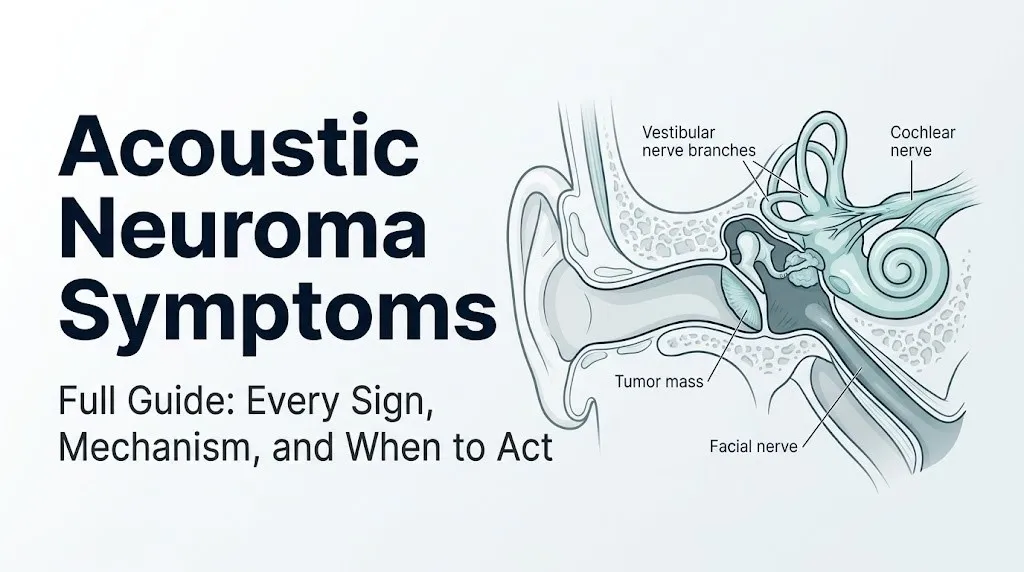
An acoustic neuroma is a benign, slowly growing tumor arising from the Schwann cells that produce myelin for the vestibular branch of cranial nerve VIII (the vestibulocochlear nerve), most commonly at the Obersteiner-Redlich zone, the anatomical transition point between central nervous system myelin (produced by oligodendrocytes) and peripheral nervous system myelin (produced by Schwann cells), located approximately 8 to 12 millimeters from the brainstem.
The molecular cause of acoustic neuroma is loss of functional merlin protein (also called schwannomin), encoded by the NF2 gene on chromosome 22q12. Merlin normally acts as a tumor suppressor by linking the actin cytoskeleton to cell surface receptor complexes and inhibiting the mTOR and Ras-MAPK cell proliferation signaling pathways. When both copies of the NF2 gene are inactivated in a Schwann cell (by sporadic somatic mutation in non-NF2 cases, or by germline plus somatic mutation in NF2 disease), merlin loss removes the brake on Schwann cell division, allowing the tumor to form.
The name “acoustic neuroma” is technically inaccurate: the tumor arises from the vestibular nerve, not the acoustic (cochlear) nerve, and it is a schwannoma (a Schwann cell tumor) not a neuroma (which implies nerve cell degeneration). The clinically preferred term is vestibular schwannoma. However, “acoustic neuroma” remains the term most widely used by patients and in general health literature.
The NIDCD reports that the vast majority of acoustic neuromas are sporadic (not inherited), arising from somatic NF2 gene mutations in a single Schwann cell. A small subset (approximately 5%) occurs in the context of NF2 disease, where the inherited germline NF2 mutation results in bilateral acoustic neuromas and additional central nervous system tumors. Prior radiation exposure to the head and neck region has been associated with an elevated risk of acoustic neuroma development in several cohort studies.
How Acoustic Neuroma Grows and Why Symptoms Develop
Acoustic neuroma grows from its origin on the vestibular nerve within the internal auditory canal medially toward the cerebellopontine angle cistern, compressing the anatomically adjacent cochlear nerve, facial nerve, trigeminal nerve, and ultimately the cerebellar and brainstem surfaces in a sequence that directly predicts the order and type of symptoms a patient will experience.
Think of the internal auditory canal as a narrow bone tunnel containing four nerve bundles bundled tightly together: the cochlear nerve in the anteroinferior position, the facial nerve (cranial nerve VII) in the anterosuperior position, the superior vestibular nerve in the posterosuperior position, and the inferior vestibular nerve in the posteroinferior position. The tumor begins in one of the vestibular nerve bundles and expands outward. Because the cochlear nerve lies immediately adjacent to the vestibular nerve origin, cochlear nerve compression occurs early in growth, producing hearing loss and tinnitus before any other cranial nerve is involved.
The growth rate of acoustic neuroma is unpredictable. Approximately 45 to 50% of acoustic neuromas demonstrate no measurable growth over a period of several years of observation, according to data from serial MRI surveillance studies published in Otology and Neurotology. Approximately 30 to 40% demonstrate slow growth averaging 1 to 2 mm per year, and a smaller subset (approximately 10 to 15%) demonstrate more rapid growth. This growth variability is part of why watchful waiting with serial MRI is a legitimate management option for small, asymptomatic or minimally symptomatic tumors in older patients.
Adults over 70 with small acoustic neuromas and stable symptoms are more often managed with active surveillance rather than immediate surgery or radiosurgery, because the natural history of slow or no growth in this age group means the risks of treatment may outweigh the risks of observation. Younger adults with NF2 or documented growing tumors typically face earlier treatment decisions.
Key Takeaway: Acoustic neuroma symptoms follow the tumor’s anatomical growth path: hearing loss and tinnitus appear first (cochlear nerve compression), then imbalance (vestibular nerve), then facial numbness (trigeminal nerve), then facial weakness (facial nerve VII), then headache and coordination loss (brainstem and cerebellum) in large tumors. The symptom you have tells you where the tumor is growing.
Acoustic Neuroma Hearing Loss: Mechanism and Pattern
Hearing loss in acoustic neuroma is a sensorineural hearing loss produced by compression of the cochlear nerve fascicles within the internal auditory canal, which disrupts the precisely timed firing patterns of spiral ganglion neurons that encode auditory signals, producing not just reduced hearing sensitivity but a characteristic disproportionate impairment of speech understanding that distinguishes it from cochlear hearing loss.
Normal hearing requires the cochlear nerve to transmit precise temporal patterns of neural firing that encode the frequency, intensity, and timing relationships of sound. Cochlear nerve compression by an acoustic neuroma disrupts this temporal coding before it significantly raises pure tone thresholds, which is why the word recognition score (WRS) (the percentage of single syllable words correctly repeated at a comfortable hearing level) is abnormally low relative to what the pure tone audiogram would predict. This retrocochlearing audiometric pattern is the audiological hallmark of acoustic neuroma.
According to the American Academy of Audiology, a word recognition score that is significantly poorer than expected from the pure tone audiogram is one of the key indicators for ordering an auditory brainstem response (ABR) test and referral to an otolaryngologist or neurotologist for retrocochlear evaluation. The ABR measures the brainstem’s electrical response to auditory clicks and can detect the neural transmission delay caused by cochlear nerve compression even when the pure tone audiogram shows only mild hearing loss.
Acoustic neuroma hearing loss is characteristically unilateral, slowly progressive over months to years, and most pronounced for high-frequency sounds in some cases but may affect all frequencies. Patients frequently report that they can hear sounds but cannot understand speech well, particularly in background noise. This speech-in-noise difficulty reflects the disrupted neural timing that is the specific consequence of cochlear nerve compression rather than cochlear hair cell damage.
People who notice that hearing in one ear is subjectively worse than the other, that conversations are harder to follow on one side, or that they’ve begun consistently turning their head to present one ear in conversations, should request formal audiometric evaluation by an audiologist rather than assuming age-related hearing change is the cause, particularly if the asymmetry is noticeable.
Acoustic Neuroma Tinnitus: Why It Sounds Different From Other Tinnitus
Acoustic neuroma tinnitus is produced by ectopic spontaneous electrical discharge in the compressed and partially demyelinated fibers of the cochlear nerve, which generates phantom auditory signals perceived as a continuous, unilateral ringing, hissing, buzzing, or rushing sound in the affected ear rather than in both ears as typically seen in noise-induced or age-related cochlear tinnitus.
The key clinical feature distinguishing acoustic neuroma tinnitus from the more common forms of tinnitus is strict unilaterality. Tinnitus from cochlear hair cell damage (noise-induced, age-related, or medication-related) typically affects both ears, even if one side is perceived as louder. Acoustic neuroma tinnitus is confined to the affected ear because the ectopic discharge originates in the compressed nerve fibers on that side. Bilateral cochlear tinnitus heard equally in both ears is much less likely to represent acoustic neuroma than persistent tinnitus heard only or predominantly in one ear.
The character of acoustic neuroma tinnitus is typically continuous rather than pulsatile. Pulsatile tinnitus (tinnitus that beats in synchrony with the heartbeat) is a red flag for a different category of conditions, including vascular tumors (glomus jugulare or glomus tympanicum tumors), arteriovenous fistulae, or venous hypertension, and should prompt evaluation by an otolaryngologist or neurotologist for a distinct set of causes from acoustic neuroma.
According to clinical series reported in Otology and Neurotology, unilateral tinnitus as the only initial symptom is present in a meaningful proportion of acoustic neuroma patients, often preceding hearing loss by months or years. Any adult with persistent, unilateral, unexplained tinnitus lasting more than three months should request audiometric evaluation and, if the audiogram shows asymmetry, further evaluation including ABR testing.
Older adults often accept unilateral tinnitus as an expected feature of aging, delaying evaluation. Because tinnitus from acoustic neuroma may be the only symptom for an extended period before hearing loss or imbalance becomes apparent, persistent unilateral tinnitus without an identified cause in any adult warrants audiological evaluation regardless of age.
Acoustic Neuroma Dizziness and Balance Problems
Acoustic neuroma causes a chronic, low-grade unsteadiness and imbalance rather than the acute spinning vertigo associated with other vestibular conditions, because the gradual compression of the superior and inferior vestibular nerve branches allows the brainstem’s vestibular nuclei to partially compensate for the developing asymmetry in resting vestibular nerve firing rates through a process called vestibular compensation.
The vestibular system works by comparing the firing rate of vestibular nerve signals from each ear and interpreting the difference as head movement or position change. Under normal conditions, the two vestibular nerves fire symmetrically at rest, and any asymmetry signals head movement to the brain. When a tumor gradually compresses one vestibular nerve, the resting firing rate on that side decreases slowly. The brainstem vestibular nuclei adapt to the new lower baseline over weeks to months, reducing the perceived vertigo but leaving the patient chronically unsteady, particularly in visually complex environments or in the dark when visual cues cannot substitute for vestibular input.
This pattern of chronic, nonspecific unsteadiness is clinically different from the episodic, severe spinning vertigo lasting minutes to hours that characterizes Meniere’s disease, or the severe continuous vertigo lasting days that characterizes vestibular neuritis. Acoustic neuroma imbalance is characteristically worse at night (when visual compensation is reduced), on uneven terrain, and when walking in visually complex environments like busy shopping areas.
The clinical significance of this distinction: a patient who reports that their dizziness or unsteadiness is episodic, severe, and associated with a definite spinning sensation lasting minutes to hours is less likely to have acoustic neuroma as the primary cause than a patient who describes chronic unsteadiness and difficulty with balance in the dark or on uneven ground.
Women with pre-existing anxiety or vestibular migraine may experience an amplified perception of their vestibular symptoms, making it clinically challenging to distinguish acoustic neuroma-related vestibular dysfunction from anxiety-driven somatic amplification of normal vestibular asymmetry. This is one reason audiometric evaluation and ABR testing rather than symptom history alone are required for diagnosis.
Key Takeaway: Acoustic neuroma dizziness is a persistent, low-grade unsteadiness that worsens in the dark or on uneven ground, not the sudden spinning vertigo of Meniere’s disease. The chronic quality reflects successful (but incomplete) brainstem compensation for gradually asymmetric vestibular nerve input.
Acoustic Neuroma Facial Numbness and Trigeminal Nerve Involvement
Facial numbness in acoustic neuroma is produced by compression of the trigeminal nerve (cranial nerve V) as the tumor expands from the internal auditory canal into the cerebellopontine angle cistern, where the trigeminal nerve’s sensory root enters the pons at a position directly superior and medial to the tumor’s growth trajectory.
The trigeminal nerve is the largest cranial nerve and carries sensory signals from the face (via its ophthalmic V1, maxillary V2, and mandibular V3 divisions) to the brainstem, as well as motor signals to the muscles of mastication. When acoustic neuroma compresses the trigeminal root or ganglion, patients experience tingling, numbness, or a sensation of facial “heaviness” in one or more divisions of the trigeminal territory. The numbness may affect the forehead (V1), cheek (V2), jaw or lower lip (V3), or any combination depending on the specific aspect of the nerve compressed.
According to clinical observational data published in Neurosurgery journal, trigeminal symptoms (facial numbness, tingling, or in some cases trigeminal neuralgia-like pain) are present in a clinically meaningful proportion of medium to large acoustic neuromas and are virtually absent in small tumors confined to the IAC. The presence of facial numbness therefore serves as a clinical indicator that the tumor has grown beyond the IAC fundus into the CPA cistern and has reached a size where trigeminal involvement occurs.
The practical implication: any patient with known acoustic neuroma who develops new facial numbness or tingling that was not present at their last evaluation should report this to their managing neurotologist or neurosurgeon, as it likely indicates tumor growth into the CPA territory and may alter the treatment recommendation.
Older adults may attribute mild facial tingling or numbness to dental problems, sinus pressure, or cardiovascular disease rather than a neurological cause, potentially delaying the recognition that the acoustic neuroma has grown into the CPA territory.
Acoustic Neuroma Facial Weakness and Cranial Nerve VII Compression
Facial weakness in acoustic neuroma results from compression of the facial nerve (cranial nerve VII), which runs in the anterosuperior compartment of the internal auditory canal just above the cochlear nerve, and whose function can be impaired by direct tumor compression as the acoustic neuroma expands within the IAC.
The facial nerve controls all voluntary and involuntary facial expression muscles through its five peripheral branches (temporal, zygomatic, buccal, marginal mandibular, and cervical). Compression of the facial nerve by acoustic neuroma can produce a spectrum of facial weakness: from subtle asymmetry of the smile visible on close observation, to incomplete eye closure, to complete ipsilateral facial paralysis (House-Brackmann Grade VI) in the most severe cases.
Facial weakness from acoustic neuroma is relatively uncommon as a presenting symptom because the facial nerve is anatomically more resistant to compression by a slowly growing tumor than the cochlear nerve. According to surgical case series published in Otology and Neurotology, preoperative facial weakness is present in only a minority of acoustic neuroma patients at the time of diagnosis but becomes a more frequent surgical risk during tumor removal, particularly for large tumors where the facial nerve is stretched over the tumor surface.
The clinical importance of facial weakness in acoustic neuroma: any new or progressive weakness of one side of the face in a person with known acoustic neuroma requires urgent evaluation by the managing neurotologist or neurosurgeon, as it may indicate that the tumor is growing rapidly or has reached a size where facial nerve surgical risk is increasing. Any acute onset of complete facial paralysis alongside other acoustic neuroma symptoms requires same-day emergency evaluation to distinguish tumor-related facial nerve compression from other causes of acute facial nerve palsy.
People with pre-existing conditions affecting facial symmetry (prior Bell’s palsy, TMJ disorder with reduced muscle use, stroke history) may have difficulty recognizing new subtle facial weakness attributable to acoustic neuroma. A photograph comparison between a recent image and an older one can sometimes help identify subtle progressive asymmetry before it is clinically obvious.
Acoustic Neuroma Headache and Symptoms of Brainstem Compression
Headache in acoustic neuroma is a late symptom that appears when the tumor has grown large enough to compress the lateral surface of the cerebellar hemisphere or to displace the brainstem, producing increased intracranial pressure or direct compression of pressure-sensitive dural and vascular structures in the posterior fossa.
Large acoustic neuromas (greater than 2.5 to 3 cm) in the cerebellopontine angle can compress the ipsilateral cerebellar hemisphere, producing ipsilateral cerebellar ataxia: a broad-based, unsteady gait with falls toward the affected side, difficulty with fine finger movements on the affected side (finger-nose testing), and slurred speech from cerebellar involvement of the speech motor system. This cerebellar ataxia is distinct from the vestibular imbalance of smaller tumors and indicates that the tumor has grown into a category where treatment is typically urgently recommended.
The most clinically dangerous scenario is compression of the fourth ventricle by a very large acoustic neuroma, which can obstruct cerebrospinal fluid (CSF) flow and produce obstructive hydrocephalus: a rapid increase in intracranial pressure producing severe headache, nausea, vomiting, visual disturbances (diplopia or blurred vision from papilledema), and altered consciousness. This is a neurological emergency.
People with diagnosed large acoustic neuromas who develop a new severe headache (particularly worse in the morning or with straining), vomiting without nausea, double vision, or any decline in consciousness or coordination must go to the emergency room immediately. These presentations can indicate obstructive hydrocephalus requiring emergency neurosurgical intervention.
Adults over 60 who develop new persistent headache, particularly in the context of any prior diagnosis of acoustic neuroma or any history of hearing and balance symptoms, should have neuroimaging performed as part of their headache evaluation rather than assuming a primary headache disorder.
Key Takeaway: Headache, ataxia (coordination problems), or any alteration in consciousness in a person with known or suspected acoustic neuroma is not a minor symptom. It signals large tumor mass with potential brainstem compression, and requires emergency room evaluation rather than a scheduled neurotologist appointment.
Early Acoustic Neuroma Symptoms: Small Tumor Stage
Early acoustic neuroma symptoms, occurring when the tumor is still confined within the internal auditory canal and measures less than approximately 1 centimeter, are typically limited to the cochlear and vestibular nerve territory and include subtle asymmetric hearing loss, unilateral tinnitus, and mild nonspecific unsteadiness that are easily attributed to other more common conditions.
The clinical problem with early acoustic neuroma is that the symptoms are indistinguishable on symptoms alone from far more common, benign conditions. Unilateral tinnitus occurs in a much larger population due to noise exposure, aging, or medications than will ever have acoustic neuroma. Mild unsteadiness is extremely common in the general adult population. The early hearing loss of acoustic neuroma, confined to one ear and often affecting speech clarity more than pure tone thresholds, may not be noticed until the person’s daily communication is already impaired.
The symptoms that should specifically raise concern for an acoustic neuroma evaluation rather than watchful observation of “normal aging changes”:
- Tinnitus that is strictly unilateral and persistent for more than three months without a clear cause
- Hearing loss in one ear that is clearly worse than the other, particularly if affecting speech understanding
- Difficulty understanding speech on the phone when using the affected ear (many patients notice they switch the phone to the other ear)
- Unsteadiness that is worse in the dark or on uneven ground, with preserved balance in bright, flat environments
- Any combination of unilateral hearing symptoms with unsteadiness in an adult over 40
According to the AAO-HNS, any adult with asymmetric sensorineural hearing loss greater than 15 dB at three consecutive frequencies or greater than 15 dB difference in word recognition score between ears should be evaluated for retrocochlear pathology including acoustic neuroma.
Late Stage Acoustic Neuroma Symptoms: Large Tumor and Brainstem Involvement
Late stage acoustic neuroma symptoms develop when the tumor has grown to a size (typically greater than 2.5 cm) that extends well into the cerebellopontine angle cistern, compressing the adjacent trigeminal nerve, facial nerve, cerebellum, and eventually the brainstem, producing a multi-neurological syndrome that goes far beyond the cochlear and vestibular symptoms of the early stage.
By the time an acoustic neuroma reaches the large tumor stage, most patients have experienced years of progressive hearing loss and imbalance. The late stage is characterized by the emergence of symptoms from neural structures compressed outside the IAC:
- Facial numbness and tingling: from trigeminal nerve compression in the CPA
- Facial weakness: from facial nerve compression that has advanced beyond tolerable levels
- Dysphagia and hoarseness: from lower cranial nerve (IX, X) compression against the brainstem in very large tumors
- Progressive gait ataxia: from cerebellar hemisphere compression
- Headache: from posterior fossa mass effect and dural stretch
- Nausea and vomiting: from increased intracranial pressure or direct brainstem compression
The transition from the early to late stage in acoustic neuroma represents a change in the urgency of treatment decision-making. Small acoustic neuromas can reasonably be managed with active surveillance (serial MRI every 6 to 12 months). Large tumors with brainstem compression symptoms are generally recommended for prompt treatment, typically surgical resection or, in selected cases, stereotactic radiosurgery, at a specialized neurosurgical center.
People who were diagnosed years ago with a “small acoustic neuroma” managed with surveillance and who now notice new symptoms (facial numbness, worsening coordination, new headache) should contact their neurotologist or neurosurgeon promptly rather than waiting for their next scheduled imaging appointment.
Sudden Hearing Loss as an Acoustic Neuroma Symptom
Sudden sensorineural hearing loss (SSNHL) occurring within the context of acoustic neuroma is a medical emergency that requires same-day evaluation, gadolinium-enhanced MRI to rule out retrocochlear pathology, and potentially urgent corticosteroid treatment within a narrow treatment window.
According to AAO-HNS clinical practice guidelines updated in 2019, sudden sensorineural hearing loss is defined as a decrease of 30 dB or more in pure tone average occurring over 72 hours or less, and warrants urgent evaluation including a complete audiological assessment and retrocochlear imaging. The guidelines specifically identify acoustic neuroma as a condition that must be excluded by gadolinium-enhanced MRI in any patient with sudden SNHL.
Approximately 10 to 15% of acoustic neuromas present with sudden hearing loss as their first recognizable symptom, according to clinical series in Otology and Neurotology. The mechanism is thought to involve vascular compromise of the internal auditory artery (a terminal vessel with no collateral supply) or intratumoral hemorrhage producing sudden pressure on the cochlear nerve.
The treatment window for sudden hearing loss is narrow. Corticosteroid therapy (administered systemically or through intratympanic injection into the middle ear cavity) is most effective when initiated within 72 hours of symptom onset and loses efficacy progressively after this period. Delayed evaluation is one of the most common preventable causes of permanent hearing loss following SSNHL.
If you or someone with you experiences sudden, unexplained hearing loss in one ear, contact an otolaryngologist for same-day evaluation or go to the nearest emergency room. Do not wait to see if the hearing returns on its own, because the 72-hour treatment window is the only pharmacological opportunity to improve outcomes.
Women who are pregnant and experience sudden unilateral hearing loss require the same urgent evaluation, though corticosteroid route and dosing decisions must be made in conjunction with their obstetrician-gynecologist given pregnancy-related considerations.
Key Takeaway: Sudden unexplained hearing loss in one ear is a same-day medical emergency. The 72-hour corticosteroid treatment window is real, and acoustic neuroma must be excluded by gadolinium-enhanced MRI. Do not wait and see.
Acoustic Neuroma vs. Meniere’s Disease: Distinguishing the Symptoms
Acoustic neuroma and Meniere’s disease (endolymphatic hydrops) share overlapping symptoms including unilateral hearing loss, unilateral tinnitus, and dizziness, but they differ in the temporal pattern, character of dizziness, and audiometric features in ways that allow clinical differentiation, though gadolinium-enhanced MRI remains the only method to definitively rule out acoustic neuroma.
| Feature | Acoustic Neuroma | Meniere’s Disease |
|---|---|---|
| Hearing loss pattern | Progressive, slowly worsening over months to years | Fluctuating, worse during attacks, partially recovers |
| Tinnitus character | Constant, continuous, non-fluctuating | Variable, often worse before and during attacks |
| Dizziness type | Chronic unsteadiness, non-episodic | Episodic severe spinning vertigo lasting 20 min to hours |
| Aural fullness | Rare | Characteristic, often precedes attacks |
| Audiometric pattern | Retrocochlearing (poor WRS disproportionate to tones) | Cochlear (low-frequency SNHL, WRS proportionate to tones) |
| ABR result | Abnormal (prolonged latency, abnormal morphology) | Normal in most cases |
| MRI finding | Tumor visible on gadolinium-enhanced MRI | Normal MRI (endolymph not visible on standard MRI) |
The most reliable clinical differentiators are the character of dizziness and the audiometric pattern. Meniere’s disease produces episodic, severe spinning vertigo lasting 20 minutes to several hours with complete resolution between attacks. Acoustic neuroma produces chronic, persistent unsteadiness rather than episodic spinning. The audiometric hallmark of Meniere’s is a low-frequency sensorineural hearing loss with a cochlear pattern (word recognition scores proportionate to pure tone thresholds), which is the opposite of the retrocochlearing pattern of acoustic neuroma.
Despite these clinical distinguishing features, definitive differentiation requires gadolinium-enhanced MRI, because a proportion of Meniere’s disease patients also have asymmetric audiometric findings that overlap with the acoustic neuroma pattern. Older adults with both conditions simultaneously (which does occur) present a particular diagnostic challenge that requires specialist evaluation.
Acoustic Neuroma Symptoms in Neurofibromatosis Type 2 (NF2)
Acoustic neuroma symptoms in neurofibromatosis type 2 (NF2) follow the same neuroanatomical progression as sporadic acoustic neuromas but occur earlier in life (often in adolescence or young adulthood), present bilaterally rather than unilaterally, and are accompanied by additional central nervous system tumors (meningiomas, spinal schwannomas, ependymomas) that create a more complex and severe overall neurological picture.
NF2 is caused by germline mutation in both copies of the NF2 gene (inherited as an autosomal dominant condition) or by spontaneous new mutation in approximately 50% of NF2 cases. The diagnostic hallmark of NF2 is bilateral vestibular schwannomas, which the National Institute of Neurological Disorders and Stroke (NINDS) uses as a defining criterion for NF2 diagnosis.
In NF2, hearing loss is typically the first symptom, developing in adolescence or early adulthood and progressing on both sides, creating the risk of bilateral profound deafness that is not a concern in sporadic unilateral acoustic neuroma. Patients with NF2 may also experience:
- Tinnitus in both ears simultaneously
- Balance problems that are more severe than in unilateral sporadic cases because both vestibular pathways are affected
- Visual symptoms from meningiomas or schwannomas affecting the optic pathway or eye movement cranial nerves
- Weakness or sensory loss in the limbs from spinal cord tumors or spinal schwannomas
Anyone with a confirmed NF2 diagnosis should be managed by a multidisciplinary team including a neurotologist, a neurosurgeon with expertise in NF2, a neuro-oncologist, an audiologist, and a genetic counselor, at a center with specific NF2 expertise. Annual hearing assessment and regular MRI surveillance of the brain and spine are standard components of NF2 monitoring.
Children of a parent with NF2 have a 50% chance of inheriting the NF2 gene mutation and should be evaluated by a geneticist and screened with audiology and MRI from adolescence, as early detection of bilateral acoustic neuromas allows more preservation of hearing function with timely treatment.
How Acoustic Neuroma Symptoms Are Diagnosed and Evaluated
Acoustic neuroma symptoms are diagnosed through a systematic clinical and audiological evaluation that progresses from pure tone audiometry and word recognition testing through auditory brainstem response (ABR) testing to gadolinium-enhanced MRI of the internal auditory canals and posterior fossa, with MRI being the definitive imaging standard that directly visualizes the tumor.
The standard diagnostic pathway for suspected acoustic neuroma:
- Pure tone audiometry and word recognition score (WRS) testing by an audiologist: identifies the pattern of hearing loss, quantifies the asymmetry between ears, and calculates the word recognition score to assess for the retrocochlearing pattern. Any asymmetric SNHL greater than 15 dB at three consecutive frequencies or WRS discrepancy greater than 15% between ears triggers further evaluation.
- Auditory brainstem response (ABR) testing: an electrophysiological test measuring the brainstem’s electrical response to auditory click stimuli. In acoustic neuroma, the ABR shows prolonged latency of wave V (the component generated by the inferior colliculus, reflecting the delay introduced by cochlear nerve compression) and interaural wave V latency differences. ABR sensitivity is approximately 95% for tumors larger than 1 cm but is lower for small tumors, which is why it has been partially superseded by gadolinium MRI as the primary diagnostic tool.
- Gadolinium-enhanced MRI of the IAC and posterior fossa: the gold standard for acoustic neuroma diagnosis. Gadolinium contrast agent accumulates in the highly vascular tumor tissue, producing a bright enhancement on T1-weighted MRI images that is visible even for very small intracanalicular tumors. The FIESTA or CISS MRI sequences (heavily T2-weighted) can also detect small tumors as filling defects in the fluid-filled IAC without gadolinium.
- Videonystagmography (VNG) or electronystagmography (ENG) with caloric testing: measures the vestibular function of each ear separately by introducing warm and cold water or air into the external ear canal and measuring the resulting eye movements. A reduced caloric response on one side (canal paresis) indicates reduced vestibular nerve function on that side and is present in most acoustic neuroma patients with significant vestibular nerve compression.
Any person whose primary care physician identifies an asymmetric audiogram should be referred to an otolaryngologist or neurotologist for ABR testing and, if warranted, gadolinium-enhanced MRI of the IAC rather than being reassured without further workup.
Acoustic Neuroma Red Flags and When to Seek Urgent Care
Certain acoustic neuroma symptoms require urgent or emergency evaluation that cannot wait for a routine specialist appointment, because they indicate either rapidly progressing tumor growth or a life-threatening complication of large tumor mass.
Symptoms requiring same-day urgent evaluation by an otolaryngologist, neurotologist, or emergency medicine physician:
- Sudden, unexplained hearing loss in one ear occurring over 72 hours or less (possible SSNHL with acoustic neuroma as a potential cause; requires same-day evaluation and potential corticosteroid treatment within the 72-hour window)
- New progressive facial weakness on one side of the face in a person with known or suspected acoustic neuroma (indicates facial nerve compression that may be worsening rapidly)
- Any new neurological symptom in a person with a known acoustic neuroma including new facial numbness, new difficulty swallowing, or new change in voice quality
Emergency Symptoms: When to Call 911 or Go to the ER
Certain symptoms associated with acoustic neuroma require immediate emergency evaluation. Do not wait to see if these resolve on their own.
Call 911 or go to the nearest emergency room immediately if you experience:
- Sudden severe headache in the posterior head or neck region in a person with known acoustic neuroma, particularly if worse when lying flat or straining (possible obstructive hydrocephalus from fourth ventricle compression, a neurological emergency)
- Progressive loss of coordination, staggering gait, or inability to walk steadily that develops over hours to days in a person with known acoustic neuroma (possible cerebellar compression from rapid tumor growth or hemorrhage)
- Any alteration in consciousness, confusion, or unusual drowsiness in a person with a large acoustic neuroma (possible raised intracranial pressure from hydrocephalus)
- Nausea and vomiting with a new severe headache in a person with acoustic neuroma who has not had these symptoms before (possible obstructive hydrocephalus)
- Sudden complete loss of hearing and severe vertigo simultaneously in a known acoustic neuroma patient (possible intratumoral hemorrhage)
These presentations can indicate obstructive hydrocephalus, brainstem compression, or intratumoral hemorrhage, all of which require emergency neurosurgical assessment in a hospital with posterior fossa neurosurgical capability, not an urgent care center or a next-day specialist appointment.
Frequently Asked Questions About Acoustic Neuroma Symptoms
What are the first symptoms of acoustic neuroma?
The first symptoms of acoustic neuroma are typically progressive unilateral hearing loss and unilateral tinnitus (ringing or buzzing in one ear), reflecting early cochlear nerve compression within the internal auditory canal.
Chronic mild unsteadiness, particularly noticeable in the dark or on uneven surfaces, is also an early symptom produced by gradual vestibular nerve compression.
According to the National Institute on Deafness and Other Communication Disorders, these early symptoms are frequently attributed to other conditions for years before an acoustic neuroma diagnosis is made.
Can acoustic neuroma cause sudden hearing loss?
Approximately 10 to 15% of acoustic neuromas present with sudden sensorineural hearing loss as their first recognizable symptom, according to clinical series in Otology and Neurotology.
Sudden hearing loss from acoustic neuroma is thought to result from vascular compromise of the internal auditory artery or intratumoral hemorrhage producing acute pressure on the cochlear nerve.
Any sudden, unexplained hearing loss in one ear requires same-day evaluation by an otolaryngologist or at the nearest emergency room, because corticosteroid treatment within 72 hours is the standard of care and acoustic neuroma must be excluded by gadolinium-enhanced MRI.
What does acoustic neuroma tinnitus sound like?
Acoustic neuroma tinnitus is typically a continuous, unilateral ringing, hissing, buzzing, or rushing sound perceived only in the affected ear, as opposed to bilateral tinnitus heard in both ears.
It is produced by ectopic spontaneous electrical discharge in compressed cochlear nerve fibers, generating phantom auditory signals from one side only.
Pulsatile tinnitus that beats rhythmically with the heartbeat is not characteristic of acoustic neuroma and suggests a different category of conditions, including vascular tumors or arteriovenous fistulae, requiring separate evaluation.
Is acoustic neuroma dizziness constant or episodic?
Acoustic neuroma dizziness is characteristically a chronic, persistent unsteadiness rather than episodic severe spinning vertigo, because the slow compression of the vestibular nerve allows the brainstem’s vestibular nuclei to partially compensate for the developing asymmetry.
This persistent, low-grade unsteadiness is distinctly different from the episodic spinning vertigo lasting 20 minutes to hours that characterizes Meniere’s disease, which is the most clinically important condition to distinguish from acoustic neuroma.
If someone is experiencing severe episodic spinning vertigo rather than chronic unsteadiness, Meniere’s disease is a more likely primary diagnosis than acoustic neuroma, though gadolinium MRI is still required to exclude acoustic neuroma definitively.
How do you know if you have an acoustic neuroma?
Acoustic neuroma is diagnosed by gadolinium-enhanced MRI of the internal auditory canals and posterior fossa, which directly visualizes the tumor as an enhancing mass within or adjacent to the internal auditory canal.
Clinical indicators that warrant audiological evaluation and possible imaging include unilateral tinnitus lasting more than three months, asymmetric sensorineural hearing loss with poor word recognition in the affected ear, and persistent unexplained unsteadiness.
An audiologist performs the pure tone audiogram and word recognition score testing that identifies the retrocochlearing audiometric pattern prompting referral to an otolaryngologist or neurotologist for auditory brainstem response testing and MRI.
When does acoustic neuroma become an emergency?
Acoustic neuroma becomes a neurological emergency when it reaches a size that compresses the fourth ventricle, producing obstructive hydrocephalus with raised intracranial pressure, manifesting as severe posterior headache, vomiting, coordination loss, or altered consciousness.
Any person with a known acoustic neuroma who develops these symptoms should call 911 or go to the nearest emergency room immediately, as obstructive hydrocephalus requires emergency neurosurgical evaluation.
Sudden complete hearing loss with severe vertigo in a person with a known acoustic neuroma also requires emergency evaluation, as this may indicate intratumoral hemorrhage.
Closing
The single most actionable fact from this article: if you have persistent unilateral tinnitus, progressive hearing difficulty in one ear, or unexplained chronic unsteadiness, request a formal audiological evaluation including a pure tone audiogram and word recognition score. That audiogram is the gateway to retrocochlear screening. If the word recognition score is disproportionately poor relative to the pure tone thresholds, that is the specific finding that prompts the next step: auditory brainstem response testing and gadolinium-enhanced MRI.
For sudden hearing loss in one ear, act the same day. Contact an otolaryngologist for urgent evaluation or go to the nearest emergency room. The 72-hour corticosteroid treatment window closes regardless of what caused the sudden hearing loss, and acoustic neuroma must be on the differential until MRI excludes it.
If you have a confirmed acoustic neuroma already being monitored, any new symptom (facial numbness, new headache, new coordination problems) means contacting your neurotologist or neurosurgeon before your next scheduled imaging, not after.