Eustachian Tube Dysfunction Symptoms: Full Guide 2026
Eustachian tube dysfunction symptoms include ear fullness, muffled hearing, tinnitus, popping or clicking sounds, ear pain, and dizziness, all produced by the failure of the Eustachian tube to adequately equalize pressure between the middle ear and the surrounding atmosphere. The specific symptoms and their severity depend on whether the tube is failing to open (obstructive ETD, the most common type) or failing to close (patulous ETD, less recognized but frequently misdiagnosed).
According to the American Academy of Otolaryngology-Head and Neck Surgery (AAO-HNS), ETD is one of the most frequently encountered ear complaints in both primary care and otolaryngology practice. The condition directly affects the middle ear pressure equalization system, and when left unmanaged over months, it can progress to complications including otitis media with effusion and, in severe cases, cholesteatoma formation, an abnormal skin growth within the middle ear that requires surgical treatment.
This article covers every recognized symptom of eustachian tube dysfunction with its specific anatomical and physiological mechanism, distinguishes obstructive from patulous ETD, explains how symptoms change with altitude and flying, addresses who is at highest risk, and gives you clear clinical thresholds for when emergency evaluation is necessary.
Eustachian Tube Dysfunction Symptoms: A Clinical Overview
Eustachian tube dysfunction symptoms are produced when the tube connecting the middle ear to the back of the throat fails to regulate middle ear pressure normally, causing a cascade of mechanical and sensory changes in the ear that range from mild intermittent fullness to persistent hearing impairment.
The symptom picture of ETD falls into two distinct clinical categories depending on which type of dysfunction is present. In obstructive ETD (the more prevalent form), the tube cannot open sufficiently to equalize pressure, producing negative middle ear pressure with its associated symptoms. In patulous ETD, the tube stays open and fails to close, producing a completely different and often opposite set of symptoms.
| Symptom | Obstructive ETD | Patulous ETD |
|---|---|---|
| Ear fullness | Yes, worse with swallowing | Yes, worse when upright |
| Muffled hearing | Yes, persistent | Intermittent, voice-dependent |
| Autophony | No | Yes, pronounced |
| Breath sounds in ear | No | Yes, audible own breathing |
| Relief with swallowing | Sometimes, temporarily | No relief; may worsen |
| Relief with dependent head position | No | Yes, bending forward relieves |
| Tinnitus | Low-pitched, variable | Synchronous with breathing |
According to the National Institute on Deafness and Other Communication Disorders (NIDCD), ear disorders affecting the middle ear and its pressure regulation are among the most prevalent causes of hearing-related complaints in both children and adults. Understanding which symptom pattern is present guides both the diagnosis and the treatment direction, since treatments effective for obstructive ETD can worsen patulous ETD.
Adults over 60 with multiple medical comorbidities may present with ETD symptoms that overlap with other ear conditions, including Meniere’s disease and sudden sensorineural hearing loss (SSNHL). These overlapping presentations require clinical evaluation by an otolaryngologist rather than self-management.
What Is Eustachian Tube Dysfunction and How Does It Work?
Eustachian tube dysfunction occurs when the tube connecting the middle ear space to the nasopharynx fails to perform its three primary functions: ventilating the middle ear, draining secretions from the middle ear toward the nasopharynx, and protecting the middle ear from nasopharyngeal sound pressure and pathogens.
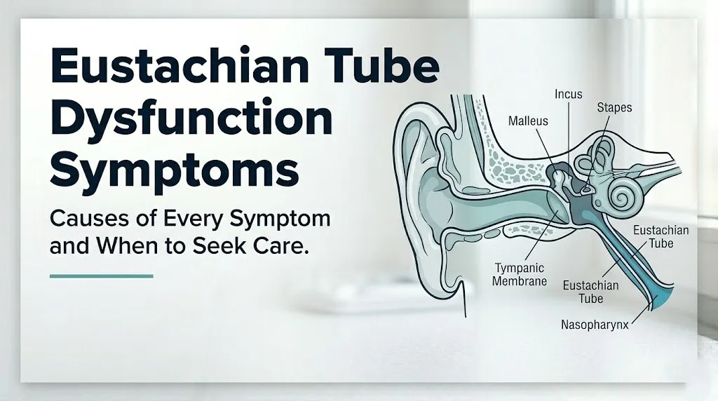
The Eustachian tube (also called the auditory tube or pharyngotympanic tube) measures approximately 35 mm in adults and runs at a roughly 45-degree angle from the tympanic ostium (ear end) down to the nasopharyngeal ostium (throat end). It consists of a bony portion (the tympanic two-thirds, which is normally open) and a cartilaginous portion (the nasopharyngeal one-third, which is normally closed at rest by elastic cartilage and the surrounding peritubal fat pad).
Think of the Eustachian tube like a pressure-release valve on a sealed container. The middle ear is the container. The valve (the tube’s cartilaginous portion) stays closed most of the time to protect the middle ear from bacteria and sound pressure fluctuations in the throat. Every time you swallow or yawn, the tensor veli palatini muscle pulls the valve open for a fraction of a second, releasing any pressure differential that has built up. When that valve stops opening reliably, pressure accumulates and symptoms begin.
The tensor veli palatini is the primary muscle responsible for active tube opening. It is innervated by the medial pterygoid nerve, a branch of the mandibular division of cranial nerve V (trigeminal nerve). This specific innervation is clinically relevant in patients with cranial nerve V disorders, temporomandibular joint dysfunction, or conditions affecting the skull base, all of which can impair tensor function and produce secondary ETD.
People with a history of cleft palate (even after surgical repair) have structurally abnormal tensor veli palatini muscles that cannot efficiently open the Eustachian tube. This population has the highest rates of chronic ETD and recurrent otitis media with effusion of any pediatric group, according to clinical research published in the International Journal of Pediatric Otorhinolaryngology.
Ear Fullness and Pressure as Symptoms of Eustachian Tube Dysfunction
Ear fullness in eustachian tube dysfunction is produced by progressive negative middle ear pressure that displaces the tympanic membrane (eardrum) inward from its normal position, creating a sensation of blockage or pressure deep in the ear canal that the person cannot relieve by touching or rubbing the external ear.
When the Eustachian tube fails to open during swallowing and yawning, the middle ear’s resident air is gradually absorbed by the mucosa lining the tympanic cavity without replacement. The result is a measurable drop in middle ear pressure below atmospheric pressure, typically ranging from negative 100 to negative 300 daPa (decapascals) in symptomatic ETD, as documented by tympanometry (a test that measures tympanic membrane compliance and middle ear pressure).
As middle ear pressure drops further below atmospheric pressure, the tympanic membrane is drawn progressively inward. This inward displacement stiffens the membrane, reducing its normal flexibility. The stiffness is what produces the sensation of fullness: the ear feels blocked because the tympanic membrane is under mechanical tension in an abnormal position.
Aural fullness in ETD has a characteristic behavior that distinguishes it from fullness caused by cerumen impaction or other causes. In ETD, the fullness is often:
- Temporarily relieved by swallowing, yawning, or the Valsalva maneuver (gently blowing against pinched nostrils), because these actions briefly force the Eustachian tube open
- Worsened by altitude changes (flying up or descending in an airplane, driving up a mountain)
- Fluctuating across the day, often better in the morning and worse after prolonged upright activity
- Associated with a popping or crackling sensation when the tube momentarily equalizes
Older adults and people with reduced jaw mobility from temporomandibular joint disorders may have less efficient swallowing-triggered tube opening, making their aural fullness more persistent and less responsive to standard Valsalva maneuver attempts.
Muffled Hearing as a Symptom of Eustachian Tube Dysfunction
Muffled hearing in eustachian tube dysfunction occurs because the inward displacement of the tympanic membrane from negative middle ear pressure reduces the membrane’s ability to vibrate efficiently in response to sound waves, impairing the transmission of acoustic energy through the ossicular chain to the oval window and cochlea.
Sound reaches the cochlea through a specific mechanical pathway: sound waves travel through the external ear canal, vibrate the tympanic membrane, transmit that vibration through the ossicular chain (the three smallest bones in the human body: the malleus, incus, and stapes), and then transfer the vibration to the fluid of the inner ear through the oval window. The efficiency of this pathway depends on the tympanic membrane’s ability to move freely in response to sound pressure.
When the tympanic membrane is held under tension by negative middle ear pressure, its compliance decreases. A stiff membrane transmits sound less efficiently than a mobile one. The result is a conductive hearing loss, typically of 20 to 30 decibels (dB) in mild to moderate ETD, which subjectively sounds like listening through a thick blanket or through water.
This is conductive hearing loss (affecting sound transmission through the outer and middle ear), not sensorineural hearing loss (affecting the cochlea or auditory nerve). This distinction matters clinically. Conductive hearing loss from ETD is generally reversible once middle ear pressure is normalized. Sensorineural hearing loss is not, and a sudden onset of hearing loss that does not improve with swallowing or the Valsalva maneuver requires urgent evaluation to rule out SSNHL.
People with pre-existing sensorineural hearing loss from noise exposure, aging, or ototoxic medications who develop ETD face particular difficulty because the two types of hearing loss compound each other. An audiologist should perform a formal pure tone audiogram to distinguish the conductive component (reversible) from any pre-existing sensorineural component (not reversible with ETD treatment).
Key Takeaway: Muffled hearing from ETD is conductive hearing loss caused by reduced tympanic membrane compliance from negative middle ear pressure. It is generally reversible. Hearing loss that does not improve with swallowing, does not fluctuate, or develops suddenly and without a cold or altitude change requires same-day evaluation to rule out sudden sensorineural hearing loss.
Tinnitus as a Symptom of Eustachian Tube Dysfunction
Tinnitus in eustachian tube dysfunction is a low-pitched, often described as a roaring, rushing, or humming sound in the affected ear, produced by the abnormal mechanical state of the tympanic membrane and middle ear system under the influence of negative middle ear pressure or middle ear fluid accumulation.
The tinnitus of ETD has specific characteristics that distinguish it from the high-pitched tinnitus typically associated with sensorineural hearing damage (from noise exposure or cochlear pathology). ETD tinnitus is usually low-frequency, fluctuating in intensity, synchronous with the heartbeat in some cases (pulsatile tinnitus), and often changes in character when the person swallows, yawns, or performs the Valsalva maneuver.
According to the American Academy of Otolaryngology-Head and Neck Surgery (AAO-HNS), tinnitus that changes character with pressure equalization maneuvers or swallowing is a reliable clinical indicator of a middle ear or Eustachian tube component to the tinnitus, rather than a purely sensorineural origin.
Pulsatile tinnitus deserves a specific safety note. While ETD can produce mild pulsatile tinnitus from the mechanical transmission of vascular pulsations through the middle ear fluid, persistent pulsatile tinnitus that is rhythmically synchronized with the heartbeat requires evaluation by an otolaryngologist and potentially vascular imaging to rule out a vascular lesion (such as a glomus tumor, carotid artery stenosis, or arteriovenous fistula) as the source. These are uncommon but important conditions that can present identically to ETD tinnitus on symptom description alone.
Older adults and people with cardiovascular disease who develop new pulsatile tinnitus should have this symptom specifically evaluated as a primary complaint rather than assuming ETD is the cause.
Popping, Clicking, and Crackling Sounds in Eustachian Tube Dysfunction
Popping, clicking, and crackling sounds in eustachian tube dysfunction are produced by the sudden equalization of pressure across the tympanic membrane when the Eustachian tube briefly opens, causing an abrupt movement of the membrane and middle ear contents that generates an audible and sometimes sensible acoustic event.
The mechanical basis of these sounds is the release of pressure differential. When negative middle ear pressure has built up because the Eustachian tube hasn’t been opening adequately, any sudden tube opening (triggered by swallowing, yawning, chewing, or a jaw movement) rapidly equilibrates that pressure difference. The tympanic membrane, held inward by negative pressure, snaps back toward its neutral position when pressure is equalized. That rapid membrane movement is the “pop.”
Crackling sounds, which are softer and more continuous than discrete pops, often reflect the movement of middle ear fluid or mucus through a partially open or narrowed Eustachian tube, similar to the sound of a straw being slowly dragged through a thick liquid. The presence of crackling during swallowing, particularly in someone with a recent upper respiratory infection, typically indicates mucus or serous fluid in the middle ear space, consistent with early otitis media with effusion (fluid in the middle ear without acute infection signs).
Quick Tip:
- Popping with relief of fullness after swallowing indicates the Eustachian tube opened successfully and middle ear pressure equalized: this is the intended normal process
- Crackling without relief of fullness suggests middle ear fluid is present: report this to a primary care physician if it persists beyond two weeks after a cold
- Children who develop crackling ear sounds after an upper respiratory infection should be evaluated by a primary care physician or pediatrician after four to six weeks if the crackling persists, as otitis media with effusion is more likely in this age group
People with previous ear surgery or tympanic membrane perforations may experience different or absent popping and crackling sounds despite ETD, since the normal pressure differential mechanism across the intact membrane is altered.
Ear Pain as a Symptom of Eustachian Tube Dysfunction
Ear pain in eustachian tube dysfunction is produced by the mechanical stress placed on the pain-sensitive fibers of the tympanic membrane and the middle ear mucosa as negative pressure draws the membrane inward, causing tension in the tympanic ring, the annular ligament, and the mucosa of the tympanic cavity.
The tympanic membrane is innervated by multiple nerve branches: the auriculotemporal nerve (from cranial nerve V3, covering the outer surface), the glossopharyngeal nerve (cranial nerve IX, covering the inner surface partially), and the vagus nerve (cranial nerve X, covering the lower part of the outer ear canal). When the membrane is displaced from its normal position by abnormal middle ear pressure, these nociceptive fibers produce a dull aching or pressure-type pain localized deep in the ear.
ETD pain differs from the pain of acute otitis media (middle ear infection) in several important ways. ETD pain is typically:
- Dull, aching, or pressure-like rather than throbbing or lancinating
- Not accompanied by fever in adults (distinguishing it from acute bacterial otitis media)
- Variable in intensity, often worsening with altitude changes and improving after successful pressure equalization
- Bilateral in many cases, particularly when ETD follows an upper respiratory infection affecting both Eustachian tubes
- Without discharge from the ear (which would suggest a perforated eardrum from acute otitis media or from chronic ear disease)
Ear pain accompanied by fever (above 100.4°F / 38°C), yellow or green discharge from the ear canal, or rapid worsening over 12 to 24 hours suggests acute otitis media rather than uncomplicated ETD. Adults with these features should contact a primary care physician for same-day evaluation, as bacterial otitis media requires antibiotic treatment in most cases in immunocompromised individuals and in cases with systemic symptoms.
Key Takeaway: ETD ear pain is dull, pressure-type, and variable, without fever or discharge. Ear pain with fever, discharge, or rapid worsening within 24 hours is not typical ETD and should be evaluated by a primary care physician the same day for possible acute otitis media.
Dizziness and Vertigo in Eustachian Tube Dysfunction
Dizziness in eustachian tube dysfunction arises from the pressure asymmetry created by unequal middle ear pressures in the two ears, which sends slightly discordant positional and pressure signals to the vestibular system, producing a mild sense of imbalance, lightheadedness, or a floating sensation rather than the true spinning vertigo of a primary vestibular disorder.
The vestibular system relies on matched input from both inner ears and the visual system to produce stable spatial orientation. When middle ear pressure differs substantially between the two sides (as occurs in unilateral ETD or when one Eustachian tube is more obstructed than the other), a slight asymmetry in the pressure transmitted to the inner ear fluid through the round window membrane and oval window pathway creates a mismatch in vestibular signal output. This is typically experienced as mild dizziness or unsteadiness rather than room-spinning vertigo.
True rotational vertigo (the room spinning around you) is not a typical feature of uncomplicated obstructive ETD. When a person with ETD-like symptoms experiences true rotational vertigo lasting minutes to hours, the differential diagnosis broadens significantly. These conditions must be distinguished from ETD:
- Benign paroxysmal positional vertigo (BPPV): Vertigo lasting seconds to a minute, triggered by specific head positions, caused by displaced otoconia in the semicircular canals
- Meniere’s disease: Episodes of rotational vertigo lasting 20 minutes to hours, with low-frequency hearing loss, tinnitus, and aural fullness as a syndrome
- Labyrinthitis or vestibular neuritis: Sudden onset continuous severe vertigo from viral infection of the vestibular nerve
The clinical significance: mild positional dizziness from ETD is not an emergency. Severe rotational vertigo with vomiting, sudden hearing loss, or neurological symptoms (double vision, facial numbness, difficulty swallowing) is an emergency requiring immediate emergency room evaluation.
Older adults who develop dizziness alongside ear fullness should be specifically evaluated by an otolaryngologist or neurologist to distinguish ETD-related imbalance from central vestibular pathology, as cerebellar strokes can present with dizziness that mimics inner ear disease.
Patulous Eustachian Tube Dysfunction Symptoms and Autophony
Patulous Eustachian tube dysfunction produces a distinctly different and often more distressing symptom set than obstructive ETD, characterized primarily by autophony (the abnormal perception of one’s own voice, chewing, and breathing echoing loudly and uncomfortably inside the ear) and is caused by the Eustachian tube remaining abnormally open rather than failing to open.
In patulous ETD, the normally closed cartilaginous portion of the Eustachian tube remains open because the peritubal fat pad that normally keeps the tube gently collapsed at rest has atrophied. Anything that transmits pressure oscillations into the now-permanently open tube produces direct acoustic and pressure stimulation of the tympanic membrane from the nasopharyngeal side. Your own voice, which resonates through your nasopharynx, travels directly into the middle ear. Your breathing, which creates pressure fluctuations in the nasopharynx, moves the tympanic membrane with each breath.
The hallmark symptoms of patulous ETD include:
- Autophony: Hearing your own voice loudly and distractingly in the affected ear, as if speaking into a hollow barrel. The voice sounds resonant, excessively loud, or reverberant compared to external sounds.
- Breath-synchronous ear symptoms: The sensation of the ear opening and closing with each breath, or hearing your own breathing (often described as like hearing through a seashell)
- Tympanic membrane movement visible on otoscopy: A clinician performing pneumatic otoscopy may observe the tympanic membrane moving inward with each exhalation and outward with each inhalation
- Relief with dependent head position: Bending the head forward (chin toward chest) or lying down reverses the venous congestion effect, temporarily re-expanding peritubal tissue and partially closing the tube, providing brief relief
The critical clinical point: patients with patulous ETD are frequently misdiagnosed with obstructive ETD and given nasal decongestants, which worsen patulous ETD by further drying out peritubal tissue. An otolaryngologist who performs nasopharyngoscopy (direct visualization of the nasopharyngeal ostium) can observe the tube remaining open during normal breathing, confirming the patulous diagnosis.
People who have lost 10 or more pounds of body weight rapidly, who are pregnant, or who have neurological conditions affecting the palatal muscles are at elevated risk for patulous ETD, according to clinical observation documented in the Laryngoscope journal.
Acute Versus Chronic Eustachian Tube Dysfunction Symptoms
Acute eustachian tube dysfunction symptoms develop over hours to days, typically in the context of an upper respiratory infection or a rapid altitude change, and in otherwise healthy adults resolve within two to six weeks as the triggering inflammation subsides, while chronic eustachian tube dysfunction symptoms persist beyond three months and carry a risk of progressive structural complications in the middle ear.
The AAO-HNS clinical practice guidelines on ETD define chronic ETD as symptoms persisting for more than three months, regardless of whether the underlying cause has resolved. Chronic ETD is clinically important because sustained negative middle ear pressure over this timeframe begins to produce structural changes in the tympanic membrane and middle ear.
| Feature | Acute ETD | Chronic ETD |
|---|---|---|
| Duration | Hours to 6 weeks | More than 3 months |
| Typical trigger | URI, allergy flare, altitude | Persistent allergy, sinus disease, anatomical |
| Tympanic membrane changes | Minimal inward displacement | Retraction, possible retraction pocket |
| Middle ear fluid | May develop transiently | Otitis media with effusion likely |
| Hearing loss | Mild, fluctuating | Moderate, more persistent |
| Risk of cholesteatoma | Very low | Elevated with pars flaccida retraction |
| Management | Self-care, brief medical | Otolaryngology referral indicated |
In chronic ETD, the pars flaccida portion of the tympanic membrane (the upper, loosely attached segment) is pulled progressively inward by sustained negative pressure into a retraction pocket. This pocket can accumulate desquamated squamous epithelium, forming a cholesteatoma, a destructive lesion that erodes the ossicular chain and mastoid bone and requires surgical removal. Cholesteatoma development is a serious complication requiring evaluation by an otolaryngologist.
Adults who have had ear fullness and muffled hearing for more than three months without improvement should request a referral from their primary care physician to an otolaryngologist, specifically asking about tympanometry and otoscopy to assess tympanic membrane position.
Key Takeaway: ETD symptoms persisting beyond three months are no longer considered self-resolving. Chronic ETD can cause progressive tympanic membrane retraction and cholesteatoma formation, making otolaryngologist evaluation necessary after the three-month threshold, not optional.
Eustachian Tube Dysfunction Symptoms Triggered by Flying and Altitude
Eustachian tube dysfunction symptoms triggered by flying are produced when the rapid atmospheric pressure changes during aircraft ascent and descent outpace the Eustachian tube’s ability to equalize middle ear pressure, producing barotrauma that ranges from mild ear fullness and popping to severe ear pain and temporary conductive hearing loss.
The mechanism is straightforward physics. During aircraft ascent, cabin pressure decreases. The middle ear, which had been in equilibrium with sea-level pressure, now has higher pressure than the cabin environment. This excess pressure pushes outward on the tympanic membrane, initially causing a sensation of fullness that typically resolves spontaneously through passive Eustachian tube venting. Descent is more problematic: cabin pressure increases rapidly, and the Eustachian tube must actively open to allow higher-pressure air into the middle ear. In a person with obstructive ETD or even temporary mucosal congestion from a cold, the tube cannot open quickly enough, and the middle ear develops sustained negative pressure with its associated pain, fullness, and hearing reduction.
To manage ETD symptoms during flying:
- Begin nasal saline irrigation or a nasal corticosteroid spray 24 to 48 hours before flying if you have known nasal congestion or allergic rhinitis
- Swallow frequently during descent, particularly in the 20 minutes before landing, to actively open the Eustachian tube via tensor veli palatini contraction
- Perform the modified Valsalva maneuver (pinch the nostrils, close the mouth, and gently blow as if blowing against resistance) during descent if swallowing alone does not relieve pressure
- Avoid sleeping during descent, as swallowing frequency decreases during sleep and pressure equalization does not occur
- Consider using filtered earplugs (flight earplugs) that slow the rate of pressure change at the tympanic membrane, giving the Eustachian tube more time to equalize
People with active upper respiratory infections should avoid flying when possible. An already-congested Eustachian tube has dramatically reduced capacity to equalize against the pressure changes of aircraft descent, increasing the risk of barotrauma, which in severe cases can cause tympanic membrane rupture.
Eustachian Tube Dysfunction Symptoms in Children
Eustachian tube dysfunction symptoms in children differ substantially from adult presentations because the pediatric Eustachian tube is shorter (approximately 18 mm compared to 35 mm in adults), more horizontal in its orientation, and has less efficient muscular opening due to the developmental immaturity of the tensor veli palatini muscle, making children far more susceptible to both obstructive ETD and its primary complication, otitis media with effusion.
Children with ETD often cannot articulate the specific symptoms of aural fullness or pressure the way adults do. Instead, parents and pediatricians observe behavioral indicators:
- Pulling or tugging at the ear (not always indicating pain; can reflect fullness)
- Turning up the television volume or asking for repetition of words more than usual
- Appearing inattentive or slow to respond to spoken instructions (from conductive hearing loss)
- Delayed speech and language development in toddlers with chronic bilateral ETD
- Fussiness or irritability following a cold that should have resolved
- Mouth breathing and snoring (suggesting adenoid hypertrophy, a primary driver of pediatric ETD)
According to the American Academy of Pediatrics (AAP), otitis media with effusion (persistent middle ear fluid from chronic ETD) is present in approximately 90% of children at some point before age 10. The AAP recommends a period of watchful waiting of 3 months from the date of fluid onset before considering pressure equalization tubes in children with bilateral effusion and hearing loss, provided there are no speech or developmental concerns that warrant earlier intervention.
Children with Down syndrome have particularly high rates of ETD and otitis media with effusion because of anatomical differences in the palate, skull base, and tensor veli palatini muscle attachment. These children should have annual hearing assessments beginning in early childhood, according to the AAP clinical guidance for Down syndrome management.
Eustachian Tube Dysfunction Symptoms in Pregnancy and Weight Changes
Eustachian tube dysfunction symptoms in pregnancy are primarily caused by the effects of elevated estrogen and progesterone on nasal mucosal blood vessel dilation and mucosal congestion, which produce edema at the nasopharyngeal ostium of the Eustachian tube that impairs tube opening, alongside the paradoxical risk of patulous ETD from pregnancy-related changes in body composition and peritubal tissue.
Pregnancy-related nasal congestion (“rhinitis of pregnancy”) affects an estimated 20 to 30% of pregnant women, according to clinical data reported in Otolaryngology-Head and Neck Surgery journal, and can produce secondary Eustachian tube mucosal congestion sufficient to cause obstructive ETD with ear fullness, muffled hearing, and pressure symptoms. These symptoms are typically bilateral and fluctuate with the degree of nasal congestion.
The paradox in pregnancy is that while mucosal congestion typically drives obstructive ETD, pregnancy-associated changes in body fat distribution and peritubal tissue can simultaneously predispose some women to patulous ETD. Women who experience rapid weight gain followed by rapid loss (including postpartum weight changes) may develop peritubal fat pad changes that produce patulous ETD symptoms after delivery.
Pregnant women experiencing new ear symptoms should report them to their obstetrician-gynecologist or midwife, who can evaluate whether nasal congestion is contributing and whether referral to an otolaryngologist is warranted. Many medications used to treat ETD (oral decongestants, certain nasal sprays) have restricted use in pregnancy, making physician-guided management necessary rather than self-treatment.
Women in the first trimester with severe ear fullness and hearing reduction should specifically have sudden sensorineural hearing loss ruled out before attributing symptoms to ETD, as SSNHL can occur in pregnancy and requires the same urgent evaluation as in the general population.
Eustachian Tube Dysfunction Symptoms With Allergies, GERD, and Sinus Disease
Eustachian tube dysfunction symptoms driven by allergic rhinitis, gastroesophageal reflux disease, and chronic sinusitis share a common final pathway: inflammation and mucosal edema at the nasopharyngeal ostium and along the Eustachian tube lining that impairs tube opening and mucociliary clearance of tube secretions.
In allergic rhinitis, IgE-mediated mast cell degranulation in the nasal and nasopharyngeal mucosa releases histamine, leukotrienes, and prostaglandins that produce mucosal edema, goblet cell hyperplasia (excess mucus production), and ciliary dysfunction. The nasopharyngeal ostium of the Eustachian tube is anatomically adjacent to the posterior nasal space, and allergen-driven mucosal swelling in this region directly narrows or obstructs the tube opening. ETD symptoms in allergic patients characteristically worsen during high pollen seasons and improve outside allergy season.
Laryngopharyngeal reflux (LPR), a form of acid reflux that reaches the throat and nasopharynx, can produce chronic mucosal irritation at the nasopharyngeal ostium through direct acid and pepsin exposure. Research published in the Annals of Otology Rhinology and Laryngology has found an association between LPR and chronic ETD in adults, with nasopharyngeal pH monitoring documenting acid events at the posterior nasopharynx in patients with unexplained chronic ear symptoms.
Chronic sinusitis produces ETD symptoms through two mechanisms: mucosal edema from the sinus-nasopharyngeal inflammatory continuum obstructing the tube ostium, and mucopurulent secretions pooling in the nasopharynx and being aspirated into or irritating the tube lining. People with recurrent ETD and a history of chronic facial pressure, post-nasal drip, and nasal congestion should be evaluated for underlying chronic rhinosinusitis by an otolaryngologist.
People with food allergies (dairy, wheat) sometimes report episodic ear fullness and ETD-like symptoms following specific food exposures. The available evidence for dietary allergy as a direct cause of ETD through non-IgE mechanisms is preliminary, and formal allergy testing with an allergist is the appropriate evaluation pathway rather than self-directed elimination diets.
How Eustachian Tube Dysfunction Symptoms Are Diagnosed
Eustachian tube dysfunction symptoms are diagnosed through a combination of clinical history, physical examination of the ear, and objective measurement of middle ear pressure and function, with the Eustachian Tube Dysfunction Questionnaire-7 (ETDQ-7) serving as the validated patient-reported outcome tool that quantifies symptom severity and tracks response to treatment.
The ETDQ-7 is a seven-item questionnaire validated in clinical research published in Otolaryngology-Head and Neck Surgery that asks patients to rate the severity of ear pressure, pain, sensation of water in the ear, cold or sinusitis symptoms affecting the ears, and noise in the ear on a scale of 1 to 7. A total score above 14.5 out of 49 is consistent with clinically significant ETD. Clinicians use this tool both for diagnosis and for monitoring treatment response over time.
The clinical diagnostic workup for suspected ETD includes:
- Otoscopy and pneumatic otoscopy: Visual examination of the tympanic membrane to assess for inward displacement, retraction pockets, fluid behind the membrane (visible as an amber tympanic membrane or fluid level), or membrane movement with positive and negative pressure applied through the otoscope
- Tympanometry: An objective measurement of tympanic membrane compliance and middle ear pressure. A Type C tympanogram (peak compliance at negative pressure) confirms negative middle ear pressure from ETD. A Type B tympanogram (flat curve with no compliance peak) confirms middle ear fluid (otitis media with effusion).
- Pure tone audiometry: Formal hearing test to quantify conductive hearing loss and rule out coexisting sensorineural hearing loss. Ordered by the otolaryngologist or audiologist.
- Nasopharyngoscopy: Endoscopic visualization of the nasopharynx and nasopharyngeal ostium to assess for adenoid hypertrophy, nasopharyngeal mass, or patulous tube (visible in the open position during quiet breathing).
- Baro-challenge testing: For suspected obstructive ETD of a mild degree, the clinician may perform a sonotubometry or tubal manometry assessment to objectively measure Eustachian tube opening function.
A primary care physician typically performs otoscopy and orders tympanometry as the initial evaluation. An otolaryngologist performs nasopharyngoscopy, audiometry, and more detailed assessment when the diagnosis is uncertain or when symptoms do not resolve with initial management.
Eustachian Tube Dysfunction Red Flags and When to Seek Emergency Care
Certain symptoms that may initially appear consistent with eustachian tube dysfunction require urgent or emergency evaluation because they can represent conditions far more serious than ETD, including sudden sensorineural hearing loss, cholesteatoma, nasopharyngeal carcinoma, and central nervous system pathology. Do not wait to see if these resolve on their own.
Red flag symptoms that require physician evaluation within 24 to 48 hours (not emergency, but urgent):
- Ear fullness and hearing reduction persisting more than six weeks without improvement
- Unilateral ear symptoms without any cold, allergy, or altitude trigger in a person over 50 (possible nasopharyngeal pathology)
- Pulsatile tinnitus (tinnitus that beats rhythmically with the heartbeat)
- Ear fullness accompanied by facial numbness or weakness on the same side
- Blood-tinged ear discharge
- Visible mass at the back of the throat on self-examination
Emergency Symptoms: When to Call 911 or Go to the ER
Certain presentations associated with ear symptoms require immediate emergency evaluation. Do not wait to see if these resolve on their own.
Call 911 or go to the nearest emergency room immediately if you experience:
- Sudden onset hearing loss in one or both ears that develops within hours and does not improve with swallowing or the Valsalva maneuver (possible sudden sensorineural hearing loss, which requires corticosteroid treatment within 72 hours for best recovery outcome, per AAO-HNS clinical practice guidelines)
- Severe rotational vertigo with vomiting, inability to stand or walk, occurring alongside hearing loss and ear fullness (possible acute labyrinthitis, vestibular neuritis, or cerebellar stroke, which requires imaging to distinguish)
- Sudden severe ear pain with complete hearing loss and a visible blood-tinged fluid from the ear (possible tympanic membrane rupture from severe barotrauma requiring emergency evaluation)
- Ear symptoms with sudden facial paralysis on the same side (possible Ramsay Hunt syndrome from herpes zoster infection of the facial nerve, which requires emergency antiviral and corticosteroid treatment)
- Ear fullness with severe headache, neck stiffness, and fever (possible meningitis, as described in infection sections above)
These presentations can indicate sudden sensorineural hearing loss, stroke, facial nerve emergency, or meningitis, and require emergency medical assessment in a hospital setting, not an urgent care appointment or a wait-and-see approach.
Frequently Asked Questions About Eustachian Tube Dysfunction Symptoms
What does eustachian tube dysfunction feel like?
Eustachian tube dysfunction typically feels like a persistent sense of pressure, fullness, or blockage deep in the ear that does not resolve by touching or cleaning the outer ear.
Most people also notice that sounds seem muffled or as if they are listening through water, and that swallowing or yawning temporarily but incompletely relieves the pressure by briefly opening the Eustachian tube.
In patulous ETD, the sensation is different: your own voice sounds excessively loud and hollow in the affected ear, and you may be able to hear your own breathing resonating inside the ear.
How do I know if I have eustachian tube dysfunction or an ear infection?
Eustachian tube dysfunction typically produces ear fullness, pressure, and muffled hearing without fever and without ear discharge, while acute otitis media (middle ear infection) usually includes ear pain that is more intense and throbbing, fever above 100.4°F, and sometimes yellow or green discharge if the eardrum ruptures.
ETD symptoms often fluctuate with swallowing or pressure changes, while infection-related pain tends to worsen progressively over 24 to 48 hours regardless of these maneuvers.
A primary care physician can distinguish the two conditions with otoscopy in a single office visit, and this evaluation is warranted if ear pain is accompanied by fever or rapidly worsening.
Can eustachian tube dysfunction cause hearing loss?
Eustachian tube dysfunction causes conductive hearing loss by reducing tympanic membrane compliance from negative middle ear pressure, typically reducing hearing by 20 to 30 decibels in moderate cases.
This type of hearing loss is generally reversible once middle ear pressure is normalized through treatment of the underlying ETD.
Hearing loss from ETD that does not fluctuate with swallowing or pressure maneuvers, or that develops suddenly without a cold or altitude change, requires same-day evaluation by an otolaryngologist to rule out sudden sensorineural hearing loss, which is not reversible without prompt treatment.
How long do eustachian tube dysfunction symptoms last?
Acute eustachian tube dysfunction following an upper respiratory infection or altitude change typically resolves within two to six weeks in healthy adults as mucosal inflammation subsides.
Chronic ETD, defined by the AAO-HNS as symptoms persisting more than three months, does not resolve spontaneously and requires medical or procedural intervention to restore normal tube function.
Children generally have a longer resolution timeline than adults because the immature Eustachian tube requires more time to clear congestion, and persistent symptoms beyond three months in a child warrant pediatrician evaluation with referral to a pediatric otolaryngologist if hearing is affected.
What makes eustachian tube dysfunction symptoms worse?
ETD symptoms characteristically worsen with rapid altitude changes (airplane flights, mountain driving, diving), which create pressure differentials that the obstructed tube cannot equalize.
Active nasal congestion from colds, allergies, or sinusitis worsens symptoms by further narrowing the nasopharyngeal ostium, preventing the already-compromised tube from opening.
Paradoxically, aggressive nose blowing with excessive force can worsen ETD by forcing nasopharyngeal contents and bacteria into the Eustachian tube; a gentle, one-nostril-at-a-time blowing technique is clinically preferable.
Is sudden ear fullness with hearing loss an emergency?
Sudden ear fullness with hearing loss that develops within hours, without a cold or recent altitude change, and that does not improve with swallowing or the Valsalva maneuver may represent sudden sensorineural hearing loss (SSNHL), which requires same-day evaluation by an otolaryngologist or emergency medicine physician.
The AAO-HNS clinical practice guidelines state that systemic corticosteroid treatment within 72 hours of SSNHL onset significantly improves the probability of hearing recovery, making time the critical factor.
Go to the emergency room the same day if you cannot reach an otolaryngologist, rather than waiting for a scheduled appointment.
Closing
The most practically useful distinction from this article: ETD symptoms that fluctuate with swallowing, yawning, or altitude changes point toward obstructive ETD and can often be managed with conservative care for up to six weeks. Symptoms that stay constant, worsen over days, or develop suddenly without a trigger need clinical evaluation.
The two-month mark matters. If ear fullness, muffled hearing, or pressure have been present for two months without clear improvement, schedule an appointment with your primary care physician specifically requesting tympanometry. That single test objectively confirms whether your middle ear pressure is abnormal, which directs the entire management pathway.
Sudden hearing loss in either ear is never a symptom to wait out at home. If your hearing drops noticeably within hours and does not recover with swallowing, go to the emergency room the same day. The treatment window for sudden sensorineural hearing loss is 72 hours, and you have the clinical knowledge now to act on that without hesitation.