What Causes Receding Gums: Causes, Stages and Treatment
Receding gums are caused by a range of factors including periodontal disease, aggressive tooth brushing, teeth grinding, genetic tissue type, tobacco use, hormonal changes, systemic conditions like type 2 diabetes, and certain medications. What causes receding gums is rarely a single factor, and understanding which mechanism is driving the recession in your specific case determines both the appropriate treatment and how urgently you need professional evaluation.
The scale of the problem is larger than most people realize. According to the National Institute of Dental and Craniofacial Research (NIDCR), a branch of the National Institutes of Health, severe periodontitis affects approximately 9% of adults in the United States, and milder forms of periodontal disease that can drive gum recession affect nearly half of adults over age 30. The Centers for Disease Control and Prevention (CDC) reports that prevalence rises to over 70% in adults aged 65 and older.
This article covers every major cause of gum recession with the specific physiological mechanism behind each one, how each cause produces tissue destruction at a structural level, which populations are most at risk, how recession is clinically classified, and what level of professional evaluation each presentation warrants.
What Causes Receding Gums
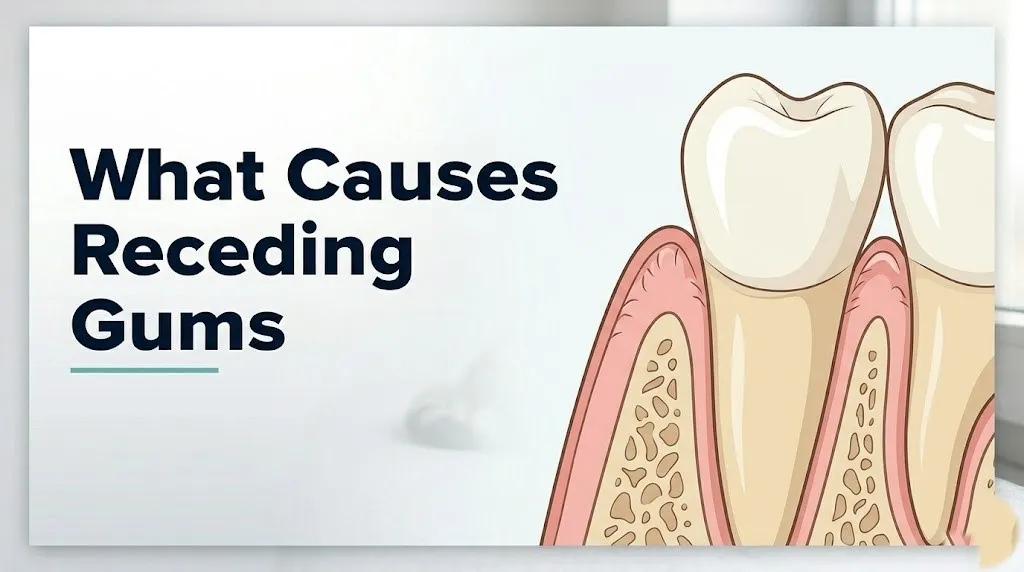
Receding gums are caused by the loss of the gingival tissue that forms the collar around each tooth, resulting in the apical (downward) migration of the free gingival margin below the cementoenamel junction (CEJ), which exposes the root cementum that was previously protected beneath the gumline.
The causes fall into two broad categories. The first is destructive inflammatory disease, where an immune response to bacterial pathogens degrades the connective tissue and bone that anchor the gum in place. The second is mechanical or traumatic tissue loss, where physical forces or tissue architectural deficiencies allow the gingival margin to shift without the infectious trigger.
Both categories produce the same visible result: teeth that look longer than they used to, a notch or groove at the base of the visible tooth crown where the root begins, and sensitivity to cold temperatures or touch at the newly exposed root surface. The root cementum contains dentinal tubules that, when exposed, allow temperature and pressure changes to reach the pulp of the tooth through fluid movement, which is the mechanism behind the sharp sensitivity that many people notice before they see the recession visually.
Key clinical features of gum recession:
- Teeth that appear longer than normal, particularly at the front of the mouth
- A visible notch or step at the gumline where the crown and root meet
- Increased sensitivity to cold liquids, air, or pressure at specific teeth
- Redness, puffiness, or bleeding along the gumline (present in inflammatory causes, often absent in mechanical causes)
- Tooth mobility in advanced cases where alveolar bone support is lost
What Is Gum Recession
Gum recession, clinically called gingival recession (ICD-10 code K06.0), is the apical displacement of the free gingival margin from its normal position at or slightly above the cementoenamel junction, resulting in exposure of the root surface.
Understanding the anatomy makes the mechanism of recession clear. The gingiva that surrounds each tooth has two distinct zones. The free gingiva forms the collar directly against the tooth and normally sits 1 to 2 millimeters above the CEJ in a healthy mouth. Below it, the attached gingiva is firmly bound to the underlying alveolar bone by dense connective tissue fibers. The width of the attached gingiva varies between individuals and between teeth within the same mouth. This variation is clinically important because teeth with narrow zones of attached gingiva have less structural resistance to recession.
The periodontal ligament consists of bundles of collagen fibers called Sharpey’s fibers, which anchor the root cementum to the surrounding alveolar bone and suspend the tooth in its socket. When periodontal disease or other causes destroy these fibers and the supporting bone, the free gingival margin loses its anchorage and migrates apically.
The American Academy of Periodontology (AAP) recognizes gingival recession as one of the most clinically important manifestations of periodontal disease and mucogingival conditions, noting that it can occur at a single tooth or across multiple teeth simultaneously depending on the causative mechanism.
Individual variation note: The width of the zone of keratinized attached gingiva varies by genetic gingival biotype. People with a thin scalloped biotype have narrower attached gingiva bands and thinner labial cortical bone plates, making them anatomically predisposed to recession regardless of oral hygiene quality.
Periodontal Disease and Gum Recession
Periodontal disease (periodontitis) is the single most common cause of gum recession, producing tissue destruction through a chronic inflammatory response driven by bacterial pathogens in the subgingival plaque biofilm that destroys the periodontal ligament, alveolar bone, and gingival connective tissue over months to years.
Think of the gingival tissue as a tent held up by a series of stakes driven into the ground. The periodontal ligament fibers are the stakes, and the alveolar bone is the ground. Periodontitis systematically pulls those stakes out of the ground, one by one. Once enough stakes are gone, the tent fabric collapses downward. That collapse is what gum recession looks like clinically.
The pathogenic mechanism begins with the establishment of a subgingival biofilm containing red complex periodontal bacteria: Porphyromonas gingivalis, Treponema denticola, and Tannerella forsythia. These bacteria produce proteases, lipopolysaccharides, and fimbriae that evade initial immune clearance and stimulate a dysregulated host inflammatory response. Macrophages and lymphocytes in the gingival tissue release interleukin-1 beta (IL-1β), tumor necrosis factor-alpha (TNF-α), and prostaglandin E2 (PGE2) in sustained quantities. These cytokines activate osteoclasts through the RANKL pathway, which resorbs the alveolar bone. Simultaneously, matrix metalloproteinases 8 and 13 (MMP-8, MMP-13) released by neutrophils and fibroblasts degrade the collagen fibers of the periodontal ligament.
The combined loss of alveolar bone support and connective tissue fiber attachment allows the junctional epithelium to migrate apically along the root surface, the periodontal pocket deepens, and the free gingival margin follows the underlying tissue destruction downward.
The Journal of Periodontology has consistently documented that untreated moderate-to-severe periodontitis produces measurable clinical attachment loss and radiographic alveolar bone loss that directly corresponds to the recession observed clinically.
Older adults and those with immune-compromising conditions including HIV infection or chemotherapy-induced immunosuppression experience accelerated periodontal bone loss because the immune response that should contain bacterial proliferation in the sulcus is weakened, allowing more extensive tissue destruction per unit time.
Does Brushing Too Hard Cause Receding Gums
Aggressive tooth brushing causes gum recession through a mechanical abrasion mechanism that physically wears away both the thin labial cortical plate of alveolar bone and the overlying gingival epithelium over time, without requiring any bacterial inflammatory component.
This is one of the most misunderstood causes of recession because the tissue loss looks identical to periodontal disease-driven recession on visual inspection, yet the treatment approach and prognosis are completely different. A periodontist distinguishes between the two primarily through the presence or absence of periodontal pocketing, bone loss on radiographic examination, and the pattern of recession across the mouth.
Traumatic brushing-induced recession tends to occur on the labial surfaces (the surfaces facing the lips) of the teeth that a right-handed person brushes with a scrubbing motion from the right side, typically the upper left and lower left canines and premolars. The recession is often localized to those specific teeth rather than generalized across the mouth. The gingival tissue in these areas shows characteristic cleft formation and a smooth, knife-edge gingival margin rather than the irregular, inflamed margin typical of periodontitis.
Hard-bristle toothbrushes concentrate force on a smaller surface area per bristle and dramatically increase abrasive load per stroke compared to soft-bristle brushes. A 2016 study published in the Journal of Clinical Periodontology found that brushing force above 150 grams consistently produced gingival abrasion in research participants over an 8-week observation period.
Quick Tip:
- Switch to a soft-bristle brush and use the Bass technique: angle bristles 45 degrees toward the gumline and vibrate gently rather than scrubbing horizontally
- Electric toothbrushes with pressure sensors alert you when brushing force exceeds a safe threshold, which is particularly useful for people who brush aggressively without realizing it
- Left-handed people typically see the mirror-image pattern: more recession on the upper right and lower right premolar and canine areas
Key Takeaway: Periodontal disease destroys gum tissue through an inflammatory cytokine cascade and osteoclast-mediated bone resorption, while aggressive brushing destroys it through direct mechanical abrasion of the labial bone plate and epithelium — the causes look identical visually but require completely different professional management approaches.
Bruxism and Gum Recession
Bruxism, the involuntary grinding or clenching of teeth during sleep or waking hours, causes gum recession indirectly through occlusal trauma, which generates lateral and compressive forces at the periodontal ligament that accelerate alveolar bone resorption and increase susceptibility to gingival tissue loss when combined with other risk factors.
Bruxism does not cause recession in the same direct way that periodontitis does. Its role is to amplify existing tissue vulnerability. Teeth subject to excessive lateral forces experience what periodontists call primary occlusal trauma when the supporting structures are healthy, and secondary occlusal trauma when pre-existing periodontitis has reduced the bone support available to absorb that force. In both cases, the excessive mechanical loading at the periodontal ligament fibers and the alveolar bone crest accelerates the remodeling and resorption of the bone in the areas of highest force concentration.
The American Academy of Periodontology notes that while occlusal trauma alone does not initiate periodontal inflammation, it acts as a co-destructive factor that increases the rate of bone loss in periodontally compromised patients by 30% to 50% compared to periodontally compromised patients without occlusal trauma, based on longitudinal clinical studies.
Bruxism in the presence of thin gingival biotype creates a compounding risk. The thin labial cortical plate that characterizes this tissue type has less structural integrity to absorb occlusal force, making bone fenestration (a window-like opening through the labial plate) more likely under chronic grinding loads.
People with temporomandibular disorders, high stress levels, or a history of stimulant medication use (including certain attention deficit hyperactivity disorder medications) have higher rates of bruxism. A board-certified periodontist or prosthodontist should assess occlusal force distribution in any patient with recession who also reports jaw pain, morning headaches, or visible tooth wear.
Thin Gingival Biotype and Recession Risk
A thin gingival biotype, also called a thin scalloped phenotype, is a genetically determined anatomical characteristic in which the labial gingival tissue and the underlying cortical bone plate are thinner than average, creating a structural predisposition to recession even in the absence of disease or trauma.
Every person’s gingiva has a biotype. At one end of the spectrum is the thick flat biotype: dense gingival tissue, broad zones of attached gingiva, thick cortical bone, and square-shaped tooth crowns. At the other end is the thin scalloped biotype: delicate translucent gingival tissue, narrow keratinized gingiva bands, thin to absent labial cortical bone, and tapered tooth crowns. Most people fall somewhere between these two poles.
The clinical significance is substantial. A thin labial cortical plate can measure less than 0.5 millimeters in some individuals, compared to 1.5 to 2 millimeters in thick biotype individuals. A plate that thin is vulnerable to resorption from even normal functional forces, let alone from brushing trauma, orthodontic movement, or the early stages of periodontal inflammation. Research published in the Journal of Periodontology has documented that patients with thin gingival biotype experience statistically higher rates of recession following orthodontic treatment, periodontal surgery, and restorative procedures compared to thick biotype patients.
| Biotype | Tissue Thickness | Cortical Bone | Recession Risk | Crown Shape |
|---|---|---|---|---|
| Thick flat | 2 mm or more | 1.5 to 2 mm thick | Lower | Square |
| Average | 1 to 2 mm | 1 to 1.5 mm thick | Moderate | Oval |
| Thin scalloped | Less than 1 mm | Less than 0.5 mm | Higher | Triangular or tapered |
A periodontist can assess gingival biotype through visual inspection, periodontal probe transparency through the tissue, or cone beam computed tomography (CBCT) imaging of the labial cortical bone thickness. This assessment matters before orthodontic treatment, implant placement, and certain restorative procedures.
Key Takeaway: Thin gingival biotype is a structural predisposition, not a disease, but it means that any additional stress on the gingival tissue, whether from brushing technique, orthodontic movement, or early periodontal disease, will produce recession faster than it would in a person with a thicker tissue type.
What Causes Gum Recession on One Tooth
Gum recession isolated to a single tooth is most commonly caused by a localized mechanical factor, such as a high frenal attachment, a labially positioned tooth, a localized brushing trauma pattern, or a single-tooth occlusal contact abnormality, rather than systemic periodontal disease.
When recession appears on one tooth and the adjacent teeth are unaffected, the cause is almost always localized rather than systemic. Periodontal disease typically produces recession in a pattern consistent with plaque accumulation areas and immune response distribution, rarely in a perfectly isolated single-tooth presentation in an otherwise healthy mouth.
Localized causes of single-tooth recession include:
- A high frenal attachment: the frenum (the fold of tissue connecting the lip or cheek to the gum) inserts too close to the gingival margin of a specific tooth, creating a pull that progressively detaches the gingiva from that tooth. The lower central incisors and lower canines are most frequently affected.
- Labial tooth malposition: a tooth that sits in front of the dental arch (labially displaced) has less supporting bone on its facial surface by anatomical necessity, making that tooth’s gingiva more recession-prone from the outset
- Localized occlusal contact abnormality: a dental crown, filling, or bite alignment issue that directs disproportionate force through one tooth can accelerate localized bone loss and recession at that specific location
- Root prominence: the root of a tooth that projects further labially than adjacent roots sits closer to the outer surface of the thin cortical plate, and any bone loss at that site produces visible recession faster
A board-certified periodontist examining single-tooth recession will use periodontal probing to measure pocket depth at all six sites around the affected tooth, compare probing depths and attachment levels to adjacent teeth, assess frenal attachment location, review radiographs for localized bone loss, and evaluate the bite to identify any occlusal force concentration on that tooth.
Smoking and Gum Recession
Tobacco smoking causes and accelerates gum recession through two simultaneous mechanisms: nicotine-induced gingival vasoconstriction that reduces blood flow and masks clinical signs of inflammation, and direct suppression of gingival fibroblast proliferation and function that impairs the tissue’s capacity to repair and maintain itself.
The vasoconstriction mechanism is particularly insidious because it makes smokers’ gums appear healthier than they are. The classic clinical signs of periodontal inflammation, redness and bleeding on probing, are reduced in smokers because the blood supply to the gingival tissue is constricted by nicotine’s action on alpha-adrenergic receptors in gingival blood vessel walls. A dentist who expects bleeding to indicate inflammation may underestimate the true severity of periodontal disease in a smoker, and the smoker themselves may feel reassured by gums that don’t bleed.
Beneath that clinically quiet surface, tissue destruction is occurring at an accelerated rate. Research published in the Journal of Clinical Periodontology has found that smokers have two to three times greater rates of clinical attachment loss and alveolar bone resorption compared to non-smokers with equivalent plaque levels, controlled for other variables. The nicotine-driven suppression of gingival fibroblast collagen production means the periodontal ligament cannot repair microtraumatic damage, and the immune-suppressant effects of tobacco combustion byproducts reduce neutrophil and macrophage efficiency in clearing the subgingival biofilm.
Former smokers retain elevated recession risk for years after cessation. The American Academy of Periodontology recommends that all current and former smokers inform their periodontist of their tobacco history, including the number of pack-years, because this information directly influences the expected treatment response and maintenance interval.
People who use smokeless tobacco (chewing tobacco, snuff) develop a specific pattern of localized recession at the vestibular area where the tobacco product habitually sits, typically the lower incisors or canine region, due to direct chemical irritation and abrasion from tobacco particles against the gingival tissue.
Hormonal Changes and Gum Recession in Women
Hormonal changes throughout a woman’s life, particularly the decline in estrogen during perimenopause and postmenopause, cause gum recession by reducing estrogen’s protective effect on gingival collagen synthesis, gingival vascularity, and the inflammatory response threshold of periodontal tissue.
Estrogen receptors are present in gingival fibroblasts, the cells responsible for producing and maintaining the collagen matrix of gingival connective tissue. When circulating estrogen levels are adequate, these receptors support collagen production, maintain gingival capillary integrity, and modulate the inflammatory response to periodontal bacteria. As estrogen declines during perimenopause and falls sharply after menopause, these protective effects diminish. Gingival collagen production decreases, the connective tissue thins, and the gingival response to even low-level bacterial challenge becomes more inflammatory.
Postmenopausal women who are not on hormone replacement therapy show statistically higher rates of periodontal bone loss and clinical attachment loss than premenopausal women of equivalent oral hygiene status, according to research published in the Journal of Periodontology. The mechanism parallels the systemic osteoporosis risk seen after menopause: the same hormonal environment that reduces systemic bone mineral density also reduces the density and structural integrity of alveolar bone.
Pregnant women experience a temporary increase in gingival inflammation called pregnancy gingivitis driven by elevated progesterone, which promotes growth of certain anaerobic periodontal bacteria and increases gingival vascular permeability. While pregnancy gingivitis is not the same as periodontitis and typically resolves postpartum, unresolved pregnancy gingivitis in women with pre-existing periodontal susceptibility can progress to recession-inducing periodontitis.
Women taking oral contraceptives containing progesterone-dominant formulations may experience similar gingival sensitivity and increased inflammatory response at the gingival level. Any woman undergoing significant hormonal changes who notices new tooth sensitivity or visible gumline changes should request a periodontal assessment from their dentist or a board-certified periodontist, and mention their current hormonal status and any hormone therapy details at that appointment.
Key Takeaway: Estrogen is a structural protector of gingival collagen and vascularity, and postmenopausal women lose that protection at the same time systemic bone density is declining, making combined periodontal and bone health assessment particularly important after menopause.
Diabetes and Gum Recession
Type 2 diabetes mellitus accelerates gum recession by impairing the neutrophil-mediated immune response in the periodontal sulcus, promoting the accumulation of advanced glycation end-products (AGEs) in gingival connective tissue, and reducing the periodontal tissue’s capacity for repair and regeneration after inflammatory damage.
The relationship between type 2 diabetes and periodontal disease is bidirectional and well-established in the clinical literature. Elevated blood glucose impairs neutrophil chemotaxis (the ability of white blood cells to migrate toward and engulf periodontal pathogens in the gingival sulcus), allowing bacterial populations to establish deeper and more destructive biofilms than would occur in a person with normal glucose regulation. The HbA1c (glycated hemoglobin) level, which reflects average blood glucose over three months, is directly correlated with periodontal severity: patients with HbA1c above 7% consistently show greater clinical attachment loss and alveolar bone resorption than patients with the same plaque levels but controlled glucose.
The CDC reports that adults with diabetes are approximately three times more likely to develop severe periodontitis than adults without diabetes. That increased periodontitis risk translates directly into a higher rate and faster progression of gum recession.
AGEs accumulate in the collagen matrix of the periodontal ligament and alveolar bone in people with chronically elevated blood glucose. These modified protein structures make the tissue more brittle, less capable of normal remodeling, and more susceptible to inflammatory degradation. The practical result is that a diabetic patient’s periodontal tissue responds more severely to the same bacterial challenge that a non-diabetic person’s immune system would contain more effectively.
Patients with type 2 diabetes should tell their periodontist their current HbA1c level, their most recent ophthalmologist and nephrologist results (as a marker of systemic diabetes control), and any recent changes in their diabetes medication, because all of these factors affect the expected treatment response for periodontal recession.
Medications That Cause Gum Recession
Several commonly prescribed medications alter gingival tissue architecture in ways that can produce recession, specifically phenytoin (an anticonvulsant), cyclosporine (an immunosuppressant used in organ transplant recipients), and calcium channel blockers including amlodipine and nifedipine (used for hypertension and cardiac conditions).
These three drug classes produce a phenomenon called drug-induced gingival enlargement, in which gingival tissue overgrowth distorts the normal gingival architecture. This might seem counterintuitive as a cause of recession, but the relationship is indirect. Gingival enlargement creates pseudopockets that trap plaque and disrupt normal oral hygiene, which promotes secondary periodontitis, which then drives bone loss and tissue destruction that results in recession when the drug is changed or discontinued, or recession in areas adjacent to the enlarged tissue where bone support is compromised.
Drug-induced gingival changes affect patients differently. Research in the Journal of Clinical Periodontology has documented that gingival enlargement occurs in approximately 50% of phenytoin users, 25% to 30% of cyclosporine users, and 10% to 20% of patients taking nifedipine specifically. The severity is strongly influenced by pre-existing plaque levels and baseline periodontal health: patients with excellent plaque control show significantly less drug-induced gingival change.
Other medications associated with gum tissue changes include:
- Anticoagulants (warfarin, heparin): increase gingival bleeding tendency, which can mask recession
- Bisphosphonates (alendronate, zoledronic acid): alter alveolar bone metabolism and can complicate periodontal surgical treatment for recession
- Chemotherapy agents: cause mucositis and immunosuppression that accelerate periodontal tissue breakdown
- Selective serotonin reuptake inhibitors (SSRIs): associated with bruxism in some patients, which can indirectly accelerate recession
Any patient starting a medication in one of these classes should inform both their prescribing physician and their dentist or periodontist so that a baseline periodontal assessment can be established and appropriate monitoring scheduled.
Key Takeaway: Drug-induced gingival enlargement from phenytoin, cyclosporine, and calcium channel blockers creates the conditions for secondary periodontitis and recession, and patients on these medications need scheduled periodontal monitoring from the time of prescription initiation.
Orthodontic Treatment and Gum Recession
Orthodontic treatment, specifically the labial (forward) movement of teeth through bracket-and-wire systems or clear aligner therapy, can cause localized gum recession by thinning or fenestrating the labial cortical bone plate as the tooth root moves toward or through that thin bony wall.
The labial cortical plate of bone that covers the facial surface of the tooth root is not uniformly thick around the entire dental arch. In many people, particularly those with narrow arches or crowded teeth, the labial plate over the lower incisors measures less than 1 millimeter. When orthodontic force moves a tooth labially (toward the lip), the root moves in the same direction as the cortical plate. If the root approaches the inner surface of that thin plate, the plate can resorb, creating a fenestration or dehiscence in the bone. Without bony support, the gingiva above it cannot be maintained, and recession follows.
Research published in the Journal of Periodontology has documented that proclination (tipping forward) of lower incisors during orthodontic treatment is associated with a measurable increase in recession prevalence at those teeth, particularly in patients with thin biotype gingiva and narrow lower arch dimensions.
This does not mean orthodontic treatment should be avoided. It means that orthodontic treatment planning should include:
- Pre-treatment CBCT imaging to document labial bone thickness at high-risk teeth
- Gingival biotype assessment before initiating treatment
- Avoidance of excessive labial proclination of lower incisors, particularly in thin biotype patients
- Periodontal consultation for patients with pre-existing thin tissue or recession before beginning treatment
- Regular periodontal monitoring throughout active orthodontic treatment
- Post-treatment periodontal assessment to identify any recession that developed during tooth movement
Adult orthodontic patients are at higher risk for orthodontic-induced recession than adolescents, because adult alveolar bone is less adaptive to tooth movement forces and adult gingival tissue has had more time to develop other recession risk factors.
Gum Recession in Young Adults
Gum recession in young adults under age 35 is most commonly caused by aggressive tooth brushing trauma, thin gingival biotype, or localized frenal attachment issues rather than periodontal disease, which typically takes decades of cumulative bacterial damage to produce substantial recession.
The popular assumption is that gum recession is a condition of older age. That is true for periodontitis-driven recession, which accumulates over years of inflammatory tissue destruction. But traumatic recession from brushing technique errors can begin as early as the teenage years, particularly in young adults who brush vigorously, use hard-bristle brushes, and have thin gingival tissue that offers little resistance to mechanical abrasion.
A 2020 observational study published in the Journal of Clinical Periodontology found that gingival recession was detectable in a substantial proportion of young adults aged 18 to 35, and that in this age group, non-inflammatory causes (brushing trauma and anatomical factors) were far more prevalent than inflammatory periodontal disease as the primary driver.
Young adults who play contact sports should know that occlusal trauma from sports-related impacts, even when protected by mouthguards, can contribute to localized periodontal ligament stress and recession susceptibility. Young adults who use vaping devices or e-cigarettes may experience gingival tissue changes similar to those seen in traditional tobacco smokers, though the long-term periodontal research on vaping is still emerging.
Gum recession in a young adult that is rapid in onset, diffuse across multiple teeth, and accompanied by general malaise, lymph node swelling, or fatigue warrants evaluation not only by a board-certified periodontist but also by a primary care physician to assess for systemic conditions including hematologic disorders, autoimmune diseases (such as lupus or Crohn’s disease affecting oral mucosa), or immunodeficiency states that can accelerate periodontal destruction at an atypically young age.
How Gum Recession Is Classified and Diagnosed
Gum recession severity is classified using either the Miller Classification of Marginal Tissue Recession (Classes I through IV) or the more recent Cairo Classification (Recession Types RT1, RT2, and RT3), and both systems directly determine which treatment options are clinically viable for a given patient.
The Miller Classification, developed in 1985, classifies recession based on the relationship between the recession depth, the mucogingival junction, and the level of bone and soft tissue in the spaces between adjacent teeth (the interdental areas). Class I recession does not extend to the mucogingival junction and has no interproximal bone loss, meaning complete root coverage is achievable with surgical grafting. Class IV recession extends beyond the mucogingival junction and has severe interproximal bone loss, meaning complete root coverage is generally not achievable.
The Cairo Classification, published in the Journal of Clinical Periodontology in 2011 and endorsed by the European Federation of Periodontology, modernized the approach by focusing on the interproximal clinical attachment level:
| Cairo Type | Definition | Surgical Coverage Potential |
|---|---|---|
| RT1 | No interproximal attachment loss at adjacent teeth | Complete root coverage expected |
| RT2 | Interproximal attachment loss less than or equal to buccal recession | Partial coverage; complete possible |
| RT3 | Interproximal attachment loss greater than buccal recession | Complete coverage not achievable |
Diagnosis is conducted by a board-certified periodontist using:
- Periodontal probing to measure recession depth at six sites per tooth (midbuccal, mesiobuccal, distobuccal, and lingual equivalents)
- Clinical attachment level (CAL) measurement from the CEJ to the base of the periodontal pocket
- Dental radiographs (periapical films) to assess alveolar bone height
- CBCT imaging for precise labial bone thickness assessment when surgical planning is anticipated
- Visual assessment of gingival biotype, keratinized tissue width, and frenal attachments
A periodontist uses the classification to counsel the patient on realistic treatment expectations before any surgical discussion begins.
Key Takeaway: The Cairo Classification tells you and your periodontist whether complete root coverage through tissue grafting is achievable for your specific recession pattern: RT1 recession has the best surgical prognosis, RT3 does not respond to complete coverage attempts, and knowing which you have before surgery prevents unrealistic expectations.
When to See a Dentist or Periodontist for Receding Gums
Any visible gum recession, new tooth sensitivity, or a visible notch at the base of a tooth warrants evaluation by a dentist as the minimum first step, with referral to a board-certified periodontist for all cases involving more than one tooth, progressive recession, recession associated with mobility or bleeding, or recession in the context of diabetes, immunosuppression, or other systemic risk factors.
Many people delay seeking evaluation because recession is painless in its early stages and appears to progress slowly. That delay is the primary reason that what could have been RT1 or RT2 recession amenable to complete surgical coverage becomes RT3 recession that cannot be fully corrected. Recession does not stabilize on its own without identifying and eliminating the causative factor.
To prepare for a productive first appointment with a periodontist:
- Write down when you first noticed the gum change and whether it has progressed, noting any visible change in tooth length or new sensitivity
- List all current prescription and over-the-counter medications, including supplements, because several medication classes directly affect gingival tissue
- Document your tobacco use history, including current and former use and years of use
- Note any relevant medical history including type 2 diabetes, autoimmune conditions, recent chemotherapy, organ transplant, or hormone therapy status
- Bring your most recent dental radiographs if your periodontist is not connected to your general dentist’s records
- Ask specifically: “Which classification does my recession fall into, and what does that mean for treatment options?”
- Ask whether a CBCT scan would add useful information before any surgical planning begins
People with diabetes should have their most recent HbA1c result available. People on bisphosphonate medications for osteoporosis must tell their periodontist before any surgical procedure, including grafting, is discussed, because bisphosphonate therapy significantly alters perioperative risk in periodontal surgery.
Can Receding Gums Be Treated
Receding gums cannot regenerate spontaneously, but surgical treatment with tissue grafting procedures can achieve partial or complete root coverage in appropriately selected patients, depending on the Cairo recession classification and the adequacy of donor tissue.
The three most evidence-supported surgical approaches for gum recession are:
- Connective tissue graft (CTG): a section of connective tissue is harvested from beneath the palatal epithelium and placed over the exposed root surface, then covered with the adjacent gingival tissue advanced coronally. The Journal of Periodontology rates this as the gold standard for achieving complete root coverage in RT1 and many RT2 recession defects.
- Free gingival graft (FGG): a section of full-thickness palatal tissue is grafted to the recession site to increase the width of keratinized gingiva. This approach prioritizes tissue stability over esthetic coverage and is particularly useful at sites with minimal residual keratinized tissue.
- Pinhole surgical technique: a minimally invasive approach in which a small instrument is inserted through a tiny hole in the gingiva to loosen and reposition the existing tissue over the recession defect without harvesting a separate graft.
Non-surgical management, while insufficient to reverse existing recession, is the necessary foundation for preventing further recession:
| Approach | Target Cause | Evidence Basis |
|---|---|---|
| Brushing technique correction (Bass method, soft bristle) | Traumatic brushing | Well-established, supported by clinical guidelines |
| Scaling and root planing (SRP) | Periodontal disease | Well-established by controlled clinical trials |
| Occlusal splint for bruxism | Bruxism-related trauma | Supported by clinical observation and controlled studies |
| Tobacco cessation | Smoking-related progression | Well-established by prospective cohort studies |
| Glycemic control (for diabetics) | Diabetes-driven progression | Well-established by clinical epidemiological data |
| Frenotomy (frenal attachment) | High frenal pull | Supported by clinical case series and controlled studies |
Receding gums are not reversible without professional intervention, and the window for the best surgical outcomes closes as recession progresses from RT1 to RT3. A board-certified periodontist should assess the current classification and help determine the appropriate timing for intervention.
When to Seek Urgent Dental or Medical Evaluation for Gum Changes
Certain presentations associated with gum recession or gum tissue changes require prompt professional evaluation within 24 to 48 hours rather than a routine scheduled appointment. Do not delay evaluation if these signs are present.
Contact a dentist or emergency dental clinic the same day if you experience any of the following:
- Facial swelling, jaw swelling, or swelling under the chin associated with a painful tooth or gum area, which can indicate a dental abscess spreading to facial fascial spaces, a potentially life-threatening condition requiring immediate drainage and antibiotic treatment
- Gum changes with high fever (above 38.5°C or 101.3°F), difficulty swallowing, or difficulty opening the mouth, which may indicate Ludwig’s angina or deep neck space infection
- Rapidly progressing gum recession over days to weeks rather than months, particularly in a young person who does not smoke and has no identified risk factor, which may indicate an underlying immunodeficiency or hematologic disorder requiring medical evaluation by a primary care physician
- Gum tissue that appears white, thickened, or has persistent sores that do not heal within two weeks, which requires evaluation to rule out oral mucosal pathology including leukoplakia or oral squamous cell carcinoma
Go to the nearest emergency room immediately if you have facial swelling that is spreading toward your eye, your neck, or is causing breathing difficulty.
These presentations require emergency medical assessment and cannot wait for a scheduled dentist appointment.
Frequently Asked Questions About Receding Gums
Can receding gums grow back on their own?
Receding gums cannot grow back on their own because gingival tissue does not spontaneously regenerate once the underlying bone support and connective tissue attachment are lost.
Without professional surgical intervention such as a connective tissue graft or free gingival graft, existing recession remains permanent, though eliminating the causative factor (treating periodontal disease, correcting brushing technique, controlling diabetes) prevents further progression.
A board-certified periodontist can assess whether your recession pattern is surgically correctable and which procedure offers the best chance of partial or complete root coverage based on your Cairo recession classification.
What is the fastest way to stop gum recession from getting worse?
The fastest way to stop recession from progressing is to identify and eliminate the specific cause through professional evaluation, because no single home measure addresses all the mechanisms that can drive recession.
Correcting brushing technique to a low-force soft-bristle Bass method is immediately actionable and addresses traumatic recession, while scaling and root planing by a periodontist addresses the inflammatory periodontal disease mechanism that causes the majority of recession cases.
A board-certified periodontist can establish which cause is driving your recession within the first appointment, giving you a specific action plan rather than a general list of precautions.
What does gum recession feel like?
Gum recession most commonly first presents as a sharp or lingering sensitivity to cold beverages, cold air, or direct touch at the base of specific teeth, produced by fluid movement through the exposed dentinal tubules of the root cementum reaching the tooth pulp.
Some people notice no sensitivity at all and discover recession only when a dentist points it out, because the rate of exposure determines whether the pulp has had time to deposit secondary dentin and partially self-insulate the exposed tubules.
Visual signs that you can notice yourself include teeth that appear longer than they used to or a visible notch or color change at the base of the visible tooth crown where the root surface begins.
Can stress cause receding gums?
Stress does not directly cause gum recession, but chronic psychological stress is associated with behaviors and physiological changes that accelerate recession through established mechanisms.
Research has found an association between chronic stress and elevated cortisol levels that suppress neutrophil function in the periodontal sulcus, reduce immune surveillance against periodontal bacteria, and increase bruxism frequency, all of which contribute to periodontal tissue destruction and recession when combined with existing risk factors.
Stress-related neglect of oral hygiene and increased tobacco or alcohol use during high-stress periods also remove protective behaviors that would otherwise slow periodontal progression.
How quickly do gums recede?
The rate of gum recession varies widely depending on the cause: periodontal disease-driven recession typically progresses at approximately 0.1 to 0.3 millimeters per year in untreated moderate periodontitis, while traumatic brushing-induced recession can occur over months in people with thin gingival biotype who brush with high force.
Recession associated with aggressive periodontitis, uncontrolled type 2 diabetes, or immunosuppression can progress significantly faster, with multiple millimeters of attachment loss occurring over weeks to months in untreated cases.
Any recession that appears to change noticeably within weeks rather than years warrants immediate evaluation by a board-certified periodontist rather than a routine scheduled check-up.
Is gum recession a sign of something serious?
Gum recession is a sign of an underlying structural or inflammatory problem that requires professional identification, and in some cases it is the first visible oral sign of a systemic condition including type 2 diabetes, autoimmune disease, or medication effects that warrant medical evaluation beyond dental treatment.
The most immediately serious association is with untreated periodontitis, which the American Academy of Periodontology and the American Heart Association have both identified as associated with elevated cardiovascular disease risk, though the evidence for a causal relationship remains under active investigation.
Single-tooth recession in a young, healthy non-smoker with good oral hygiene is less likely to indicate a systemic problem, but diffuse, rapidly progressing recession in someone without the standard risk factors always warrants a medical workup alongside the dental evaluation.
Understanding Your Recession Means Knowing Its Cause
Gum recession is not an inevitable part of aging that you simply accept. Every case has a specific anatomical, inflammatory, mechanical, or systemic cause, and that cause determines both the treatment approach and the expected outcome. The readers who do best are the ones who go to their periodontist armed with the specific question: “Which mechanism is driving my recession, and what classification does it fall into?”
If you’ve noticed your teeth looking longer, felt sensitivity at the gumline, or been told at a check-up that your gums are receding, make an appointment with a board-certified periodontist rather than waiting for another annual cleaning to see whether it changes. Recession in Cairo RT1 is surgically correctable with excellent outcomes. The same recession left untreated for two more years may progress to RT3, where complete correction is no longer achievable.
What you know now about the mechanism behind your recession, whether it is inflammatory, mechanical, hormonal, or systemic, is the clinical foundation you need to have a specific, productive conversation with the right provider.