What Causes Trigger Finger? Reasons, Risks, and Relief
Trigger finger is caused by a narrowing of the first annular pulley, a tight band of tissue at the base of the finger, that prevents the underlying flexor tendon from gliding smoothly during finger movement. This narrowing creates a mechanical mismatch between the tendon and the channel it must pass through, producing the clicking, catching, or locking sensation that gives the condition its name.
According to the National Institute of Arthritis and Musculoskeletal and Skin Diseases (NIAMS), trigger finger is one of the most common causes of hand pain and disability in adults, with diabetes mellitus increasing lifetime risk by as much as tenfold compared to the general population. Women develop the condition at a rate two to six times higher than men, according to epidemiological data published in the Journal of Hand Surgery.
This article covers every clinically documented cause of trigger finger, from the cellular-level changes that thicken the A1 pulley to the systemic diseases that accelerate tendon sheath damage, including why some fingers are more vulnerable than others and exactly when professional evaluation is warranted.
What Is Trigger Finger and What Causes It?
Trigger finger is a condition in which inflammation and structural thickening of the fibrous flexor tendon sheath at the base of the finger, specifically at the first annular (A1) pulley, prevents the flexor tendon from moving freely through its canal, causing the finger to snap, click, or lock during bending and straightening.
The finger’s ability to bend and straighten depends on a precisely coordinated gliding system. Two tendons, the flexor digitorum superficialis and the flexor digitorum profundus, run along the palm side of each finger inside a protective sheath. This sheath is held against the bone by a series of reinforced bands called pulleys. The A1 pulley sits directly over the metacarpophalangeal (MCP) joint, at the base of the finger where it meets the palm.
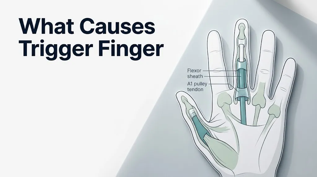
When the A1 pulley thickens and its internal diameter narrows, the tendon must force its way through a canal that is no longer the right size. A nodule can form on the tendon itself, making the mismatch even worse. Think of it like trying to slide a swollen garden hose through a pipe fitting that hasn’t changed size: the hose catches, sticks, then suddenly pops through with force.
The clinical result is the characteristic triggering sensation. In mild cases, the finger clicks but moves freely. In severe cases, the finger locks in a bent position and must be manually straightened using the other hand.
Key clinical distinction: Trigger finger is technically named stenosing tenosynovitis, where “stenosing” refers to the narrowing of the canal, “teno” refers to the tendon, and “synovitis” refers to inflammation of the synovial lining that lubricates tendon movement. All three processes contribute to the condition in different proportions depending on the underlying cause.
Trigger Finger Causes: The Full Clinical Picture
The causes of trigger finger fall into three broad categories: mechanical overuse, systemic metabolic or inflammatory disease, and idiopathic origin, meaning no clear cause can be identified despite thorough evaluation.
Epidemiological data from the Journal of Bone and Joint Surgery shows that approximately 28% of people with diabetes develop trigger finger at some point during their lifetime, making metabolic disease the single most important identifiable systemic cause. Repetitive occupational gripping is the most frequently reported environmental cause among working-age adults without a diagnosed systemic disease.
The remaining cases are classified as idiopathic. These individuals have no identified occupational exposure, no systemic disease, and no prior injury, yet the A1 pulley thickens and narrows over time. Researchers believe low-grade chronic mechanical stress during ordinary daily hand use, combined with individual variation in tendon sheath biology, drives idiopathic cases.
Summary of Cause Categories:
| Cause Category | Examples | Estimated Proportion |
|---|---|---|
| Systemic metabolic disease | Diabetes mellitus, hypothyroidism, amyloidosis | Approximately 25 to 30% |
| Systemic inflammatory disease | Rheumatoid arthritis, gout, psoriatic arthritis | Approximately 10 to 15% |
| Repetitive mechanical overuse | Occupational gripping, prolonged tool use | Approximately 20 to 25% |
| Idiopathic (no identified cause) | Unknown biological predisposition | Approximately 30 to 40% |
| Post-traumatic | Direct injury to the palm or finger base | Fewer than 5% |
For people in the 40 to 60 age range, idiopathic and metabolic causes account for the majority of cases. For people with diagnosed rheumatoid arthritis, the inflammatory pathway dominates.
What Causes a Trigger Finger to Develop?
A trigger finger develops when the structural integrity of the A1 pulley breaks down faster than the body can repair it, causing progressive thickening of the pulley’s fibrocartilaginous rim and reduction in the internal diameter of the tendon canal.
At the tissue level, this breakdown is driven by fibroblast proliferation and excess deposition of collagen type I within the pulley. Histopathological studies published in the Journal of Hand Surgery have confirmed that A1 pulleys removed during surgical release consistently show fibrocartilaginous metaplasia, meaning the pulley tissue has undergone a cellular transformation that makes it stiffer, thicker, and less flexible than healthy pulley tissue.
Concurrent changes occur in the synovial lining of the tendon sheath. The synovial cells that produce lubricating fluid become inflamed, reducing lubrication and increasing friction between the tendon and the canal wall. This friction accelerates mechanical wear on the tendon surface, sometimes producing the nodule that makes triggering mechanically worse.
For people with a diagnosed inflammatory condition such as rheumatoid arthritis, this process is supercharged by cytokine activity. Tumor necrosis factor alpha (TNF-alpha) and interleukin-1 beta (IL-1 beta) promote synovial hypertrophy and pannus formation, adding bulk to the tendon sheath from the inside.
People with carpal tunnel syndrome are at elevated risk of developing trigger finger simultaneously, because both conditions reflect a shared predisposition to tendon and sheath pathology in the hand. Clinicians at the Mayo Clinic note that up to 11% of people presenting with trigger finger also have concurrent carpal tunnel syndrome.
Key Takeaway: Trigger finger develops when the A1 pulley thickens through fibroblast-driven collagen deposition and fibrocartilaginous metaplasia, narrowing the tendon canal and causing the flexor tendon to catch during movement.
Stenosing Tenosynovitis: The Condition Behind Trigger Finger
Stenosing tenosynovitis is the formal clinical name for trigger finger, describing the simultaneous narrowing (stenosing) of the tendon canal and inflammation (synovitis) of the synovial lining that sheathes the flexor tendon.
The word “stenosing” is key. Unlike acute tendinitis, which involves sudden inflammation after overuse, stenosing tenosynovitis is a chronic structural process. The sheath does not simply become inflamed and then return to normal. It progressively narrows, and without intervention, that narrowing typically worsens over months to years.
The American Academy of Orthopaedic Surgeons (AAOS) describes stenosing tenosynovitis as fundamentally a mismatch between the size of the tendon or its associated nodule and the diameter of the A1 pulley through which it must travel. This mismatch produces three mechanical events: friction, catching, and locking, each representing a more advanced stage of structural incompatibility.
The synovitis component adds an inflammatory layer to what is otherwise a mechanical problem. The synovial sheath that envelops the flexor tendon secretes synovial fluid to reduce friction. When the sheath becomes inflamed, that fluid production is disrupted, lubrication fails, and the tendon moves through the canal against resistance rather than in a friction-free glide.
Stenosing vs. non-stenosing tenosynovitis:
| Feature | Stenosing Tenosynovitis (Trigger Finger) | Non-Stenosing Tenosynovitis |
|---|---|---|
| Structural change to pulley | Yes, fibrous thickening | No, primarily inflammatory |
| Tendon canal narrowed | Yes | Not typically |
| Clicking or locking | Yes | No |
| Self-resolution pattern | Uncommon without intervention | More common |
| Systemic disease association | Strong | Variable |
For older adults over 65, stenosing tenosynovitis may progress more rapidly because the reduced cellular turnover rate in aging tendon tissue limits the body’s capacity to remodel and repair the thickened pulley. A primary care physician or board-certified hand surgeon should evaluate any trigger finger that has persisted beyond 6 weeks without improvement.
A1 Pulley Anatomy and How It Fails
The A1 pulley is a 8 to 10 millimeter band of fibrous tissue located at the base of each finger, directly over the metacarpophalangeal joint, and it is the single most important anatomical structure in the development of trigger finger.
The hand contains five flexor tendon pulleys on each finger, labeled A1 through A5, plus the palm-level oblique pulleys for the thumb. The A1 pulley acts as the first and most mechanically stressed entry point for the flexor tendon as it travels from the palm into the finger. Because it sits directly at the MCP joint, it is subjected to the highest compressive forces during gripping activities.
Healthy A1 pulley tissue is dense, organized collagen. The internal surface is lined with synoviocytes that produce lubricating fluid to minimize tendon friction. When mechanical stress exceeds the tissue’s capacity to self-repair, or when systemic disease accelerates collagen remodeling, the pulley undergoes fibrocartilaginous metaplasia. Cartilage-like cells replace the normal fibrous architecture, the pulley thickens, and its internal diameter shrinks.
Research published in the Journal of Hand Surgery in 2019 confirmed that A1 pulleys excised from patients with trigger finger show a statistically measurable reduction in internal diameter compared to cadaveric control samples, with mean canal diameter in trigger finger pulleys measuring approximately 30 to 40% narrower than in unaffected age-matched controls.
The A1 pulley failure is progressive unless the underlying cause is addressed. Splinting reduces the mechanical stress on the pulley. Corticosteroid injection reduces local inflammation and can temporarily restore canal space. Surgical or percutaneous A1 pulley release directly solves the anatomical problem by cutting the restricting band.
People with smaller hand anatomy, a characteristic more common in women than men, have proportionally smaller A1 pulley diameters at baseline. This anatomical difference is one reason women develop trigger finger more often than men, even without occupational heavy gripping or systemic disease.
Repetitive Motion and Occupational Trigger Finger Causes
Repetitive gripping, sustained tool use, and prolonged hand vibration are the most frequently cited occupational causes of trigger finger, and the mechanism is straightforward: cumulative mechanical microtrauma to the A1 pulley exceeds the pulley’s capacity to repair itself between loading cycles.
Every time the hand grips a tool, handle, or instrument, the flexor tendon is pulled proximally through the A1 pulley under load. A single grip generates relatively minor compressive force on the pulley. Thousands of repetitions per workday over months or years generate cumulative damage to the pulley’s collagen architecture. This is the same principle as stress fractures in bone: the individual load is tolerable, but the accumulated load overwhelms the tissue’s repair capacity.
Occupations with documented elevated trigger finger risk include:
- Industrial machine operators and factory workers who grip tools repetitively
- Musicians, particularly guitarists, pianists, and string instrument players
- Farmers and agricultural workers who perform sustained gripping tasks
- Hairdressers and barbers who use scissors repeatedly throughout the day
- Construction workers who use vibrating hand tools such as pneumatic drills
According to data cited by the AAOS, occupational hand disorders including stenosing tenosynovitis are among the most commonly reported musculoskeletal work-related injuries in the United States, accounting for a substantial proportion of lost workdays in manual labor industries.
Hand vibration is a specific occupational risk factor beyond simple repetitive gripping. Vibration transmitted through the handle of a power tool creates a rapid microtrauma cycle in the palmar structures including the A1 pulley. Workers who use vibrating tools for more than four hours per day have a measurably higher incidence of trigger finger compared to matched controls who grip without vibration.
For workers who perform repetitive gripping and also have diabetes, the two risk factors compound each other, producing trigger finger that is more bilateral, more severe, and more resistant to conservative management than either factor alone would produce.
Key Takeaway: Cumulative mechanical microtrauma to the A1 pulley from repetitive gripping or hand vibration is the dominant occupational cause of trigger finger, with risk compounding sharply in workers who also have diabetes mellitus.
Trigger Finger and Diabetes
Diabetes mellitus is the most well-established systemic disease cause of trigger finger, and the mechanism involves a process entirely different from simple inflammation: it is the chemical modification of tendon and pulley collagen by glucose itself.
Persistently elevated blood glucose levels cause glucose molecules to attach non-enzymatically to collagen proteins in a process that produces advanced glycation end-products (AGEs). AGEs cause abnormal cross-linking between collagen fibers, making the collagen in the A1 pulley stiffer, less elastic, and more prone to thickening. The result is a pulley that narrows progressively regardless of whether the person does repetitive gripping work.
According to a systematic review published in the Journal of Hand Surgery, the lifetime prevalence of trigger finger in people with type 2 diabetes is approximately 20 to 28%, compared to 2 to 3% in the general adult population. This dramatically higher rate reflects the systemic effects of AGEs on connective tissue throughout the hand.
Diabetic trigger finger has several clinically distinct features compared to idiopathic trigger finger:
- Multiple fingers are commonly affected simultaneously in diabetic patients
- Both hands are more often involved bilaterally
- The condition tends to be more resistant to corticosteroid injection as a standalone treatment
- People with poorly controlled diabetes (reflected in higher glycated hemoglobin / HbA1c levels) have higher rates of recurrence after treatment
- Trigger thumb is more common in diabetic patients than in the general population
For people with type 1 diabetes, the Journal of Bone and Joint Surgery has reported associations between diabetic hand syndromes (including trigger finger, limited joint mobility, and Dupuytren contracture) and longer disease duration and cumulative glycemic exposure.
Any person with diabetes who develops trigger finger should inform their primary care physician, because the presence of diabetic hand disease may signal the need for closer glycemic control and assessment of other microvascular complications.
Trigger Finger and Rheumatoid Arthritis
Rheumatoid arthritis causes trigger finger through a fundamentally different mechanism than diabetes or mechanical overuse: synovial pannus formation directly invades the tendon sheath from the inside, adding bulk and inflammation to the canal the flexor tendon must pass through.
Rheumatoid arthritis is an autoimmune condition in which the immune system attacks synovial tissue throughout the body, including the synovial lining of the flexor tendon sheaths in the hand. The immune attack stimulates synovial hyperplasia, meaning the synovial cells multiply abnormally, creating a layer of inflammatory tissue called pannus. This pannus fills the tendon canal, reduces the available gliding space, and compresses the flexor tendon from the sheath wall inward.
Unlike A1 pulley thickening in mechanical trigger finger, the pannus in rheumatoid-associated trigger finger can also erode the tendon itself. The Annals of the Rheumatic Diseases has published multiple histopathological studies showing that rheumatoid pannus produces matrix metalloproteinases, enzymes that degrade tendon collagen directly. This creates a risk not just of locking, but of complete tendon rupture in patients with longstanding rheumatoid hand disease.
Trigger finger in the setting of rheumatoid arthritis should be evaluated by both a board-certified rheumatologist and a board-certified hand surgeon because managing the underlying autoimmune disease is as important as treating the local mechanical problem. Corticosteroid injection provides temporary relief, but systemic disease-modifying antirheumatic drugs (DMARDs) targeting TNF-alpha or IL-6 pathways may be required to reduce pannus formation and protect the tendon long-term.
People with rheumatoid arthritis who notice new finger locking or catching should report it promptly at their next rheumatology appointment, or sooner if locking is accompanied by warmth, swelling, or new joint deformity, as these may indicate active disease flare.
Trigger Finger Causes in Women
Trigger finger causes in women follow the same fundamental anatomical mechanism as in men, but women develop the condition at two to six times the rate of men, and the reasons are both anatomical and hormonal.
Anatomically, women tend to have proportionally smaller A1 pulley internal diameters relative to their flexor tendon size, compared to men matched for hand length. A smaller starting canal diameter means less structural reserve before normal tendon size causes impingement. This anatomical difference is present from birth and does not require any systemic disease or occupational exposure to eventually produce symptoms.
Hormonally, estrogen influences tendon collagen remodeling. Postmenopausal women, who experience a rapid decline in circulating estrogen, show accelerated changes in tendon mechanical properties, including reduced collagen synthesis and altered collagen cross-linking. Research cited by the National Institute of Arthritis and Musculoskeletal and Skin Diseases identifies the peak incidence of trigger finger in women between the ages of 40 and 60, a range that overlaps directly with the perimenopausal and early postmenopausal period.
Women are also more likely than men to develop trigger finger in the context of autoimmune disease, because rheumatoid arthritis, thyroid disease, and other connective tissue disorders that cause or contribute to trigger finger are all more prevalent in women.
What this means clinically:
- Women who develop trigger finger without diabetes, heavy manual labor, or known inflammatory disease should not assume the cause is simply “overuse”
- A woman in her late 40s or 50s developing bilateral trigger finger should have thyroid function testing (thyroid-stimulating hormone / TSH) ordered, as hypothyroidism is both more common in perimenopausal women and a documented cause of trigger finger
- Women with trigger finger who are taking aromatase inhibitors for breast cancer treatment face elevated risk because these medications reduce estrogen to very low levels and are associated with musculoskeletal tendon complaints, including trigger finger
Key Takeaway: Women develop trigger finger at up to six times the rate of men because of anatomically smaller A1 pulley diameters at baseline and hormonal influences on tendon collagen remodeling, with peak incidence aligning with the perimenopausal period.
What Causes Trigger Finger in the Middle Finger?
Trigger finger in the middle finger is caused by the same A1 pulley narrowing mechanism as in other fingers, but the middle finger has specific biomechanical loading characteristics that make its pattern of occupational and repetitive-use trigger finger distinct from the ring finger or thumb, which are the most commonly affected digits.
The middle finger (third digit) plays a central role in power grip mechanics. During full-hand gripping, such as holding a steering wheel, a hammer handle, or a racket, the middle finger generates some of the highest flexor tendon forces of any digit because of its central position and length. This elevated mechanical loading makes the middle finger’s A1 pulley vulnerable in occupations that require sustained, forceful gripping without adequate rest intervals.
Among musicians, the middle finger is frequently involved in trigger finger cases because of repetitive, high-speed flexion-extension cycles during playing. Guitarists and pianists particularly are represented in case series published in hand surgery literature.
Middle finger trigger finger compared to other digits:
| Feature | Middle Finger | Ring Finger | Thumb |
|---|---|---|---|
| Overall frequency | Moderately common | Most common | Second most common |
| Primary cause pattern | Occupational / musicians | Idiopathic / metabolic | Diabetic / pediatric (different condition) |
| Biomechanical loading pattern | Central power grip | Medial grip assistance | Pinch and lateral grip |
| Bilateral involvement | Less common than ring | Variable | Common in diabetic patients |
For people asking specifically about middle finger trigger finger, the cause is most often related to repetitive forceful grip activities rather than systemic disease, though diabetes and rheumatoid arthritis can affect any digit. A hand surgeon or orthopedic surgeon can confirm the diagnosis with physical examination and assess whether ultrasound imaging is warranted to evaluate the A1 pulley and underlying tendon.
What Causes a Trigger Thumb?
Trigger thumb is caused by the same stenosing tenosynovitis mechanism as trigger finger, but the anatomy of the thumb involves the flexor pollicis longus (FPL) tendon and the A1 pulley of the thumb, which is located just proximal to the metacarpophalangeal joint of the thumb, in the thenar eminence region.
The thumb is the most mechanically loaded digit in the hand. Pinch grip, key grip, and tip-to-tip grip all concentrate force through the FPL tendon and its A1 pulley. Because the thumb’s flexor mechanism is used in virtually every gripping and manipulating task, its A1 pulley accumulates mechanical stress continuously throughout daily life, not only during heavy manual labor.
Trigger thumb in adults is strongly associated with diabetes mellitus. The Cleveland Clinic notes that adults with diabetes who develop thumb or finger triggering should expect more aggressive or bilateral presentation than non-diabetic patients and may require multiple corticosteroid injections or surgical release.
There is a separate and distinct pediatric condition called congenital trigger thumb, which is not discussed at length here because it has a different etiology (a developmental nodule on the FPL tendon called a Notta’s node) and a different clinical course. Pediatric trigger thumb requires evaluation by a pediatric orthopedic surgeon or pediatric hand specialist.
Adult trigger thumb distinct features:
- Clicking or locking at the interphalangeal joint of the thumb during extension
- Pain in the thenar eminence (fleshy base of the thumb on the palm)
- Morning stiffness of the thumb that loosens as the day progresses
- Palpable nodule in the thenar eminence at the level of the A1 pulley in many cases
- Strong association with diabetes mellitus, making blood glucose evaluation relevant in any new-onset adult trigger thumb
Key Takeaway: Trigger thumb involves the flexor pollicis longus tendon and thumb A1 pulley, carries a particularly strong association with diabetes mellitus in adults, and should prompt glucose testing in patients without a prior diabetes diagnosis.
Trigger Finger Risk Factors
The risk factors for trigger finger fall into three categories: individual biological characteristics, systemic disease status, and occupational or behavioral exposures.
Biological risk factors:
- Female biological sex (two to six times higher incidence than male)
- Age between 40 and 60 years (peak incidence range across all trigger finger types)
- Smaller baseline A1 pulley internal diameter (partly anatomically determined)
- History of carpal tunnel syndrome or de Quervain tenosynovitis (indicating shared tendon sheath pathology)
- Personal or family history of Dupuytren contracture (associated with palmar fascia biology that overlaps with A1 pulley disease)
Systemic disease risk factors:
- Type 1 or type 2 diabetes mellitus (by far the strongest single systemic risk factor)
- Rheumatoid arthritis
- Hypothyroidism
- Amyloidosis (protein deposition disease affecting connective tissue)
- Gout (uric acid crystal deposition in tendon sheaths)
- Psoriatic arthritis
Occupational and behavioral risk factors:
- Repetitive gripping for more than 4 hours per workday
- Prolonged use of vibrating hand tools
- Sustained forceful pinch grip activities
- Recent direct blunt trauma to the palm at the level of the MCP joint
The AAOS identifies diabetes mellitus and female sex as the two most consistently documented independent risk factors across large clinical series. Any combination of these factors compounds risk additively, meaning a 50-year-old woman with type 2 diabetes who works in a factory handling tools has a substantially higher lifetime risk than any single risk factor would produce.
People who have already had trigger finger treated in one finger face elevated risk of developing it in adjacent fingers, because the biological predisposition (whether metabolic, anatomical, or occupational) that caused the first case generally persists.
Trigger Finger in Older Adults
Trigger finger in older adults follows the same fundamental mechanical and metabolic pathway as in younger adults, but several age-specific factors change both the clinical presentation and the appropriate management approach.
Aging tendon tissue undergoes progressive changes in collagen composition, including a shift toward less organized collagen architecture, reduced tenocyte (tendon cell) density, and diminished capacity for structural repair after mechanical stress. These changes mean that the A1 pulley in an older adult accumulates damage faster and repairs it more slowly than in a younger person exposed to the same loading pattern.
The prevalence of systemic conditions that cause trigger finger also increases with age. Diabetes, hypothyroidism, amyloidosis, and gout all have rising prevalence across the seventh and eighth decades of life. Older adults who present with trigger finger are therefore more likely to have an identifiable systemic cause than younger adults, and appropriate systemic evaluation is warranted.
Age-specific clinical differences in trigger finger:
| Feature | Adults 40 to 60 | Adults Over 65 |
|---|---|---|
| Most common cause pattern | Mixed: idiopathic, metabolic, occupational | Metabolic and systemic disease dominant |
| Multiple finger involvement | Less common | More common |
| Response to corticosteroid injection | Generally good (60 to 70% success rate) | Slightly lower response rate; higher recurrence |
| Surgical tolerance | High | Requires careful pre-operative assessment |
| Associated carpal conditions | Carpal tunnel coexistence common | Higher likelihood of coexisting hand pathology |
Older adults with trigger finger who also have type 2 diabetes should work with their primary care physician to assess glycemic control, because achieving better blood glucose management (measured by HbA1c) can slow the progression of AGE-mediated tendon damage and reduce the recurrence risk after treatment.
People over 70 considering surgical A1 pulley release should discuss the full anesthetic and wound healing implications with a hand surgeon, as outpatient procedures performed under local anesthetic are generally preferred in this age group.
Trigger Finger and Other Systemic Diseases
Beyond diabetes and rheumatoid arthritis, several other systemic diseases cause trigger finger through distinct pathophysiological mechanisms that are worth understanding separately.
Hypothyroidism produces trigger finger through glycosaminoglycan accumulation in connective tissues throughout the body, including the flexor tendon sheaths. Glycosaminoglycans are long-chain polysaccharide molecules that, when deposited in excess within the A1 pulley, physically thicken its walls and narrow the tendon canal. The U.S. National Library of Medicine / MedlinePlus identifies trigger finger as one of the musculoskeletal manifestations of hypothyroidism, and treatment of the underlying thyroid deficiency with levothyroxine can partially reverse tendon sheath changes in mild to moderate cases.
Gout causes trigger finger through uric acid crystal (monosodium urate) deposition in the tendon sheath. Crystal accumulation creates a mechanically obstructive mass within the sheath, similar to a nodule, that impairs tendon gliding. Gouty trigger finger typically presents with more acute pain and local warmth than idiopathic trigger finger. Any trigger finger with acute inflammatory signs should prompt uric acid level testing.
Amyloidosis deposits abnormal amyloid proteins in connective tissues throughout the body. When amyloid infiltrates the flexor tendon sheaths, the resulting thickening can cause trigger finger as one of multiple hand manifestations. Amyloidosis-related trigger finger often presents with bilateral involvement, carpal tunnel syndrome, and a thickened, firm texture to the palmar soft tissues.
According to the Mayo Clinic, any patient presenting with bilateral trigger finger involving multiple digits, particularly with coexisting carpal tunnel syndrome and no apparent occupational exposure, should be evaluated for an underlying systemic condition including thyroid disease, diabetes, and amyloidosis.
People taking fluoroquinolone antibiotics (such as ciprofloxacin or levofloxacin) should be aware that this drug class is associated with tendinopathy and tendon damage, and may contribute to trigger finger presentation in individuals with baseline tendon sheath vulnerability.
Key Takeaway: Hypothyroidism, gout, and amyloidosis all cause trigger finger through distinct connective tissue deposition mechanisms, and bilateral trigger finger without occupational exposure should trigger systemic disease evaluation.
Trigger Finger Grading and Diagnosis
Trigger finger is diagnosed clinically through physical examination and graded using the Quinnell classification system, a five-stage scale that guides treatment decisions based on severity.
A clinician examining a patient for trigger finger will assess passive and active range of motion of the affected finger, palpate the A1 pulley region for tenderness and nodule formation, and ask the patient to perform a full fist closure and extension to observe triggering in real time. No blood test or imaging is required for a straightforward diagnosis.
The Quinnell grading system:
| Grade | Description | Clinical Implication |
|---|---|---|
| Grade 0 | No triggering; normal tendon movement | No treatment needed |
| Grade 1 | Uneven movement; click felt but no true catching | Activity modification; splinting |
| Grade 2 | Active triggering; finger catches but actively corrects | Splinting; corticosteroid injection considered |
| Grade 3 | Passive triggering; finger locks and requires other hand to straighten | Corticosteroid injection; surgical evaluation if unresponsive |
| Grade 4 | Locked finger; fixed flexion deformity | Surgical or percutaneous A1 pulley release |
Ultrasound imaging is increasingly used as a complementary diagnostic tool. A 2020 study published in the Journal of Hand Surgery confirmed that ultrasound can measure A1 pulley thickness and identify tendon nodules with accuracy comparable to MRI at a fraction of the cost and without radiation exposure. MRI is reserved for atypical presentations or when concurrent pathology such as a ganglion cyst or tumor is suspected.
Blood work is not routinely ordered for isolated trigger finger but is appropriate when systemic disease is suspected. A fasting blood glucose or HbA1c, thyroid-stimulating hormone (TSH) level, rheumatoid factor, anti-CCP antibody, and uric acid level should be considered based on the clinical picture and any associated systemic symptoms the patient reports.
Younger adults (under 40) presenting with trigger finger without occupational exposure or systemic disease should be evaluated for early inflammatory arthropathy, because trigger finger can be an early musculoskeletal manifestation of rheumatoid arthritis or psoriatic arthritis before joint swelling becomes obvious.
When to See a Doctor for Trigger Finger
You should see a primary care physician or board-certified hand surgeon for trigger finger when the finger has locked, is locking repeatedly, is painful at rest, or when symptoms have persisted for more than 4 to 6 weeks without improvement.
Many people initially manage trigger finger at home by reducing gripping activities, wearing a ring splint that keeps the affected finger in a neutral position overnight, and applying ice to the A1 pulley region for 10 to 15 minutes after activities that aggravate symptoms. These conservative measures can relieve mild Grade 1 or Grade 2 triggering. They do not address Grade 3 or Grade 4 locking and should not be continued indefinitely if symptoms are not improving.
Specific circumstances that warrant professional evaluation:
- The finger locks in a bent position and cannot be straightened without using the other hand
- Pain in the palm at the base of the affected finger is present even at rest
- Symptoms have not improved after 4 to 6 weeks of activity modification and splinting
- Multiple fingers on one or both hands are involved
- The affected finger shows warmth, redness, or swelling beyond mild puffiness
- Trigger finger is developing in the setting of newly diagnosed or poorly controlled diabetes
- Trigger finger is accompanied by symptoms suggesting rheumatoid arthritis (morning joint stiffness lasting more than one hour, swelling in multiple small joints)
- The patient has had prior trigger finger treated in the same or adjacent finger and notices recurrence
A primary care physician is the appropriate first contact for most patients. They can confirm the diagnosis, order relevant systemic disease screening blood work, and refer to a board-certified hand surgeon or board-certified orthopedic surgeon with hand surgery specialization for corticosteroid injection or surgical consultation if conservative management fails.
Emergency Symptoms: When to Call 911 or Go to the ER
Certain presentations involving the hand and fingers require immediate emergency evaluation. Do not wait to see if these resolve on their own.
Call 911 or go to the nearest emergency room immediately if you experience:
- Finger or hand pain accompanied by fever, chills, or shaking, as these may indicate septic tenosynovitis, a rapidly spreading bacterial infection of the tendon sheath that can destroy the tendon within hours and requires emergency surgical irrigation
- Severe redness, warmth, and swelling tracking up the palm toward the wrist, as this pattern suggests spreading infection (ascending tenosynovitis) rather than simple trigger finger
- A finger that has become completely immobile and is held in a fixed bent position following a direct blow to the hand or palm, which may indicate a tendon rupture rather than trigger finger
- Any hand injury with a penetrating wound near the palm, followed by locking or catching of a finger, because puncture wounds to the palm can seed bacteria directly into the tendon sheath
These presentations indicate conditions distinct from routine stenosing tenosynovitis. Septic tenosynovitis in particular requires emergency surgery, not an urgent care appointment.
Frequently Asked Questions About Trigger Finger
What causes trigger finger to suddenly get worse?
Trigger finger typically worsens when inflammatory activity in the tendon sheath increases, when the A1 pulley thickens enough to progress from mild narrowing to near-complete tendon impingement, or when a tendon nodule enlarges.
Sudden worsening can also occur after a period of intensive gripping activity, after a direct impact to the palm, or in the context of worsening glycemic control in a person with diabetes.
If a trigger finger that was previously manageable suddenly locks completely and cannot be straightened actively, evaluation by a hand surgeon should occur within a few days, not weeks.
Can trigger finger go away on its own without treatment?
Some cases of mild trigger finger, particularly Grade 1 and early Grade 2, improve with activity modification and rest, though spontaneous complete resolution without any intervention is less common than improvement with conservative management.
Research cited by the American Academy of Orthopaedic Surgeons suggests that approximately 50% of patients with mild trigger finger experience symptom improvement with splinting alone over 6 weeks, but Grade 3 or Grade 4 locking almost never resolves without corticosteroid injection or surgical release.
People with diabetes or rheumatoid arthritis should not expect spontaneous resolution, as the underlying systemic process driving tendon sheath changes persists.
What causes trigger finger in both hands at the same time?
Bilateral trigger finger is most commonly caused by a systemic condition affecting connective tissue throughout the body, with diabetes mellitus, hypothyroidism, and rheumatoid arthritis being the most frequently identified causes.
Idiopathic bilateral trigger finger does occur but is less common than systemic disease-driven bilateral involvement.
Any person who develops trigger finger in both hands simultaneously should be evaluated by a primary care physician with blood testing for fasting glucose or HbA1c, TSH level, and rheumatoid factor as a starting point.
Is trigger finger caused by too much typing or phone use?
Typing and smartphone use are low-force, high-repetition activities that may contribute to trigger finger in individuals with baseline tendon sheath vulnerability, but they are not among the primary documented occupational causes.
The strongest occupational risk factors involve forceful, sustained gripping rather than light repetitive typing. However, any sustained posture that holds the fingers in partial flexion for prolonged periods can increase friction within the A1 pulley region.
People who type extensively and develop trigger finger alongside carpal tunnel syndrome should be evaluated for an underlying systemic predisposition rather than attributing both conditions solely to keyboard use.
What causes a trigger finger that clicks but doesn’t lock?
Clicking without locking corresponds to Grade 1 or Grade 2 trigger finger in the Quinnell classification, where the A1 pulley has narrowed enough to create tendon friction and intermittent catching, but not so much that the tendon is fully impinged.
The click is produced by the tendon or its nodule snapping through the narrowed A1 pulley during active finger movement, similar to the snap of a rubber band flicked over a fingernail.
Grade 1 and 2 triggering can often be managed with activity modification and ring splinting. It warrants medical evaluation if symptoms persist beyond 6 weeks without improvement or begin to progress toward locking.
Can trigger finger be a sign of something serious?
Trigger finger itself is not a dangerous condition, but it can be an early sign of an undiagnosed systemic disease, most commonly diabetes mellitus, hypothyroidism, rheumatoid arthritis, or amyloidosis.
A trigger finger that develops without obvious occupational cause in a person over 40 should prompt their primary care physician to assess for these systemic conditions, particularly if multiple fingers or both hands are involved.
Trigger finger that presents with fever, spreading redness, or progressive warmth in the hand is potentially septic tenosynovitis, which is a surgical emergency, and should be evaluated in an emergency department the same day.
What Trigger Finger Is Really Telling You
Most people who develop trigger finger are experiencing a localized mechanical problem: the A1 pulley at the base of their finger has thickened enough to impede the flexor tendon that must pass through it. The cause varies, ranging from cumulative occupational gripping stress to the collagen-altering effects of diabetes to autoimmune synovial disease in rheumatoid arthritis. The mechanism, however, is consistent: a narrowed canal cannot accommodate a tendon that has not changed in size.
The most actionable insight from the clinical evidence is this: trigger finger rarely appears in isolation from its cause. When multiple fingers are involved, when both hands are affected, or when symptoms develop without heavy manual labor exposure, systemic disease evaluation is not optional. Diabetes and hypothyroidism in particular are common, treatable, and frequently undiagnosed in the same age group most likely to develop trigger finger.
If your finger is clicking, catching, or locking, a primary care physician can confirm the diagnosis and order appropriate blood work in a single visit. If the finger has already locked and cannot be straightened without manual assistance, a hand surgeon should evaluate it this week, not next month. The A1 pulley does not self-repair once structural narrowing has progressed to that degree.