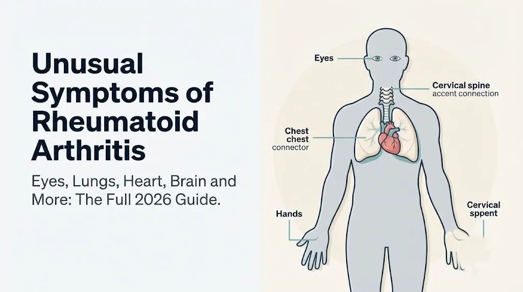
Unusual Symptoms of Rheumatoid Arthritis: Full 2026 Guide
The unusual symptoms of rheumatoid arthritis extend far beyond stiff, swollen joints, affecting the eyes, lungs, heart, brain, blood, skin, and nervous system through a systemic inflammatory process driven by cytokines that circulate through the entire body. These atypical manifestations affect a substantial portion of people with RA and are frequently misattributed to unrelated conditions, causing delays in treatment that allow preventable organ damage to progress.
The National Institute of Arthritis and Musculoskeletal and Skin Diseases (NIAMS) estimates that approximately 1.3 million Americans live with rheumatoid arthritis, and research published in Arthritis and Rheumatology has found that extra-articular manifestations occur in up to 40 percent of RA patients over the course of their disease, with patients who test positive for rheumatoid factor (RF) or anti-cyclic citrullinated peptide antibodies (anti-CCP) facing substantially higher risk.
This article covers every clinically recognized unusual symptom of RA, the specific immune and inflammatory mechanism producing each one, how disease characteristics like seropositivity and disease duration change individual risk, which unusual symptoms require urgent versus routine evaluation, and the critical distinction between symptoms caused by RA itself and symptoms caused by the medications used to treat it.
Unusual Symptoms of Rheumatoid Arthritis: Why RA Affects the Whole Body
The unusual symptoms of rheumatoid arthritis occur because RA is a systemic autoimmune disease, not a joint-specific condition, and the inflammatory mediators it generates circulate through the bloodstream to affect virtually every organ containing vascular tissue.
The core mechanism works like this: synovial macrophages and fibroblast-like synoviocytes in the inflamed joint lining produce massive quantities of tumor necrosis factor-alpha (TNF-alpha), interleukin-6 (IL-6), and interleukin-1 beta (IL-1 beta). These cytokines were designed to localize immune responses to infection sites. In RA, they are produced in excess and escape into systemic circulation. Once circulating, they activate endothelial cells in blood vessels throughout the body, increase vascular permeability, and recruit immune cells to tissues that were never the original target.
Think of RA’s systemic inflammation like a malfunctioning factory alarm. The alarm is supposed to trigger a local response at one location. Instead, it triggers every alarm in the building simultaneously. Every organ system that hears the alarm, meaning every organ with blood vessels carrying those cytokines, mounts an inflammatory response.
Anti-CCP antibodies and RF form immune complexes that deposit in blood vessel walls and serosal membranes (the thin layers surrounding the lungs, heart, and other organs). These deposits activate the complement cascade, generating the complement fragments C3a and C5a, which recruit neutrophils and drive local inflammatory tissue damage far from any joint.
The American College of Rheumatology (ACR) recognizes this systemic dimension in the 2010 ACR/EULAR classification criteria for RA, which include inflammatory markers like C-reactive protein (CRP) and erythrocyte sedimentation rate (ESR) as core criteria, reflecting that RA is fundamentally a systemic inflammatory disease from diagnosis. Patients with higher CRP and ESR at baseline have higher rates of extra-articular manifestations throughout their disease course.
How RA Seropositivity Predicts Unusual Symptoms
Seropositive RA (testing positive for rheumatoid factor and/or anti-CCP antibodies) is one of the strongest individual predictors of unusual extra-articular symptoms, because the antibodies themselves are the drivers of the immune complex-mediated tissue damage that produces non-joint manifestations.
Seropositivity means that the immune system has generated specific autoantibodies: RF (an antibody that targets the Fc portion of IgG immunoglobulin) and anti-CCP2 (an antibody targeting proteins that have undergone citrullination, a post-translational amino acid modification that occurs in inflamed tissue). Both antibody types form circulating immune complexes that deposit in tissues throughout the body.
Research published in Annals of Internal Medicine found that seropositive RA patients have two to three times the risk of extra-articular manifestations compared to seronegative patients, including substantially higher rates of rheumatoid nodules, vasculitis, pleuritis, and pericarditis. Seronegative RA patients (approximately 20 to 30 percent of all RA patients) still experience joint inflammation but have lower rates of the unusual systemic manifestations described throughout this article.
| Serostatus | RF and Anti-CCP | Extra-Articular Manifestation Risk | Common Unusual Symptoms |
|---|---|---|---|
| Seropositive (double positive) | Both positive | Highest (2-3x baseline) | Nodules, vasculitis, ILD, pericarditis |
| Seropositive (single positive) | One positive | Moderately elevated | Nodules, pleuritis |
| Seronegative | Both negative | Lower but not zero | Fatigue, anemia, milder systemic symptoms |
Men with RA, despite having lower overall RA incidence, show higher rates of certain extra-articular manifestations including pulmonary nodules and vasculitis compared to women with RA of equivalent disease duration, according to clinical cohort data reviewed in Arthritis and Rheumatology. This sex-based difference in extra-articular pattern is not fully explained but may relate to differences in hormonal modulation of the immune response.
Rheumatoid Nodules as an Unusual RA Symptom
Rheumatoid nodules are firm, flesh-colored or pale subcutaneous lumps that form in pressure areas and internal organs in seropositive RA patients, representing one of the most visually distinctive unusual symptoms of the disease.
These nodules form through a specific pathological process: macrophages cluster around a central zone of fibrinoid necrosis (tissue death from immune complex deposition and complement activation) in the dermis or subcutaneous tissue. The macrophages differentiate into epithelioid histiocytes and form a palisading granuloma structure around the necrotic center. Overlying fibroblasts deposit collagen, giving the nodule its firm texture. The lesions appear most often over bony prominences: the olecranon bursa at the elbow, the dorsum of the fingers, the Achilles tendon, and the back of the scalp.
According to the Arthritis Foundation, rheumatoid nodules occur in approximately 20 to 30 percent of people with seropositive RA and are rare in seronegative patients. Their presence is considered a marker of more aggressive systemic disease. Nodules can also form internally in the lung parenchyma (pulmonary nodules), on heart valves (cardiac nodules affecting the mitral valve), and in the eye’s sclera (scleromalacia perforans in severe cases).
A clinically important nuance: methotrexate, the most widely used first-line DMARD for RA, is paradoxically associated with accelerated rheumatoid nodule formation in some patients despite effectively suppressing joint inflammation. This is called methotrexate-induced nodulosis and is supported by observational clinical data. If your nodules increase in number or size after starting methotrexate, report this to your board-certified rheumatologist at the next appointment, specifying when the nodules began relative to the medication start date.
Eye Symptoms of Rheumatoid Arthritis: Scleritis and Episcleritis
Eye symptoms are among the most recognizable unusual manifestations of rheumatoid arthritis, with the two most clinically distinct conditions being scleritis (inflammation of the white scleral layer of the eye) and episcleritis (inflammation of the episcleral tissue superficial to the sclera).
The anatomical distinction matters enormously. Episcleritis is relatively superficial and produces redness and mild discomfort in one or both eyes. It is typically self-limiting and often the first eye manifestation to appear in RA. The mechanism involves immune complex deposition in the small episcleral vessels, producing local complement activation and neutrophil recruitment. Episcleritis in RA patients usually resolves within weeks and does not threaten vision.
Scleritis is far more serious. It involves the full thickness of the sclera, the structural “shell” of the eye. The inflammatory process produces severe, boring eye pain that is often described as the worst eye pain the patient has ever experienced, distinctly different from the mild discomfort of episcleritis. The sclera appears deep red to violet. The pain worsens at night and with eye movement. Scleritis carries a real risk of scleral perforation (scleromalacia perforans) in severe cases, because sustained inflammation can destroy the collagen matrix of the scleral tissue.
According to Mayo Clinic, scleritis occurs in approximately 1 percent of RA patients but represents a serious complication requiring urgent evaluation by an ophthalmologist. Any RA patient with new severe eye pain, particularly with a deep reddish-violet discoloration of the sclera rather than the surface-level bright redness of episcleritis, should see an ophthalmologist the same day, not a primary care physician the following week.
Postmenopausal women with RA have higher overall rates of ocular manifestations due to the combined effects of reduced estrogen (which normally has protective effects on lacrimal gland function) and RA-related immune dysregulation affecting tear production.
Key Takeaway: Episcleritis in RA is typically self-limiting and produces surface-level eye redness with mild discomfort. Scleritis produces deep, severe, boring eye pain with a violet-red eye color and requires same-day ophthalmology evaluation because scleral perforation is a real risk in severe cases.
Lung Symptoms of Rheumatoid Arthritis: ILD and Pleuritis
Lung involvement is one of the most serious unusual symptoms of rheumatoid arthritis, with interstitial lung disease (ILD) and pleuritis representing the two primary pulmonary manifestations, each with a distinct mechanism and clinical urgency.
RA-associated ILD occurs when sustained cytokine-driven inflammation (primarily TNF-alpha and IL-6 acting on lung interstitial fibroblasts) activates fibroblast collagen production in the spaces between the alveoli (air sacs). Over months to years, this collagen accumulation progressively replaces functional lung tissue with scar tissue. The two main histological patterns are usual interstitial pneumonia (UIP), which carries a less favorable prognosis and resembles idiopathic pulmonary fibrosis, and nonspecific interstitial pneumonia (NSIP), which tends to respond better to immunosuppressive treatment.
Research published in Arthritis and Rheumatology found that clinically evident ILD occurs in approximately 5 to 10 percent of RA patients, though subclinical ILD detected on high-resolution computed tomography (HRCT) is present in up to 30 to 40 percent. The most common early symptoms are exertional breathlessness (dyspnea on exertion) and a dry cough that does not respond to standard cough treatments. Men with seropositive RA and a history of smoking have the highest ILD risk.
Pleuritis occurs through a distinct mechanism: immune complex deposition in the pleural membranes (the two-layered serous membrane surrounding each lung) activates complement, causing local inflammation and fluid accumulation between the pleural layers (pleural effusion). Patients describe pleuritis as a sharp, stabbing chest pain that worsens with deep breathing or coughing. The pleural effusion of RA characteristically shows low glucose and low complement levels on fluid analysis, distinguishing it from infection-related effusion.
The critical distinction no competitor addresses: RA-related ILD and methotrexate-induced pneumonitis can be clinically indistinguishable at presentation. Both produce cough, breathlessness, and ground-glass opacities on HRCT. The distinction requires careful timing analysis (methotrexate pneumonitis typically develops within the first year of therapy and may present acutely), bronchoalveolar lavage cell differential, and often a multidisciplinary discussion between the rheumatologist and pulmonologist. Any RA patient taking methotrexate who develops new respiratory symptoms should have methotrexate held while the evaluation proceeds.
Heart Symptoms of Rheumatoid Arthritis: Pericarditis and Cardiovascular Risk
Cardiac involvement in RA produces two distinct categories of unusual symptoms: acute inflammatory cardiac disease (primarily pericarditis) and accelerated atherosclerosis producing elevated cardiovascular event risk.
Pericarditis in RA occurs when immune complex deposition in the pericardium (the fibrous sac surrounding the heart) activates complement and drives local neutrophilic inflammation. The pericardium becomes thickened and inflamed, producing the characteristic sharp, stabbing chest pain of pericarditis that improves when the patient leans forward (this position reduces pressure on the inflamed pericardial surfaces). Fluid can accumulate in the pericardial space (pericardial effusion). In severe cases, the effusion compresses the heart chambers, reducing cardiac output in a life-threatening condition called cardiac tamponade.
According to Mayo Clinic, pericardial involvement is present in up to 50 percent of RA patients on autopsy studies, though clinically symptomatic pericarditis affects approximately 10 percent of patients during their disease course. Pericarditis is more common in patients with active, poorly controlled disease and in seropositive patients.
Separately, RA drives accelerated atherosclerosis through chronic systemic inflammation. IL-6 promotes hepatic production of fibrinogen and CRP, both of which contribute to arterial plaque instability. TNF-alpha directly activates endothelial cells, promoting adhesion molecule expression and foam cell formation in arterial walls. Research published in the New England Journal of Medicine has documented that RA patients have a 50 to 60 percent higher risk of myocardial infarction compared to the general population, independent of traditional cardiovascular risk factors.
Women with RA face a particularly elevated cardiovascular risk because traditional cardiovascular risk calculators underestimate their true risk. The American College of Rheumatology recommends using a 1.5 multiplication factor when applying standard cardiovascular risk scores to RA patients to better capture their elevated inflammatory burden.
Rheumatoid Arthritis Cervical Spine Involvement and Myelopathy Risk
Cervical spine involvement is one of the most serious and underrecognized unusual symptoms of rheumatoid arthritis, because RA can destabilize the upper cervical spine in a way that directly threatens the spinal cord, producing a potentially catastrophic neurological complication.
The specific anatomical mechanism involves the atlantoaxial joint (C1-C2), the joint between the first and second cervical vertebrae. This joint is stabilized primarily by the transverse ligament of the atlas, a band of fibrous tissue that holds the odontoid process (the upward projection of C2) in position behind the arch of C1. RA synovitis can affect this joint just as it affects peripheral joints. The pannus tissue invades the transverse ligament, erodes the odontoid process, and progressively loosens the constraint holding C1 and C2 together.
When the atlantoaxial joint becomes unstable (a condition called atlantoaxial subluxation), any significant neck movement can cause the loosened C1 to slip forward over C2, compressing the cervical spinal cord and the lower brainstem. The resulting cervical myelopathy produces progressive hand weakness, difficulty with fine motor tasks, unsteady gait (ataxia), loss of hand grip, and in severe cases, bowel and bladder dysfunction. This can progress to quadriplegia if unrecognized.
According to Mayo Clinic, atlantoaxial subluxation occurs in up to 25 percent of RA patients with long-standing disease, though not all cases produce neurological symptoms. Patients with known RA undergoing general anesthesia are at particular risk during intubation, when forceful neck extension can trigger subluxation. Anesthesiologists managing RA patients should be informed of the diagnosis specifically because of this risk.
Any RA patient who develops new neck pain radiating to the back of the head (suboccipital pain), weakness or numbness in the hands or legs, or unsteady walking should contact their board-certified rheumatologist immediately and have cervical spine imaging (flexion-extension lateral X-ray or MRI) performed on an urgent basis.
Key Takeaway: RA can destabilize the C1-C2 joint through synovial inflammation of the atlantoaxial joint, and the resulting cervical myelopathy produces hand weakness, gait changes, and bladder dysfunction. Any RA patient with new neurological symptoms in the arms or legs needs urgent cervical spine imaging, not a routine rheumatology appointment.
RA Vasculitis: An Unusual and Serious Complication
Rheumatoid vasculitis is a rare but serious complication in which RA-driven immune complex deposition directly damages the walls of small and medium blood vessels throughout the body, producing ischemic injury in the organs and tissues those vessels supply.
The mechanism involves the deposition of immune complexes (RF, anti-CCP, and complement components) within the vascular endothelium of small arteries and arterioles. Complement activation generates C5a, which recruits neutrophils that release proteolytic enzymes, damaging the vessel wall. The damaged vessel wall becomes inflamed and may thrombose or develop fibrinoid necrosis, cutting off blood supply to downstream tissue.
Skin manifestations are the most visible: small, punched-out ulcers typically on the lower legs, dark necrotic lesions at the digital nail folds (a feature called nail fold infarcts), and palpable purpura (raised, non-blanching purple spots from red blood cell extravasation through damaged vessel walls). When vasculitis affects the nerves’ blood supply (the vasa nervorum, the small vessels feeding peripheral nerves), it produces an acutely painful neuropathy called mononeuritis multiplex, characterized by sudden-onset asymmetric weakness and sensory loss that jumps unpredictably between nerve territories.
The American College of Rheumatology considers rheumatoid vasculitis a marker of severe, advanced RA that has been inadequately controlled. It is more common in men, in patients with high RF titers, and in those with long disease duration and poor disease control.
Treatment of rheumatoid vasculitis requires aggressive immunosuppression under the care of a board-certified rheumatologist, often including high-dose corticosteroids and biological agents (cyclophosphamide in severe cases). Skin ulcers or digital necrosis in a patient with known RA should not be attributed to poor circulation or venous insufficiency without first excluding vasculitis through dermatological consultation and skin biopsy.
Neurological Symptoms of Rheumatoid Arthritis
Rheumatoid arthritis produces neurological symptoms through three distinct mechanisms: peripheral nerve entrapment at inflamed joints, vasculitis affecting the vasa nervorum, and direct cytokine effects on central nervous system function.
Entrapment neuropathy is the most common neurological manifestation. The most familiar example is carpal tunnel syndrome (CTS), in which synovitis of the wrist’s flexor tendon sheaths compresses the median nerve within the carpal tunnel (the fibro-osseous canal formed by the carpal bones and the transverse carpal ligament). RA-associated CTS produces the classic CTS symptoms: numbness, tingling, and burning pain in the thumb, index finger, middle finger, and lateral half of the ring finger, often worst at night. According to the Arthritis Foundation, carpal tunnel syndrome occurs in approximately 5 to 8 percent of people with RA and may be the first neurological symptom to appear, sometimes preceding obvious wrist joint involvement.
Tarsal tunnel syndrome (compression of the posterior tibial nerve at the ankle by synovitis of the flexor tendon sheath) produces equivalent symptoms in the sole of the foot and is less commonly recognized as an RA manifestation.
Mononeuritis multiplex from vasculitis (described in the previous section) produces sudden, severe, asymmetric weakness and numbness that differs from entrapment neuropathy in its acuity and multi-focal distribution. Entrapment neuropathies develop gradually and affect predictable nerve territories. Vasculitic neuropathy develops within hours and affects nerves in a pattern that does not correspond to a single anatomical entrapment site.
Quick Tip:
- Distinguish RA-related CTS from non-RA CTS by noting whether wrist joint swelling or tenosynovitis (tendon sheath swelling) accompanies the hand symptoms
- RA patients with bilateral CTS have higher synovitis burden than those with unilateral CTS, according to clinical observational data
- Older adults with RA have elevated risk of cervical myelopathy producing upper and lower extremity neurological symptoms that can initially be attributed to peripheral neuropathy; these patients need cervical spine imaging before a peripheral neuropathy diagnosis is accepted
Unusual Blood and Immune Symptoms of RA: Felty’s Syndrome and Anemia
Hematological (blood-related) unusual symptoms of RA include anemia of chronic disease (the most common blood abnormality in RA) and the rare but distinctive Felty’s syndrome, which combines RA with splenomegaly and severe neutropenia.
Anemia of chronic disease in RA is produced by a specific mechanism involving hepcidin, a peptide hormone produced by the liver in response to IL-6 signaling. Hepcidin blocks ferroportin, the iron export channel on macrophages and intestinal cells, trapping iron inside these cells rather than releasing it for red blood cell production in the bone marrow. The result is a hypoproliferative anemia with normal or elevated iron stores but reduced serum iron available to erythroid precursors. This is distinct from iron deficiency anemia (where iron stores are depleted) and is typically a mild to moderate normocytic, normochromic anemia that correlates with RA disease activity. According to the NIAMS, anemia of chronic disease occurs in approximately 30 to 60 percent of RA patients with active disease.
Felty’s syndrome is a rare but serious triad occurring in fewer than 1 percent of RA patients, consisting of seropositive RA, splenomegaly (enlarged spleen), and severe neutropenia (dangerously low neutrophil count). The enlarged spleen traps and destroys neutrophils and other blood cells. The resulting neutropenia impairs the ability to fight bacterial infections, leading to recurrent, potentially life-threatening infections. Felty’s syndrome almost exclusively occurs in patients with long-standing seropositive RA, usually after 10 or more years of disease, and is more common in White patients compared to other racial groups.
Research published in Arthritis and Rheumatology found that patients with Felty’s syndrome have a higher risk of lymphoma over their disease course, making long-term monitoring by the treating rheumatologist essential.
RA-Related Sjogren’s Overlap and Mouth and Eye Dryness
Dryness of the eyes and mouth as unusual symptoms in RA often indicates a specific condition called secondary Sjogren’s syndrome, in which RA-driven autoimmune inflammation extends to the lacrimal glands (tear-producing glands located above each eye) and salivary glands.
The mechanism involves lymphocytic infiltration of the exocrine glands. CD4+ T cells and B cells accumulate in the lacrimal and salivary gland tissue, progressively replacing functional secretory acinar cells with inflammatory infiltrate. Tear production falls, producing keratoconjunctivitis sicca (dry eye disease), which causes a gritty, burning, or sandy sensation in the eyes. Salivary production falls, producing xerostomia (dry mouth), which causes difficulty swallowing dry foods, altered taste, accelerated dental decay, and painful oral fungal infections (oral candidiasis) due to the loss of saliva’s antimicrobial properties.
According to Mayo Clinic, secondary Sjogren’s syndrome occurs in approximately 30 percent of patients with RA. The eye and mouth dryness in RA-associated Sjogren’s overlap is typically milder than in primary Sjogren’s syndrome but can cause substantial quality-of-life impairment that is underreported because patients often assume dryness is a side effect of medications rather than a disease manifestation.
The Schirmer’s test (a strip of filter paper placed under the lower eyelid to measure tear production over 5 minutes) and minor salivary gland biopsy are the standard diagnostic tests for confirming lacrimal and salivary gland involvement. Women with RA are more likely to develop secondary Sjogren’s overlap than men, reflecting the generally higher autoimmune burden in female RA patients, and postmenopausal women have the highest risk because declining estrogen independently reduces tear production.
Key Takeaway: Persistent dry eyes and dry mouth in an RA patient are not simply medication side effects until Sjogren’s overlap has been excluded. This is a clinically distinct autoimmune complication requiring its own evaluation by a rheumatologist or ophthalmologist, separate from standard RA management.
Cognitive Dysfunction and Brain Fog in Rheumatoid Arthritis
Cognitive dysfunction in rheumatoid arthritis, frequently described by patients as “RA fog” or “brain fog,” is a real, physiologically grounded unusual symptom produced by the direct effects of RA’s systemic inflammatory cytokines on central nervous system function.
The primary mechanism involves IL-6 and TNF-alpha crossing the blood-brain barrier via active transport through endothelial cytokine transporters. Once inside the central nervous system, these cytokines activate microglia (the brain’s resident immune cells) and interfere with neurotransmitter synthesis and release. IL-6 specifically reduces the availability of serotonin and dopamine precursors in the brain by diverting the amino acid tryptophan toward the kynurenine pathway rather than toward serotonin synthesis. TNF-alpha impairs synaptic plasticity and reduces brain-derived neurotrophic factor (BDNF) in hippocampal neurons, a structure critical for memory formation and retrieval.
Research published in Arthritis Care and Research found that RA patients score lower on standardized cognitive assessments in the domains of processing speed, working memory, and executive function compared to age-matched controls without inflammatory disease. The cognitive impairment correlates with CRP levels and disease activity scores, supporting the cytokine mechanism rather than medication effects as the primary driver.
Patients typically describe RA fog as difficulty finding words mid-sentence, slowed information processing, difficulty holding multiple thoughts simultaneously, and short-term memory lapses. These symptoms worsen during disease flares when systemic cytokine levels peak.
Older adults with RA are at elevated risk for RA-related cognitive symptoms because they already have age-related reduction in cognitive reserve. The differential diagnosis in this population must exclude medication effects from low-dose corticosteroids (which can impair memory at higher doses) and methotrexate (which rarely causes cognitive symptoms at standard RA doses but may contribute in patients with reduced folate levels).
Depression and Mental Health as Unusual Symptoms of RA
Depression is an unusual symptom of rheumatoid arthritis that most patients and many clinicians do not initially attribute to the disease itself, yet it is produced by the same systemic cytokine biology driving every other extra-articular manifestation.
The mechanism linking RA to depression runs through several overlapping pathways. IL-6 diverts tryptophan metabolism away from serotonin synthesis toward the kynurenine pathway, reducing central serotonin availability. Elevated TNF-alpha activates the hypothalamic-pituitary-adrenal (HPA) axis, driving chronic cortisol elevation and disrupting the normal negative feedback regulation of the stress response. Chronic corticosteroid excess (both medication-derived and endogenous HPA dysregulation) alters hippocampal structure and contributes to depressive neurochemistry. Chronic pain from joint inflammation independently activates pain-processing circuits in the brain that overlap with emotional regulation circuits, contributing to mood dysregulation.
A meta-analysis published in Arthritis Care and Research found that depression affects approximately 38 to 42 percent of RA patients, compared to approximately 7 percent of the general population. This rate is not explained by reaction to a chronic diagnosis alone; studies controlling for disease duration and functional impairment still show elevated depression rates in RA, consistent with biological rather than purely psychological causation.
If you are experiencing persistent low mood, loss of interest in activities, sleep disruption, or feelings of hopelessness alongside your RA symptoms, raise this specifically with your board-certified rheumatologist at your next appointment. Bring a written list of when these symptoms began relative to RA flares and medication changes, because the pattern helps distinguish RA-driven depression from medication side effects (corticosteroids and some DMARDs are associated with mood changes).
If you are in crisis or experiencing thoughts of self-harm or suicide, contact the 988 Suicide and Crisis Lifeline by calling or texting 988 at any time. This service is free, confidential, and available 24 hours a day, 7 days a week.
Unusual RA Symptoms Caused by Medications, Not the Disease
Some of the most confusing unusual symptoms in RA patients are not caused by the disease but by the medications used to treat it, and distinguishing between the two is one of the most clinically consequential challenges in RA management.
Methotrexate is the most widely used first-line DMARD for RA and produces several unusual symptoms that can be mistaken for RA manifestations. Methotrexate-induced pneumonitis (lung inflammation) produces cough, breathlessness, and fever that is clinically difficult to distinguish from RA-related ILD. The critical distinguishing feature is timing and acuity: methotrexate pneumonitis tends to develop within the first year of therapy, can progress rapidly over days to weeks, and often presents with fever and an acute inflammatory pattern on HRCT rather than the progressive fibrosis pattern of RA-ILD. Methotrexate also causes liver toxicity (elevated liver enzymes and, with long-term use, hepatic fibrosis) and can produce mouth sores (oral mucositis) that look similar to Sjogren’s-related oral problems.
Hydroxychloroquine (an antimalarial DMARD used in mild RA and combination regimens) carries a specific risk of retinal toxicity with long-term use. The drug accumulates in the retinal pigment epithelium and can produce a distinctive “bull’s-eye” pattern of macular damage on ophthalmological examination. The American Academy of Ophthalmology recommends annual retinal screening for patients who have taken hydroxychloroquine for more than 5 years or at high cumulative doses.
Biologic DMARDs (TNF inhibitors such as etanercept, adalimumab, infliximab) can produce paradoxical inflammatory conditions: psoriasis-like skin rashes (paradoxical psoriasis), new-onset inflammatory bowel disease, and in rare cases, demyelinating neurological conditions resembling multiple sclerosis. These are not RA manifestations. They are medication-related and require switching to a different biologic class.
| Medication | Unusual Symptom | How to Distinguish from RA | Action Required |
|---|---|---|---|
| Methotrexate | Interstitial lung changes | RA-ILD progresses slowly; methotrexate pneumonitis is acute with fever | Hold methotrexate; urgent pulmonology evaluation |
| Methotrexate | Oral mucositis | RA does not directly cause mouth sores; Sjogren’s causes dryness, not ulcers | Add folate supplementation; discuss with rheumatologist |
| Hydroxychloroquine | Retinal toxicity | Bull’s-eye maculopathy on retinal exam; RA itself rarely causes macular damage | Annual ophthalmology screening |
| TNF inhibitors | Paradoxical psoriasis | RA causes rheumatoid nodules, not psoriatic plaques | Switch to non-TNF biologic class |
Unusual Early Symptoms of Rheumatoid Arthritis Before Joint Pain Begins
A group of unusual symptoms can precede the classic joint swelling and morning stiffness of RA by months to years, and recognizing them as potential prodromal (pre-clinical) RA signs may shorten the diagnostic delay that allows early joint damage to progress.
The most clinically documented pre-RA prodromal symptom is persistent fatigue that is disproportionate to activity level and unresponsive to rest. This fatigue is mechanistically distinct from ordinary tiredness: it is driven by IL-6-mediated activation of the hypothalamic sleep-wake regulatory centers and elevated CRP triggering sickness behavior pathways in the brain. Research from the NIAMS and supported by prospective cohort data suggests that fatigue may precede joint symptoms in RA by 6 months to 2 years in some patients.
Unexplained low-grade fever (temperature between 37.5 and 38.5 degrees Celsius, or 99.5 to 101.3 degrees Fahrenheit) is another prodromal unusual symptom, driven by IL-1 beta stimulating prostaglandin E2 production in the hypothalamus. This produces a persistent, low-level fever that is often attributed to undiagnosed infection.
Early RA can also present as diffuse musculoskeletal pain without clear joint swelling, making it initially indistinguishable from fibromyalgia, polymyalgia rheumatica, or early lupus. The distinguishing feature that begins to differentiate early RA is the symmetrical involvement of small joints (proximal interphalangeal joints of the fingers, metacarpophalangeal joints) and the characteristic morning stiffness lasting more than 45 minutes.
A primary care physician should refer any patient with persistent symmetrical small joint pain, morning stiffness lasting more than 45 minutes, unexplained fatigue, and positive RF or anti-CCP blood test results to a board-certified rheumatologist for formal RA evaluation without delay, because early initiation of disease-modifying therapy improves long-term joint outcomes significantly.
When Unusual RA Symptoms Require Emergency Evaluation
Certain unusual symptoms associated with rheumatoid arthritis require immediate emergency evaluation. Do not wait to see if these resolve on their own.
Emergency Symptoms: When to Call 911 or Go to the ER
Call 911 or go to the nearest emergency room immediately if you experience:
- Sudden severe neck pain with new arm or leg weakness, hand numbness, difficulty walking, or loss of bladder or bowel control in a person with known RA, because this combination suggests atlantoaxial subluxation with cervical spinal cord compression requiring emergency neurosurgical evaluation
- Progressive shortness of breath with a falling blood pressure, rapid heart rate, or visible neck vein distension, because this triad suggests cardiac tamponade from a pericardial effusion, a life-threatening compression of the heart requiring emergency pericardiocentesis
- Sudden onset severe eye pain with a deep violet-red eye color in an RA patient, because this suggests acute necrotizing scleritis with risk of scleral perforation, requiring same-day ophthalmology evaluation at a minimum and emergency evaluation if visual acuity is changing
- New chest pain with fever, worse with lying flat and improved by leaning forward, in a patient with active RA, because this suggests acute pericarditis that may be associated with pericardial effusion requiring urgent cardiac evaluation
- Sudden asymmetric limb weakness with severe pain shooting along a nerve distribution in an RA patient with known vasculitis or high RF titers, because this suggests acute vasculitic mononeuritis multiplex with nerve ischemia requiring emergency rheumatology assessment
- Signs of severe infection (high fever, rigors, confusion) in an RA patient with Felty’s syndrome or on immunosuppressive therapy, because neutropenia in these patients makes bacterial sepsis a rapidly fatal emergency
These presentations can indicate atlantoaxial subluxation, cardiac tamponade, acute vasculitic neuropathy, or infectious sepsis in an immunosuppressed patient, and require emergency medical assessment, not a phone call to the rheumatology office.
Frequently Asked Questions About Unusual Symptoms of Rheumatoid Arthritis
What are the unusual symptoms of rheumatoid arthritis?
Unusual symptoms of rheumatoid arthritis include eye inflammation (scleritis, episcleritis), interstitial lung disease, pleuritis, pericarditis, rheumatoid nodules under the skin, peripheral neuropathy, cervical spine instability, vasculitis, Felty’s syndrome, secondary Sjogren’s syndrome, cognitive dysfunction, and depression.
These extra-articular symptoms are produced by the same systemic cytokines (TNF-alpha, IL-6, IL-1 beta) that drive joint inflammation, acting on blood vessels and organ tissue throughout the body.
Seropositive RA patients (those testing positive for rheumatoid factor or anti-CCP antibodies) have two to three times the risk of developing these unusual manifestations compared to seronegative patients, according to research in Annals of Internal Medicine.
Can rheumatoid arthritis cause brain fog?
Yes, rheumatoid arthritis causes brain fog through the direct effects of IL-6 and TNF-alpha on the central nervous system, including diversion of tryptophan away from serotonin synthesis and impairment of hippocampal synaptic plasticity.
Research published in Arthritis Care and Research found that RA patients score lower than age-matched controls on measures of processing speed, working memory, and executive function, with cognitive performance correlating with CRP levels and disease activity.
The cognitive symptoms typically worsen during disease flares and may improve with effective suppression of RA disease activity, though the evidence for symptom reversal with treatment is primarily from observational data rather than randomized trials.
Does rheumatoid arthritis affect the lungs?
Rheumatoid arthritis affects the lungs through two primary mechanisms: interstitial lung disease (ILD), in which cytokine-driven inflammation causes progressive lung fibrosis, and pleuritis, in which immune complex deposition in the pleural membranes causes chest pain and fluid accumulation.
Clinically evident ILD occurs in approximately 5 to 10 percent of RA patients, though subclinical ILD detectable on high-resolution CT is present in up to 30 to 40 percent, according to data reviewed in Arthritis and Rheumatology.
Any RA patient who develops new exertional breathlessness or a persistent dry cough should be evaluated by a pulmonologist, with a specific focus on distinguishing RA-related ILD from methotrexate-induced pneumonitis before the treatment approach is determined.
What are the early unusual symptoms of rheumatoid arthritis?
Early unusual symptoms of rheumatoid arthritis include persistent disproportionate fatigue, low-grade fever, diffuse musculoskeletal pain without obvious joint swelling, and morning stiffness lasting more than 45 minutes, which can precede classic joint involvement by months to years.
These prodromal symptoms are driven by the same cytokines (IL-6, IL-1 beta) that will later produce joint synovitis, acting on the hypothalamus and brain before synovial inflammation becomes clinically apparent.
A primary care physician should refer patients with persistent unexplained fatigue, symmetrical small joint pain, and morning stiffness to a board-certified rheumatologist for RA evaluation, particularly if RF or anti-CCP blood tests are positive.
Is RA-related depression different from regular depression?
RA-related depression has a biological mechanism distinct from purely psychological depression, involving IL-6-mediated tryptophan diversion away from serotonin synthesis and TNF-alpha-driven dysregulation of the hypothalamic-pituitary-adrenal axis.
A meta-analysis published in Arthritis Care and Research found depression rates of 38 to 42 percent in RA patients, far exceeding the general population rate of approximately 7 percent and consistent with a biological rather than purely situational cause.
Effective control of RA disease activity with appropriate DMARD therapy can reduce the inflammatory drivers of RA-associated depression, though many patients benefit from concurrent evaluation by a psychiatrist or psychologist for medication and behavioral treatment alongside rheumatological management.
When should an RA patient go to the emergency room?
An RA patient should call 911 or go to the emergency room immediately for sudden severe neck pain with new arm or leg weakness (possible atlantoaxial subluxation), progressive shortness of breath with falling blood pressure (possible cardiac tamponade), or sudden asymmetric limb weakness with nerve-distribution pain (possible vasculitic mononeuritis multiplex).
These are all recognized life-threatening complications of rheumatoid arthritis that progress rapidly and require emergency medical intervention rather than an urgent rheumatology appointment.
RA patients on immunosuppressive therapy who develop high fever with rigors or confusion should also go to the emergency room immediately, as neutropenia from Felty’s syndrome or medication-related immunosuppression can make bacterial sepsis a rapidly fatal emergency.
Rheumatoid arthritis is a systemic disease, and understanding that its unusual symptoms are not coincidental or unrelated findings but predictable extensions of the same inflammatory biology driving your joint disease changes how to approach them clinically. Every unusual symptom discussed here has a named mechanism, and every one of those mechanisms flows from the same source: an overactive immune system producing cytokines that were never designed to stay local.
Your most practical next step is to keep a written symptom log that captures any new or changing symptoms outside your joints, noting when they started and whether they correlate with RA flares or medication changes. Bring that log to every rheumatology appointment. These details are what allow your board-certified rheumatologist to distinguish disease progression from medication effects, new complications from pre-existing conditions, and watchful waiting from urgent evaluation.
The unusual symptoms described in this article are not reasons to panic. They are reasons to be a thorough, well-informed reporter of your own clinical picture.