Symptoms of Bacterial Infection in Esophagus: 2026 Guide
The symptoms of bacterial infection in the esophagus include painful swallowing (a condition called odynophagia), difficulty moving food through the throat (dysphagia), chest pain behind the breastbone, fever, and in severe cases, an inability to eat or drink at all. These symptoms arise when bacteria invade and inflame the layered tissue lining the esophagus, triggering an immune response that produces mucosal edema, ulceration, and sometimes abscess formation within the esophageal wall.
Bacterial esophagitis is substantially less frequent than fungal or viral esophageal infections, but it is not a minor condition. According to the National Institute of Diabetes and Digestive and Kidney Diseases (NIDDK), infectious esophagitis of any type predominantly affects people with impaired immune function, and bacterial forms carry a risk of serious complications including esophageal perforation and mediastinitis that can become rapidly life-threatening without treatment.
This article covers the full clinical picture: the anatomical reason each symptom occurs, the specific bacteria involved and how each produces a different pattern, how bacterial esophagitis differs from fungal infection, which populations face the highest risk, how the diagnosis is made, and which symptoms require emergency evaluation rather than a scheduled appointment.
Symptoms of Bacterial Infection in Esophagus: The Clinical Overview
The defining symptoms of bacterial infection in the esophagus are painful swallowing, difficulty swallowing, midsternal chest pain, and systemic signs of infection including fever and malaise, which together reflect inflammatory damage to the esophageal mucosal lining and its underlying layers.
Not everyone develops all of these symptoms simultaneously. In early or mild bacterial esophagitis, the presentation may be limited to discomfort when swallowing solid foods and a vague burning sensation behind the sternum. As the infection progresses and mucosal damage deepens, swallowing liquids becomes painful, chest pain intensifies, and systemic symptoms including fever and reduced appetite emerge.
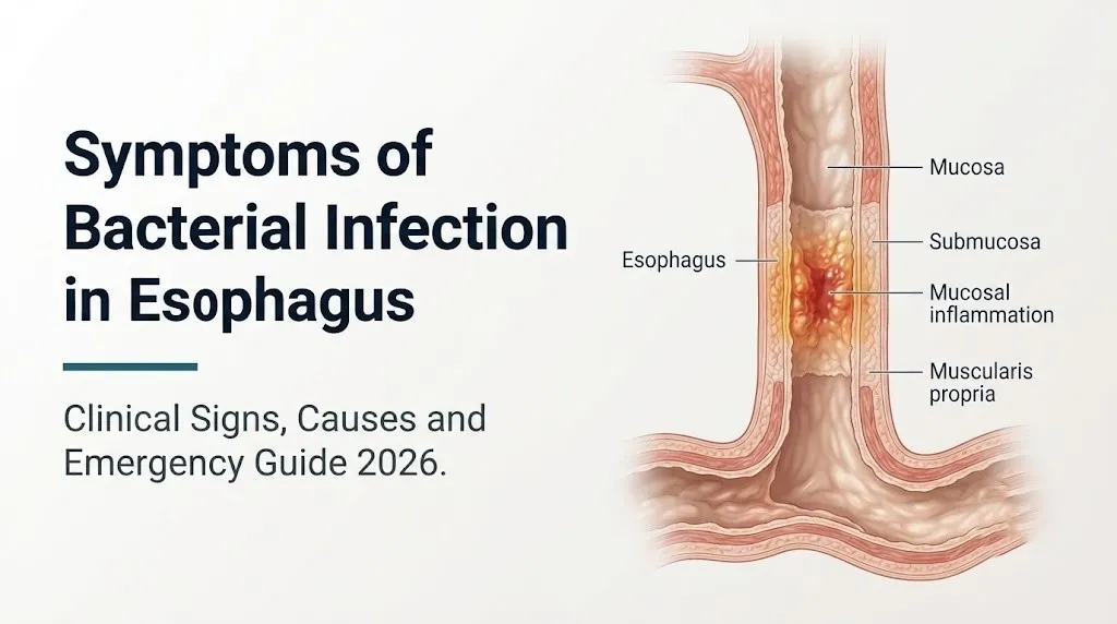
The severity of symptoms is directly proportional to the depth of bacterial invasion. Superficial infection limited to the esophageal mucosa (the stratified squamous epithelium) produces discomfort and mild dysphagia. Invasion into the lamina propria and submucosa triggers a stronger inflammatory cascade and more intense pain. Transmural infection reaching the muscularis propria and adventitia creates conditions for abscess formation and perforation.
Key symptoms of bacterial esophageal infection in order of clinical significance:
- Odynophagia (pain specifically during the act of swallowing): the most consistent and clinically prominent symptom
- Dysphagia (sensation of food sticking or not passing normally): typically worse with solid foods early, then progressing to liquids
- Midsternal or retrosternal chest pain (behind the breastbone, often confused with cardiac pain)
- Fever: present in moderate-to-severe bacterial esophagitis; absent or low-grade in mild cases
- Nausea and reduced appetite: secondary to pain on swallowing and systemic inflammatory response
- Weight loss: seen in prolonged or severe cases where eating becomes too painful to maintain adequate intake
- Regurgitation of food or liquids: occurs when mucosal edema significantly narrows the esophageal lumen
People taking proton pump inhibitors (PPIs) for chronic gastroesophageal reflux disease (GERD) may have partially masked symptoms, as acid suppression reduces one contributing factor to mucosal irritation even while the bacterial infection progresses. This masking effect can delay recognition and evaluation.
What Is Bacterial Esophagitis and Who Gets It
Bacterial esophagitis is an infection of the esophageal wall caused by bacterial pathogens, occurring almost exclusively in individuals with disrupted local mucosal defenses, systemic immune impairment, or structural abnormalities of the esophagus.
The esophagus is not sterile. The oral cavity contains a complex microbiome including gram-positive cocci, anaerobes, and gram-negative species, and saliva continuously delivers these organisms to the esophageal surface. Under normal immune conditions, the intact stratified squamous epithelium of the esophageal mucosa, supported by secretory IgA from submucosal glands and resident immune surveillance cells, prevents colonization from progressing to invasive infection.
Bacterial esophagitis develops when one or more of these defense mechanisms fails. The American College of Gastroenterology identifies immunosuppression as the primary predisposing factor for infectious esophagitis of all types, including bacterial.
The populations at highest risk:
- People with HIV/AIDS, particularly those with CD4 T-lymphocyte counts below 200 cells per microliter
- Solid organ transplant recipients on immunosuppressive regimens including calcineurin inhibitors (tacrolimus, cyclosporine) and high-dose corticosteroids
- Hematopoietic stem cell transplant recipients, particularly during the neutropenic period after conditioning chemotherapy
- People receiving systemic corticosteroids for autoimmune or inflammatory conditions
- Patients undergoing chemotherapy with agents that produce mucositis (5-fluorouracil, cisplatin, methotrexate)
- People receiving radiation therapy to thoracic fields that include the esophagus
- Older adults with pre-existing esophageal motility disorders or structural abnormalities
- People with severe malnutrition, which impairs both cellular and humoral mucosal immunity
Healthy immunocompetent adults very rarely develop bacterial esophagitis. When a person without any known immune deficiency presents with symptoms suggestive of esophageal infection, the evaluation should include assessment for undiagnosed HIV infection, diabetes mellitus, or occult malignancy as possible contributing causes.
Esophageal Anatomy: Why the Esophagus Is Vulnerable to Bacterial Infection
The esophagus is a 25-centimeter muscular tube extending from the lower border of the cricopharyngeus muscle (at the upper esophageal sphincter) to the gastroesophageal junction, and its layered architecture explains both why it normally resists bacterial infection and why it becomes vulnerable when those layers are disrupted.
From the inside out, the esophageal wall consists of four concentric layers. The innermost layer is the mucosa, composed of nonkeratinized stratified squamous epithelial cells that form a physically tough, tightly adherent barrier against microbial penetration. Beneath it lies the lamina propria, a connective tissue layer containing blood vessels, lymphatics, and the immune cells (dendritic cells, macrophages, and IgA-secreting plasma cells) that form the front line of mucosal defense. The submucosa contains Meissner’s nerve plexus (a component of the enteric nervous system governing sensory signaling), submucosal glands, and deeper vascular structures. The muscularis propria contains the muscle layers driving peristalsis, and the outermost layer, the adventitia, is a loose connective tissue covering that interfaces with the mediastinal space.
Think of the healthy esophageal wall like a layered protective door. The squamous epithelium is the locked outer surface. The lamina propria is the security system behind it. Under normal conditions, bacteria from the oral cavity touch the door, find it locked, and pass through without entry. When the epithelial barrier is breached by radiation, chemotherapy, ulceration from severe GERD, or direct mucosal injury, the door opens, and bacterial invasion proceeds inward layer by layer.
The absence of a serosal layer (which covers most of the gastrointestinal tract and provides an additional containment barrier) is an anatomically important vulnerability. The esophagus is protected only by the loose adventitia rather than the tougher visceral peritoneum. This means that transmural infection can spread directly into the mediastinum with fewer anatomical barriers to contain it.
According to the Merck Manual of Diagnosis and Therapy, the lack of a serosal covering makes esophageal perforations particularly prone to rapid, life-threatening spread of infection into the mediastinal space.
Painful Swallowing (Odynophagia) From Bacterial Esophageal Infection
Odynophagia, the sensation of pain during the act of swallowing, is the most clinically consistent symptom of bacterial esophageal infection and occurs when bacterial inflammation sensitizes the dense sensory nerve network within the esophageal submucosa.
The esophageal wall is innervated by visceral afferent nerve fibers traveling within the vagus nerve (cranial nerve X) and the thoracic sympathetic chain. Under normal conditions, swallowing is painless because peristaltic muscle contraction and bolus transit do not activate these nociceptors. When bacterial infection inflames the lamina propria and submucosa, two processes combine to produce pain. First, tissue prostaglandin E2 (produced by cyclooxygenase-2 in inflamed mucosal cells) lowers the activation threshold of local nociceptors. Second, substance P, released from sensitized submucosal nerve endings, amplifies pain signaling. Every swallow then mechanically deforms inflamed, hypersensitized tissue, triggering a discrete burst of pain.
The character of odynophagia in bacterial esophagitis is typically described as sharp, burning, or stabbing pain occurring immediately with swallowing, located behind the sternum or in the mid-chest region, and lasting for several seconds after the swallow is completed. It differs from the burning discomfort of GERD, which is not specifically triggered by the act of swallowing and is more diffuse and positional.
According to the American Journal of Gastroenterology, odynophagia severe enough to prevent adequate oral intake is an indication for urgent endoscopic evaluation regardless of suspected cause, because it cannot be distinguished from potentially serious infectious or malignant processes without direct visualization and tissue sampling.
Older adults may report odynophagia differently than younger patients. In older individuals, the sensation is often described as a “catch” or pressure rather than sharp pain, partly because age-related changes in esophageal sensory innervation can alter the qualitative character of nociceptive signals. This atypical presentation can lead to delayed evaluation in this age group.
Dysphagia and Chest Pain: How Bacterial Esophagitis Affects Swallowing
Dysphagia, the sensation of food or liquid not passing normally through the esophagus, occurs in bacterial esophagitis when inflammatory edema narrows the effective luminal diameter of the esophageal tube and when pain reflexively inhibits the normal swallowing sequence.
The esophageal lumen normally ranges from approximately 15 to 25 millimeters in diameter during swallowing. Bacterial infection produces inflammatory edema in the lamina propria and submucosa through increased vascular permeability driven by histamine and IL-1 beta release from mast cells and macrophages at the infection site. This edema physically reduces the lumen diameter, creating the sensation of food sticking or passing with effort. Early bacterial esophagitis typically produces dysphagia with solid foods only. As edema progresses, dysphagia extends to soft foods and eventually liquids.
Chest pain in bacterial esophagitis is referred pain. The esophagus shares visceral afferent pain pathways with the heart through the thoracic sympathetic chain, with both organs transmitting pain signals through the T1 to T5 spinal cord levels. The brain cannot reliably distinguish whether incoming pain signals originate in the esophageal wall or the cardiac muscle, producing chest pain that can closely mimic angina or myocardial ischemia. This overlap is clinically important: esophageal chest pain does not reliably respond to nitroglycerin, does not radiate to the left arm in a classic pattern, and is often worse during swallowing rather than during physical exertion.
Patients with pre-existing coronary artery disease who develop bacterial esophagitis face a particularly challenging diagnostic scenario because both cardiac ischemia and esophageal infection can produce midsternal chest pain at rest. A board-certified gastroenterologist and cardiologist may need to evaluate these patients concurrently to avoid missing either diagnosis.
Key Takeaway: Odynophagia (pain specifically during swallowing) is the signature symptom of bacterial esophagitis and is produced by prostaglandin E2 and substance P sensitizing submucosal nociceptors; it is distinct from GERD burning and warrants medical evaluation within 3 to 5 days of onset, or sooner if accompanied by fever or inability to eat.
Fever, Systemic Signs, and Bacterial Esophagitis
Fever accompanying esophageal symptoms is a clinically important finding that distinguishes bacterial esophagitis from non-infectious causes of esophageal discomfort and indicates that the infection is producing systemic inflammatory activation beyond the local mucosal level.
When bacterial pathogens invade the esophageal lamina propria, resident macrophages and epithelial cells release pyrogenic cytokines, primarily interleukin-1 beta and interleukin-6. These cytokines reach the hypothalamus through the bloodstream and act on the hypothalamic thermoregulatory center, stimulating prostaglandin E2 production in the preoptic area, which raises the hypothalamic set point for body temperature and produces fever. In bacterial esophagitis, fever typically ranges from 38 to 39.5 degrees Celsius and may be accompanied by chills and rigors in more severe cases.
Additional systemic signs that accompany fever in bacterial esophagitis:
- Malaise and fatigue from systemic cytokine release
- Reduced appetite beyond what swallowing pain alone explains, from systemic IL-6 effects on appetite-regulating centers
- Night sweats, particularly when Mycobacterium tuberculosis is the causative organism
- Unintentional weight loss in prolonged cases
- Elevated white blood cell count with neutrophil predominance on complete blood count (CBC) in acute bacterial infection
- Elevated C-reactive protein (CRP) and erythrocyte sedimentation rate (ESR) reflecting the acute-phase response
The presence of high fever (above 39 degrees Celsius) with rigors in a patient with esophageal symptoms should raise clinical concern for bacteremia (bacterial entry into the bloodstream from the infected esophageal tissue), which represents a complication requiring intravenous antibiotic therapy and potentially hospitalization.
Immunocompromised patients, particularly those on high-dose corticosteroids or with neutrophil counts below 500 cells per microliter, may not mount a fever response despite active bacterial infection. The National Institute of Diabetes and Digestive and Kidney Diseases notes that the absence of fever in immunosuppressed patients does not exclude serious infection; clinical suspicion based on symptoms alone must guide evaluation in this population.
Specific Bacterial Pathogens That Infect the Esophagus
The specific bacteria responsible for esophageal infection differ fundamentally in how they invade esophageal tissue, which pathogens affect which patient populations, and what symptom patterns each organism tends to produce.
Mycobacterium tuberculosis causes esophageal tuberculosis, typically by direct extension from infected mediastinal lymph nodes or by hematogenous spread. The organism produces granulomatous inflammation in the esophageal wall: epithelioid macrophages and giant cells form granulomas with central caseous necrosis. This process creates ulcerating lesions and fistulas between the esophagus and adjacent mediastinal structures. Symptoms include progressive dysphagia, odynophagia, and constitutional symptoms (weight loss, night sweats, low-grade fever) that often develop over weeks to months rather than acutely.
Beta-hemolytic Streptococcus species, including Streptococcus pyogenes (Group A), can cause acute pyogenic bacterial esophagitis, typically presenting with rapid-onset severe odynophagia, high fever, and throat pain that extends into the mid-esophageal region. This presentation overlaps significantly with severe pharyngitis, and the distinction requires endoscopic visualization.
Actinomyces israelii, a gram-positive anaerobic filamentous bacterium, produces esophageal actinomycosis characterized by the formation of fibrous, wood-hard masses and sinus tracts within the esophageal wall. Dysphagia is the predominant symptom, often mimicking esophageal malignancy on imaging.
Treponema pallidum can infect the esophagus in tertiary syphilis, producing gummatous lesions: rubbery, necrotic inflammatory masses that cause progressive dysphagia and esophageal narrowing. This presentation is now rare but remains clinically important in populations with untreated syphilis.
The Gastrointestinal Endoscopy journal has published case series documenting that bacterial esophagitis from organisms other than the above can occur in severely neutropenic patients, including infections with gram-negative enteric organisms such as Klebsiella pneumoniae and Pseudomonas aeruginosa, typically producing diffuse mucosal ulceration and a septic clinical picture.
Bacterial Esophagitis vs Fungal Esophagitis: How Symptoms Differ
Bacterial and fungal esophagitis produce overlapping symptoms because both cause mucosal injury and inflammation in the same esophageal tissue layers, but specific features in the clinical presentation, endoscopic appearance, and patient population profile help distinguish them when clinical distinction is possible without biopsy.
Candida albicans (the most common fungal cause of esophagitis) and bacterial pathogens both produce odynophagia and dysphagia. However, candidal esophagitis characteristically produces white plaque-like lesions adherent to the esophageal mucosa that are highly distinctive on endoscopy, while bacterial esophagitis more commonly produces discrete ulcers, erosions, or areas of mucosal erythema without the characteristic white plaques.
Comparing key features:
| Feature | Bacterial Esophagitis | Candidal Esophagitis |
|---|---|---|
| Most common patient | Severely neutropenic, transplant, TB patients | HIV with CD4 below 200, corticosteroid users |
| Onset | Often acute (days) or subacute (weeks for TB) | Subacute to chronic |
| Odynophagia | Prominent, often severe | Present but often less severe |
| Oral thrush | Not present | Present in approximately 75% of cases |
| Fever | More common in bacterial | Less common; usually low-grade if present |
| Endoscopic appearance | Ulcers, erosions, erythema, possible exudate | White adherent plaques on erythematous mucosa |
| Biopsy finding | Bacteria invading epithelium; neutrophilic infiltrate | Pseudohyphae and spores on Periodic acid-Schiff stain |
The absence of oral thrush in a patient with odynophagia and dysphagia makes candidal esophagitis less likely, though not impossible. The American College of Gastroenterology clinical practice guideline for esophagitis notes that empirical antifungal therapy without endoscopy may be appropriate for HIV-positive patients with oral thrush and typical candidal esophagitis symptoms, but should not be the approach when oral findings are absent or when the clinical picture does not fit, because bacterial and viral causes require entirely different treatment.
Key Takeaway: Bacterial esophagitis is more likely than fungal esophagitis when fever is high, oral thrush is absent, the patient is severely neutropenic rather than HIV-positive, and symptoms developed acutely over days rather than gradually over weeks; but only endoscopy with biopsy can definitively distinguish them.
Early vs Advanced Bacterial Esophagitis Symptoms
The clinical presentation of bacterial esophagitis follows a predictable progression from early, localized mucosal inflammation to advanced disease involving deeper esophageal layers and potentially the surrounding mediastinal space, with each stage producing a distinct and escalating symptom profile.
In the early stage, bacterial infection is largely confined to the esophageal epithelium and superficial lamina propria. Symptoms at this point typically include mild-to-moderate odynophagia with solid foods, vague retrosternal discomfort, and possibly low-grade fever. The patient can usually maintain some oral intake, and the discomfort, though notable, does not completely prevent eating.
As infection deepens into the submucosa and muscularis propria, the inflammatory response intensifies. Mucosal edema increases, producing progressive dysphagia that extends to soft foods and liquids. Pain becomes more severe and persists between swallowing attempts rather than occurring only during swallowing. High fever and chills develop as the infection crosses into the submucosal vascular plexus, allowing bacterial products to enter the bloodstream. Weight loss becomes apparent as eating becomes prohibitively painful.
Acute vs. chronic presentation comparison:
| Feature | Acute Bacterial Esophagitis | Chronic/Subacute Bacterial Esophagitis (e.g., TB) |
|---|---|---|
| Onset | Days | Weeks to months |
| Pain severity | Severe, acute | Progressive, moderate initially |
| Fever | High (38.5 to 39.5°C) | Low-grade or absent |
| Dysphagia pattern | Rapid progression solid to liquids | Gradual, initially solids only |
| Constitutional symptoms | Acute malaise, chills | Weight loss, night sweats, fatigue |
| Complication risk | Abscess, perforation (faster) | Stricture, fistula (longer course) |
| Endoscopic finding | Ulcers, erythema, exudate | Ulcers, masses, fistula tracts |
In advanced disease, esophageal perforation can occur when bacterial necrosis penetrates the full thickness of the esophageal wall. This produces sudden, severe chest or back pain, fever with rigors, and the inability to swallow even saliva. This is a medical emergency with a mortality rate that rises rapidly with delayed surgical management.
Bacterial Esophagitis in People With HIV or AIDS
People living with HIV face a distinctly elevated risk of bacterial esophagitis that correlates directly with their CD4 T-lymphocyte count, the primary marker of cellular immune function, because CD4-bearing T helper cells are central to coordinating both mucosal and systemic antibacterial immune responses.
At CD4 counts above 500 cells per microliter, HIV-positive individuals face a risk of esophageal infections similar to the general population. As CD4 counts fall below 200 cells per microliter, the risk of all opportunistic infections including infectious esophagitis increases substantially. At CD4 counts below 100 cells per microliter, the mucosal immune barrier of the esophagus is severely compromised, with markedly reduced IgA secretion, impaired dendritic cell antigen presentation, and inadequate cytotoxic T-lymphocyte responses against invading pathogens.
Bacterial esophagitis in HIV-positive individuals presents a clinical challenge because multiple simultaneous infections of the esophagus are possible, a condition called polymicrobial infectious esophagitis. An HIV-positive patient may have concurrent Candida albicans esophagitis, herpes simplex virus esophagitis, and bacterial superinfection at the same time, producing a complex overlapping symptom picture.
According to the Centers for Disease Control and Prevention, people living with HIV who present with new-onset odynophagia or dysphagia should not be assumed to have candidal esophagitis based on symptoms alone; endoscopic evaluation with biopsy and culture is the standard of care for confirming the specific pathogen or pathogens responsible, particularly when empirical antifungal therapy has not produced expected symptom improvement within 3 to 5 days.
HIV-positive patients also have higher rates of esophageal complications from bacterial infection, including abscess formation and stricture development, because impaired immune function allows deeper tissue invasion before adequate containment. A board-certified infectious disease physician and a board-certified gastroenterologist should collaborate in managing esophageal infections in this population.
Bacterial Esophagitis After Radiation, Chemotherapy, or Transplant
Patients receiving radiation therapy to thoracic fields, cytotoxic chemotherapy, or undergoing solid organ or hematopoietic stem cell transplantation develop bacterial esophagitis through three distinct immunological and anatomical mechanisms, each of which changes the timing, severity, and clinical character of the infection.
Radiation-induced esophagitis occurs when ionizing radiation delivered to the thoracic field damages the rapidly dividing squamous epithelial cells of the esophageal mucosa directly, causing acute radiation mucositis. This radiation-induced mucosal barrier disruption creates the substrate for secondary bacterial infection. Symptoms of radiation esophagitis typically begin 2 to 3 weeks into a radiation treatment course and include progressive odynophagia and dysphagia that intensify through treatment completion. Secondary bacterial infection superimposed on radiation mucositis produces fever and a more severe and rapidly worsening pain pattern than radiation injury alone.
Chemotherapy-induced mucositis is produced by cytotoxic agents that target rapidly dividing cells, including esophageal epithelial cells. Agents particularly associated with esophageal mucositis include 5-fluorouracil, methotrexate, doxorubicin, and platinum-based drugs. During the nadir period (typically 7 to 14 days after chemotherapy administration), when circulating neutrophil counts are at their lowest, the combined barrier disruption and neutropenia creates maximal vulnerability to bacterial esophageal infection.
In transplant recipients, the immunosuppressive regimen itself is the primary risk factor. Calcineurin inhibitors (tacrolimus and cyclosporine) suppress T-cell-mediated immunity, reducing the esophageal mucosal immune response to bacterial pathogens. Corticosteroids used as adjunct immunosuppression impair neutrophil function and reduce mucosal IgA secretion. The highest-risk period is the first six months after transplantation when immunosuppression is most intense.
The American Journal of Gastroenterology recommends that any transplant recipient or chemotherapy patient who develops new-onset odynophagia or dysphagia with fever be evaluated by a board-certified gastroenterologist for urgent endoscopy within 24 to 48 hours, given the risk of rapid deterioration in immunocompromised hosts.
Key Takeaway: In transplant recipients and chemotherapy patients with new odynophagia and fever, do not wait for a scheduled appointment; contact the managing oncologist, transplant physician, or a board-certified gastroenterologist the same day because immunocompromised hosts can progress from early bacterial esophagitis to esophageal perforation and mediastinitis faster than immunocompetent patients.
Esophageal Abscess and Perforation: Serious Complications of Bacterial Infection
Esophageal abscess and esophageal perforation are the two most serious complications of bacterial esophageal infection and represent escalations from localized mucosal disease to life-threatening structural destruction of the esophageal wall.
An esophageal abscess forms when bacterial necrosis within the esophageal wall creates a contained pocket of purulent material. The abscess may remain within the esophageal wall (intramural abscess) or extend into the immediately surrounding periesophageal tissue. Symptoms of an esophageal abscess include severe, constant retrosternal or chest pain that is no longer limited to swallowing, high fever with rigors, increasing dysphagia, and sometimes a palpable or radiographically visible mediastinal mass. The pain is often described as the worst chest pain the patient has experienced and differs qualitatively from the swallowing-triggered pain of early esophagitis.
Esophageal perforation occurs when bacterial necrosis penetrates the full thickness of the esophageal wall, including through the adventitia into the mediastinum. This allows esophageal contents (saliva, food, gastric reflux), air, and bacteria to enter the mediastinal space directly, producing mediastinitis, a rapidly progressive and life-threatening infection of the connective tissue compartment between the heart, great vessels, and lungs.
The mortality rate associated with esophageal perforation is directly related to the time elapsed from perforation to surgical management. Research published in the Annals of Surgery has documented that mortality from esophageal perforation treated within 24 hours of onset ranges from approximately 10 to 25%, while perforation treated after 24 hours carries mortality rates exceeding 50% in some series. This is why the symptom change from localized swallowing pain to sudden severe constant chest or back pain in a patient with known or suspected esophageal infection constitutes a surgical emergency.
A late complication of severe bacterial esophagitis that does not perforate is esophageal stricture: fibrous scar tissue formation in the healing esophageal wall that permanently narrows the lumen, producing chronic dysphagia requiring endoscopic dilation.
How Bacterial Esophagitis Is Diagnosed
Bacterial esophagitis is diagnosed through a combination of clinical evaluation, endoscopic visualization of the esophageal mucosa, and tissue sampling for pathological analysis and microbiological culture, because no symptom pattern alone is specific enough to distinguish bacterial from fungal or viral infection.
The standard diagnostic pathway:
- Clinical history and examination: the physician documents duration, character, and progression of swallowing pain; current medications including immunosuppressants, antibiotics, and corticosteroids; immune status (HIV test, recent chemotherapy, transplant history); presence of fever; and any oral findings (thrush, lesions)
- Laboratory workup: complete blood count (CBC) with differential to assess neutrophil count; CRP and ESR as acute-phase markers; HIV serology if not previously tested; blood cultures if bacteremia is suspected
- Upper gastrointestinal endoscopy (esophagogastroduodenoscopy/EGD): the standard diagnostic procedure for infectious esophagitis, allowing direct visualization of mucosal ulcers, erosions, plaques, masses, or fistulas; biopsies are taken from affected areas during the procedure
- Esophageal biopsy: tissue samples are sent for histopathology (to identify the cellular pattern of inflammation and any organisms visible on special stains), bacterial culture and sensitivity testing, fungal culture, and PCR testing for viral pathogens (herpes simplex virus and cytomegalovirus)
- CT scan of chest and neck: ordered when esophageal abscess, perforation, mediastinal involvement, or fistula is suspected; CT imaging can identify periesophageal gas, mediastinal widening, and abscess collections not visible on endoscopy
- Barium esophagram: occasionally used to identify fistula tracts, strictures, or gross perforation; generally avoided if perforation is actively suspected because barium extravasation into the mediastinum worsens injury
According to Gastrointestinal Endoscopy, the journal of the American Society for Gastrointestinal Endoscopy, tissue biopsy with culture is the definitive diagnostic standard for infectious esophagitis, and treatment decisions should be based on culture-confirmed pathogen identification and antibiotic sensitivity results wherever possible rather than on empirical therapy alone, particularly in immunocompromised patients.
What to Tell Your Doctor About Esophageal Infection Symptoms
Describing your symptoms precisely to a physician accelerates the diagnostic process and helps the clinician distinguish bacterial esophagitis from the more common conditions that share its presentation, including GERD, eosinophilic esophagitis, and pill-induced esophageal injury.
To describe your symptoms accurately to a primary care physician or gastroenterologist:
- Describe where the pain is located: Is it behind the sternum, in the mid-chest, at the level of the throat, or between the shoulder blades? Location helps identify the esophageal segment most affected.
- Describe when the pain occurs: Is it specifically triggered by swallowing (suggesting odynophagia from active mucosal disease) or is it constant (suggesting deeper involvement or abscess)?
- Describe what makes it worse: Solid foods only, soft foods, or liquids as well? Progressive difficulty with liquids suggests worsening luminal narrowing.
- State your immune status: HIV diagnosis and most recent CD4 count, any recent chemotherapy or radiation, transplant history, current immunosuppressive medications, long-term corticosteroid use
- Report any fever: its exact temperature if measured, presence of chills or rigors, how long fever has been present
- Report weight change: how many pounds, over how many weeks
- List all medications: particularly antibiotics (which alter esophageal microbiome), bisphosphonates (which can cause pill esophagitis mimicking infection), and NSAIDs (which can cause esophageal ulceration)
Bring a list of all current medications, recent laboratory results if available, and any prior endoscopy reports or imaging results to the appointment. If you are immunocompromised, contact your managing physician (HIV specialist, oncologist, or transplant physician) before or alongside a gastroenterology referral, as coordination between specialists affects treatment decisions.
Emergency Symptoms: When to Call 911 or Go to the ER
Certain symptoms associated with bacterial infection in the esophagus require immediate emergency evaluation. Do not wait to see if these resolve on their own.
Call 911 or go to the nearest emergency room immediately if you experience:
- Sudden severe chest or back pain that is constant, not limited to swallowing, and different in character from previous esophageal symptoms (suggests esophageal perforation with mediastinal spread)
- Complete inability to swallow even your own saliva combined with severe chest pain and high fever (suggests esophageal abscess or complete luminal obstruction)
- High fever above 39.5 degrees Celsius with rigors (uncontrollable shaking chills) in a patient with known esophageal infection (suggests bacteremia or sepsis)
- Rapid heart rate (above 100 beats per minute at rest), low blood pressure, or feeling faint combined with chest pain and fever (suggests septic shock from mediastinitis)
- Coughing up blood or vomiting blood combined with esophageal pain (suggests esophageal mucosal hemorrhage from severe ulceration)
- Sudden worsening of pain that was previously stable, particularly if the quality changes from swallowing-triggered to constant (suggests progression from mucosal infection to transmural abscess or perforation)
- Neck swelling or tenderness combined with swallowing pain and fever (suggests extension of esophageal infection into the retropharyngeal or deep neck space)
These presentations can indicate esophageal perforation, mediastinitis, or septic shock, all of which are surgical and medical emergencies requiring immediate hospital-level intervention, not an urgent care appointment or a phone call to your regular physician.
Recovery and What to Expect From Bacterial Esophagitis Treatment
Recovery from bacterial esophagitis depends on the specific causative organism, the depth of esophageal wall involvement at the time treatment begins, and the underlying immune status of the patient, with outcomes ranging from complete resolution within weeks in mild cases to prolonged management of complications in severely immunocompromised individuals.
The treatment approach is determined by the causative pathogen identified through biopsy and culture:
- Tuberculosis esophagitis: treated with standard four-drug anti-tuberculosis regimens (isoniazid, rifampin, pyrazinamide, ethambutol) for a minimum of six months, consistent with CDC tuberculosis treatment guidelines; symptom improvement typically begins within 4 to 8 weeks of starting therapy
- Streptococcal esophagitis: treated with penicillin-class or cephalosporin antibiotics based on culture sensitivity results; acute symptoms typically improve within 7 to 14 days with appropriate therapy
- Actinomyces esophagitis: treated with prolonged high-dose penicillin therapy, often for 6 to 12 months, because the fibrous tissue surrounding actinomycotic lesions limits antibiotic penetration
- Syphilitic esophagitis: treated with penicillin G per standard syphilis treatment protocols; esophageal gummas typically resolve with systemic treatment
Recovery timeline by severity:
| Disease Severity | Expected Recovery | Complications to Monitor |
|---|---|---|
| Mild (superficial mucosal only) | 2 to 4 weeks with appropriate antibiotics | Relapse if immune status not improved |
| Moderate (submucosal involvement) | 4 to 8 weeks | Residual stricture requiring dilation |
| Severe (abscess or near-perforation) | Weeks to months with possible hospitalization | Esophageal stricture, fistula, mediastinitis |
| Post-perforation | Months, may require surgery | Mortality risk; permanent esophageal damage |
For patients whose underlying immune deficiency cannot be corrected (such as HIV-positive patients not yet on antiretroviral therapy, or patients requiring ongoing immunosuppression after transplant), long-term suppressive strategies and close monitoring by a board-certified infectious disease physician are standard practice to prevent recurrence.
Key Takeaway: Recovery from bacterial esophagitis is achievable with correct pathogen identification and targeted antibiotic therapy, but the outcome depends heavily on how quickly treatment begins and whether the underlying immune deficiency is being addressed; a board-certified gastroenterologist and infectious disease physician working together produce the best outcomes in complex cases.
Frequently Asked Questions About Symptoms of Bacterial Infection in Esophagus
What does a bacterial infection in the esophagus feel like?
A bacterial infection in the esophagus most commonly feels like sharp or burning pain during swallowing, located behind the breastbone in the mid-chest region.
As the infection progresses, it produces difficulty moving food through the throat, a feeling of food sticking or not passing normally, and constant chest discomfort between meals.
Fever, malaise, and reduced appetite accompany the swallowing symptoms in moderate-to-severe cases, distinguishing bacterial esophagitis from non-infectious causes of esophageal discomfort.
Can a bacterial esophagus infection cause chest pain?
Yes, bacterial esophageal infection consistently causes chest pain because the esophagus shares visceral pain pathways with the heart through the thoracic sympathetic chain, producing referred pain in the mid-chest that can closely resemble cardiac pain.
This esophageal chest pain is typically worse during swallowing rather than during physical exertion, which is one clinical feature that helps differentiate it from cardiac ischemia.
Any new chest pain combined with fever and swallowing difficulty warrants same-day evaluation by a primary care physician to arrange appropriate testing, including an electrocardiogram if cardiac cause cannot be confidently excluded.
How do you know if you have a bacterial esophageal infection?
Bacterial esophageal infection cannot be confirmed or excluded based on symptoms alone; upper gastrointestinal endoscopy with biopsy and tissue culture is the definitive diagnostic test.
Symptoms that strongly suggest the diagnosis include odynophagia (pain specifically with swallowing) combined with fever in a patient with impaired immune function or a history of thoracic radiation.
A primary care physician can arrange an urgent referral to a board-certified gastroenterologist for endoscopic evaluation when the symptom pattern is consistent with esophageal infection.
Is bacterial esophagitis serious?
Bacterial esophagitis is a serious condition that does not resolve without medical treatment and carries a risk of life-threatening complications including esophageal abscess, perforation, and mediastinitis if left untreated.
In immunocompromised patients, bacterial esophagitis can progress rapidly from early mucosal infection to transmural involvement within days, making prompt evaluation and treatment time-sensitive.
With appropriate antibiotic therapy directed at the confirmed causative organism, most patients with bacterial esophagitis recover fully, though severe cases or those with delayed treatment may develop lasting esophageal narrowing (stricture) requiring ongoing management.
Who is most at risk for bacterial infection in the esophagus?
People at highest risk for bacterial esophageal infection are those with significantly impaired immune function, including HIV-positive individuals with CD4 counts below 200 cells per microliter, solid organ transplant recipients on immunosuppressive therapy, and patients during the neutropenic nadir after chemotherapy.
Patients receiving thoracic radiation therapy are also at elevated risk because radiation-induced mucositis disrupts the protective squamous epithelial barrier of the esophagus, creating the substrate for secondary bacterial infection.
Healthy adults with normal immune function very rarely develop bacterial esophagitis, and new-onset symptoms in an otherwise healthy person should prompt evaluation for an undiagnosed underlying immune deficiency.
When should I go to the ER for an esophagus infection?
Go to the nearest emergency room immediately if swallowing pain suddenly changes from pain-with-swallowing to constant severe chest or back pain, which suggests esophageal perforation spreading into the mediastinum.
High fever above 39.5 degrees Celsius with rigors, rapid heart rate, and worsening chest pain in a person with known or suspected esophageal infection also requires emergency evaluation for bacteremia or septic shock.
These symptoms cannot be safely evaluated at an urgent care center or scheduled outpatient appointment; they require emergency medicine physician assessment and immediate imaging and surgical consultation.
What to Do With This Information
Pain specifically triggered by swallowing, not the diffuse burn of reflux but a discrete sharp or burning sensation each time you swallow, is the symptom that should move you to act. Paired with fever or a known immune deficiency, it warrants same-day contact with your primary care physician, HIV specialist, oncologist, or transplant physician, who can arrange urgent endoscopic evaluation rather than watchful waiting.
The most actionable step: if you or someone you care for is immunocompromised and develops new-onset painful swallowing with fever, treat it as medically urgent from the first day, not after a week of hoping it improves. A board-certified gastroenterologist performing upper endoscopy with biopsy is the only clinician who can confirm the diagnosis and guide targeted treatment.
Sudden constant chest or back pain, an inability to swallow saliva, fever with rigors, or rapid heart rate in the context of esophageal symptoms means Call 911 or go to the nearest emergency room immediately. That specific combination of signs does not wait