Hammer Toes Causes: Why Your Toes Bend and Curl Under
Hammer toes causes include a specific tendon imbalance at the metatarsophalangeal joint, chronic compression from tight footwear, underlying foot structural differences, systemic diseases including diabetes and rheumatoid arthritis, and hereditary neuromuscular conditions that progressively weaken the intrinsic foot muscles. Each cause operates through a distinct anatomical mechanism, and identifying the correct one determines whether the deformity can be managed conservatively or will require surgical correction.
According to the American Orthopaedic Foot and Ankle Society (AOFAS), hammertoe is one of the most common lesser toe deformities seen in clinical practice, affecting millions of adults in the United States. Women develop the condition at approximately four to five times the rate of men, a disparity directly linked to footwear design rather than any fundamental biological difference in foot anatomy.
This article covers every clinically recognized cause of hammertoe, the exact anatomical and physiological mechanism behind each one, the populations most affected, how the deformity progresses over time from a reversible to a fixed structural problem, and the specific warning signs that require medical evaluation rather than watchful waiting.
Hammer Toes Causes: What the Condition Is and Why It Develops
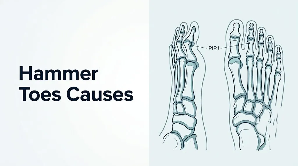
Hammer toes causes produce deformity by disrupting the precise tendon and muscle balance that normally keeps the lesser toes straight, forcing the proximal interphalangeal joint (PIPJ) into a sustained flexed position that progressively becomes structural.
The term “hammertoe” describes a flexion deformity at the PIPJ, the middle joint of the second, third, or fourth toe. The name reflects the appearance: the middle joint buckles upward while the tip of the toe presses downward, resembling the head of a hammer. The second toe is most commonly affected, followed by the third and fourth.
Understanding hammertoe requires knowing which joints are involved. Each lesser toe has three joints: the metatarsophalangeal joint (MTPJ) where the toe meets the foot, the proximal interphalangeal joint (PIPJ) in the middle of the toe, and the distal interphalangeal joint (DIPJ) near the tip. A true hammertoe involves the PIPJ specifically. This distinguishes it from a mallet toe (DIPJ flexion only) and a claw toe (MTPJ hyperextension combined with PIPJ and DIPJ flexion). The distinction matters because the tendons driving each deformity differ, and the treatment options differ accordingly.
Three categories of causes drive hammertoe formation across the population:
- Mechanical causes: footwear, foot structure, and biomechanical imbalance
- Systemic disease causes: diabetes, rheumatoid arthritis, and other inflammatory or metabolic conditions
- Neurological and genetic causes: Charcot-Marie-Tooth disease, stroke, and hereditary foot muscle weakness
The American Academy of Orthopaedic Surgeons (AAOS) recognizes that most hammertoes in adults develop gradually over years, with the initial flexible deformity progressing to a fixed structural contracture if the underlying cause is not addressed.
What Does a Hammertoe Look Like and How Is It Classified
A hammertoe appears as an upward buckle at the middle joint of a lesser toe, creating an arch-like shape with the toe tip pressing downward toward the ground.
Clinically, hammertoes are classified as either flexible or rigid, and this single distinction has more practical consequence than any other feature of the condition. A flexible hammertoe can be passively straightened by pressing the bent PIPJ back to a neutral position with a finger. The joint moves freely under external pressure. This indicates that the underlying deformity is still driven primarily by dynamic muscle and tendon imbalance, and the joint capsule and surrounding soft tissues have not yet contracted permanently.
A rigid hammertoe cannot be passively straightened. The PIPJ is fixed in flexion by contracted soft tissues: the plantar PIPJ joint capsule, the shortened flexor digitorum brevis (FDB) tendon, and in advanced cases, bony remodeling and articular cartilage changes within the joint itself. Surgery is typically required to correct a rigid hammertoe, making the flexible-versus-rigid classification the most clinically actionable piece of information a person with this condition can have.
| Classification | Passive Correction Possible | Typical Duration | Management Approach |
|---|---|---|---|
| Flexible | Yes, with finger pressure | Months to a few years | Conservative: footwear, splinting, exercises, orthotics |
| Semi-rigid | Partial correction only | Several years | Conservative plus podiatric evaluation for surgical planning |
| Rigid | No passive correction possible | Many years | Surgical correction by board-certified podiatric or orthopedic surgeon |
The dorsal surface of the buckled PIPJ is the site where shoes create friction, leading to corn formation (heloma durum), a hardened, painful buildup of keratin at the pressure point. On the plantar surface of the toe tip, a callus develops where abnormal ground pressure concentrates.
Adolescents and young adults with hammertoe are more likely to have a hereditary structural or neuromuscular cause rather than a footwear-driven acquired one, making family and developmental history particularly relevant in this age group.
The Tendon Imbalance Mechanism: How Hammertoe Actually Forms
Hammertoe forms through a specific and predictable tendon imbalance: when the metatarsophalangeal joint is forced into hyperextension, the intrinsic foot muscles lose their mechanical ability to extend the proximal interphalangeal joint, leaving the long toe flexors unopposed to create the bent deformity.
To understand this, picture the toe as a puppet controlled by two competing groups. The extensor group (extensor digitorum longus, extensor digitorum brevis, lumbrical muscles, and interosseous muscles) pulls the toe upward and keeps the PIPJ extended. The flexor group (flexor digitorum longus and flexor digitorum brevis) bends the PIPJ and DIPJ downward. Normally, these groups are balanced, holding the toe flat.
The critical variable is the MTPJ position. The lumbrical muscles arise from the flexor digitorum longus (FDL) tendons and insert into the extensor hood mechanism over the MTPJ and PIPJ. When the MTPJ is in neutral or slightly flexed position, the lumbricals contribute to PIPJ extension, helping balance the FDL’s flexion force. But when the MTPJ is hyperextended, the extensor hood is pulled proximally, placing the lumbricals at a mechanical disadvantage where they can no longer effectively extend the PIPJ.
Think of it like a tent: the tent poles (lumbricals and interossei) keep the canvas (extensor hood) taught when the tent is set up correctly. Tip the frame backward (MTPJ hyperextension), and the canvas sags in the middle (PIPJ flexion). The FDL, now unopposed at the PIPJ, wins the tug-of-war.
According to biomechanical research published in Foot and Ankle International, simulated MTPJ hyperextension in cadaveric specimens reproducibly produced PIPJ flexion deformity, confirming this mechanism experimentally. The FDB, which inserts on the middle phalanx, also contributes to PIPJ flexion and shortens progressively as the deformity becomes established.
Older adults over 70 are particularly susceptible to this mechanism because age-related atrophy of the intrinsic foot muscles (lumbricals and interossei) further reduces the stabilizing force available to resist FDL-driven PIPJ flexion, meaning less MTPJ hyperextension is required to produce the imbalance.
Key Takeaway: Hammertoe forms specifically because MTPJ hyperextension from any cause renders the lumbrical and interosseous muscles mechanically ineffective at extending the PIPJ, leaving the flexor digitorum longus to dominate and bend the middle toe joint downward, which is why the MTPJ position, not just the bent PIPJ, is the central mechanical problem.
Footwear as a Cause of Hammertoes
Footwear is the most modifiable cause of hammertoe, producing the condition by forcing the MTPJ into chronic hyperextension through two distinct mechanisms: heel height and toe box compression.
High-heeled footwear shifts the body’s weight forward onto the metatarsal heads, increasing the pressure on the forefoot with each step. As the heel rises, the metatarsals are angled downward relative to the ground, and to maintain foot-ground contact, the lesser toes must hyperextend at the MTPJ. This is exactly the mechanical position that disables the lumbricals and hands control of the PIPJ to the FDL. Hours of daily wear in heels two inches or higher sustains this deforming position for thousands of repetitions with every step.
Narrow toe box footwear operates through a different but equally effective mechanism. When the toe box compresses the lesser toes laterally, there is no room for the toes to lie flat. The toes are forced to flex at the PIPJ to fit within the available space. Over months and years, this sustained flexed position stretches the dorsal PIPJ capsule, shortens the plantar FDB tendon, and creates the progressive capsular contracture that converts a functional flexion posture into a structural deformity.
The American Podiatric Medical Association (APMA) states that footwear that does not allow toes to lie flat and unstressed is a leading contributing factor to lesser toe deformities including hammertoe, claw toe, and mallet toe.
Women who have worn narrow, high-heeled footwear for decades beginning in their 20s and 30s frequently present with rigid hammertoes by their 50s, a timeline that reflects the years required for capsular contracture to progress from flexible to irreversible. Occupational footwear with narrow steel toe caps (common in construction and industrial settings) can produce the same toe-compressive mechanism in men who wear this footwear daily.
Quick Tip:
- The toe box test: standing in your shoe, check whether you can wiggle all five toes freely without any toe pressing against the shoe material. If the second toe presses against the shoe top or sides, the box is too narrow or low.
- Heel height above 2 inches measurably increases forefoot pressure and MTPJ hyperextension load during walking.
- Ballet dancers who spend years in pointe positions face a distinct hyperextension stress on the second MTPJ that places them at elevated hammertoe risk, particularly in the second toe.
Foot Structure Causes: Flat Feet, High Arches, and Long Second Toe
Intrinsic foot structural characteristics can independently cause hammertoe by creating chronic biomechanical loading patterns at the lesser toes, even in people who wear appropriate footwear.
Morton’s toe (a second toe that extends beyond the length of the great toe) is one of the most clinically recognized structural contributors. When the second toe is longer than the first, it contacts the front of any closed shoe with every step and during standing, creating a repetitive end-to-end compressive force on the second toe. This compressive load pushes the second MTPJ into hyperextension and the PIPJ into flexion, exactly replicating the mechanical pathway described in the tendon imbalance section, but driven by foot architecture rather than footwear choice alone.
Pes cavus (high arch foot) concentrates plantar loading on the metatarsal heads and the heel, reducing the foot’s normal shock-absorbing surface area. The elevated longitudinal arch places the intrinsic foot muscles in a shortened, mechanically disadvantaged position and increases the tendency for the MTPJ to adopt a hyperextended posture during the stance phase of walking. According to a review published in Clinical Biomechanics, pes cavus is strongly associated with claw toe and hammertoe deformity because the intrinsic muscle disadvantage and metatarsal head overload both promote the imbalanced MTPJ-hyperextended position.
Pes planus (flat foot) contributes through a different pathway. As the medial longitudinal arch collapses, the forefoot pronates, placing lateral shear stress on the lesser toes and creating asymmetric MTPJ loading that predisposes to PIPJ deformity, particularly in the second and third toes.
People with pes cavus have an elevated risk not only of hammertoe but also of underlying neurological conditions driving the high arch, including Charcot-Marie-Tooth disease. A new or progressive high arch in an adult warrants neurological evaluation by a primary care physician to rule out an underlying peripheral neuropathy.
Bunions as a Cause of Hammertoe Deformity
A bunion (hallux valgus) causes hammertoe by physically displacing the second toe laterally, crowding it against the third and disrupting the normal tendon alignment at the second MTPJ.
Hallux valgus (ICD-10: M20.1) is a deformity in which the great toe deviates toward the second toe. As the great toe drifts laterally, it encroaches on the space occupied by the second toe. The second toe is pushed laterally, sometimes overriding or underriding the great toe. This lateral displacement rotates the second MTPJ out of its normal alignment, altering the lines of pull of the extensor digitorum longus and the lumbrical muscles. The result is progressive PIPJ flexion deformity that develops secondarily to the primary hallux valgus deformity.
The plantar plate at the second MTPJ is particularly vulnerable in this setting. As the second MTPJ is chronically hyperextended by the laterally displacing great toe, the plantar plate, which is the primary stabilizer against MTPJ dorsiflexion, is progressively stretched and may eventually rupture. Plantar plate rupture at the second MTPJ produces sudden worsening of the hammertoe deformity and crossover toe deformity (the second toe crossing over or under the great toe). A 2019 study published in Foot and Ankle International found that plantar plate tears at the second MTPJ were present in a high proportion of adults presenting with symptomatic second toe hammertoe and crossover deformity.
Adults with a diagnosed bunion who notice the second toe beginning to overlap or drift toward the third toe should request evaluation by a board-certified podiatric surgeon or board-certified orthopedic surgeon specializing in foot and ankle surgery. Addressing the hallux valgus before the second toe develops a rigid hammertoe is significantly simpler surgically and produces better functional outcomes than correcting both simultaneously after both deformities are established.
Key Takeaway: A bunion is not just a great toe problem; the lateral drift of the great toe physically crowds and displaces the second toe, creating a secondary hammertoe deformity through plantar plate stress and MTPJ malalignment that worsens in parallel with the bunion if neither is treated.
Diabetes and Hammertoe: The Intrinsic Minus Foot Mechanism
Diabetes causes hammertoe through diabetic peripheral neuropathy, which progressively denervates the intrinsic foot muscles, producing the specific pattern of muscle imbalance called the intrinsic minus foot that directly drives lesser toe flexion deformities.
The intrinsic foot muscles, the lumbricals and interossei, are small muscles with short, fine nerve branches that are among the first motor structures affected by length-dependent diabetic peripheral neuropathy. In diabetic peripheral neuropathy, chronic hyperglycemia produces advanced glycation end-products (AGEs) that damage the myelin sheaths and axons of peripheral nerves, beginning in the longest nerve fibers first (the distal-to-proximal pattern). The motor branches to the intrinsic foot muscles are among the earliest casualties.
As the lumbricals and interossei lose their motor innervation, the balance between intrinsic and extrinsic toe muscles is lost. The FDL and FDB, innervated by the medial and lateral plantar nerves through longer but more protected nerve trunks, remain functional longer. The result is exactly the intrinsic-minus imbalance described in the tendon mechanism section: the FDL and FDB pull the PIPJ into flexion with no lumbrical/interosseous counterforce. This produces hammertoe, claw toe, and related lesser toe deformities collectively in people with poorly controlled or long-standing diabetes.
According to the National Institute of Diabetes and Digestive and Kidney Diseases (NIDDK), diabetic peripheral neuropathy affects approximately 50 percent of people with diabetes over the course of their lifetime, making it a major population-level driver of foot deformity and foot complications.
The added danger in people with diabetes is that hammertoe creates pressure points (the dorsal PIPJ corn, the plantar toe tip callus) that, in the setting of sensory neuropathy, can ulcerate without the person feeling pain. Diabetic foot ulcers at hammertoe pressure points that become infected can progress to deep soft tissue infection or osteomyelitis. People with diabetes who notice any skin breakdown, wound, or sore at a hammertoe pressure point must seek evaluation by their primary care physician or board-certified podiatric surgeon the same day, regardless of pain level.
Rheumatoid Arthritis and Inflammatory Joint Causes of Hammertoe
Rheumatoid arthritis (RA) causes hammertoe through a distinct inflammatory mechanism that directly destroys the joint structures and surrounding soft tissues of the lesser toe MTPJs, producing deformity through a pathway entirely different from biomechanical imbalance.
In RA, the immune system generates an autoimmune attack primarily targeting the synovial membrane of joints throughout the body. In the foot, the MTPs of the lesser toes are among the earliest and most frequently involved joints. The synovial membrane proliferates into a destructive, invasive tissue called synovial pannus, driven by inflammatory cytokines including TNF-alpha, IL-6, and IL-1 beta. This pannus erodes the articular cartilage of the MTPJ, destroys the supporting ligaments and plantar plate, and progressively destabilizes the joint.
As the plantar plate and MTPJ collateral ligaments are destroyed by pannus, the lesser toes lose their primary stabilizers against dorsal dislocation. The extensor digitorum longus, now unopposed by the damaged plantar plate, pulls the MTPJ into hyperextension. This recreates the same intrinsic muscle mechanical disadvantage described in the tendon mechanism section, but through structural ligamentous destruction rather than dynamic muscle weakness. The PIPJ is driven into flexion by the FDL. Simultaneously, the great toe develops hallux valgus in most people with longstanding RA, further crowding the lesser toes laterally.
According to a review published in Arthritis and Rheumatology, forefoot deformity including hammertoe, hallux valgus, and lesser toe subluxation affects the majority of people with longstanding rheumatoid arthritis and is a primary driver of pain and disability in this population.
People with RA who develop new foot pain, particularly at the ball of the foot and the lesser toes, should ask their board-certified rheumatologist to assess foot and ankle involvement at their next appointment and to consider referral to a board-certified podiatric surgeon or orthopedic foot and ankle surgeon for evaluation of deformity and protective footwear.
Neurological Causes of Hammertoe: Charcot-Marie-Tooth Disease and Stroke
Charcot-Marie-Tooth disease (CMT) and stroke cause hammertoe through neurological mechanisms that are distinct from each other and from all the musculoskeletal causes described in previous sections, making correct identification of the neurological cause critical for both foot and systemic management.
Charcot-Marie-Tooth disease (ICD-10: G60.0) is the most common inherited peripheral neuropathy, affecting approximately 1 in 2,500 people worldwide according to the National Institute of Neurological Disorders and Stroke (NINDS). CMT is caused by mutations in genes encoding peripheral myelin proteins or axonal structural proteins (most commonly PMP22 gene duplication in CMT type 1A). The result is progressive peripheral nerve degeneration affecting both motor and sensory fibers in a length-dependent pattern, with the intrinsic foot muscles among the earliest and most severely affected motor targets.
As the intrinsic foot muscles waste from CMT-related denervation, the classic intrinsic minus foot deformity develops: the foot adopts a high arch (pes cavus) configuration because the intrinsic muscles that help maintain the arch are lost, while the extrinsic flexors that cross multiple joints remain active. The FDL and FDB dominate the lesser toes, producing progressive hammertoe, claw toe, and digital contractures. CMT-related hammertoe typically presents in adolescence or early adulthood and is often bilateral and symmetric, which distinguishes it from acquired hammertoe.
Stroke causes hammertoe through a completely different mechanism: upper motor neuron spasticity in the FDL and FDB following damage to the corticospinal tract. Unlike the denervation of CMT (which reduces muscle tone), stroke-related spasticity increases FDL and FDB muscle tone through dysregulated excitatory motor signaling from a damaged motor cortex or corticospinal pathway. The result is persistent, involuntary FDL and FDB contraction that drives the lesser toes into fixed flexion deformity, often developing within weeks to months of the stroke.
A young adult or child developing progressive bilateral high arches with hammertoe deformity and sensory changes should be evaluated by a board-certified neurologist with nerve conduction velocity (NCV) study and EMG, and if CMT is suspected, by genetic testing, as the underlying condition affects the entire peripheral nervous system and has implications beyond the feet.
Key Takeaway: Neurological causes of hammertoe, whether CMT from inherited peripheral neuropathy or stroke from upper motor neuron spasticity, operate through mechanisms completely different from footwear or structural causes, and their identification changes the entire management approach because treating only the foot without addressing the underlying neurological condition will not prevent progressive deformity.
Genetic and Hereditary Causes of Hammertoe
A hereditary predisposition to hammertoe exists independently of acquired causes, reflecting the inheritance of specific foot structural characteristics and intrinsic muscle architecture that make some people’s toes more vulnerable to developing the deformity under ordinary loading conditions.
Several foot structural characteristics with strong hereditary components contribute to hammertoe risk. Morton’s toe (a second metatarsal longer than the first, producing a second toe that extends beyond the great toe) has a familial pattern. The foot arch type, whether pes cavus or pes planus, has significant genetic determinants. The relative lengths and geometric proportions of the metatarsals and phalanges, which influence how mechanical load distributes across the forefoot, are largely inherited. Together, these structural factors create the biomechanical environment in which even normal activities can progressively drive MTPJ hyperextension and PIPJ flexion deformity.
Beyond structural inheritance, some families carry genetic variants affecting the composition and mechanical properties of ligament and joint capsule collagen. People with generalized joint hypermobility (including those with Ehlers-Danlos syndrome hypermobile type, which has a strong hereditary component) have plantar plates and MTPJ collateral ligaments with reduced tensile stiffness. This means their MTPJ is more easily driven into hyperextension by the same footwear or activity loads that would not produce deformity in a person with normal ligament stiffness.
Clinical observation consistently shows that a family history of hammertoe, specifically a parent or sibling with the condition, is one of the strongest predictors of developing it. This pattern holds even when footwear habits are similar across family members, suggesting that the underlying foot architecture is the determining variable.
People who know they have a family history of hammertoe should ask their primary care physician about referral to a board-certified podiatric surgeon for evaluation of their toe alignment during early adulthood, before deformities progress to a rigid stage, and for guidance on footwear and orthotic options that reduce the mechanical loading driving the inherited structural imbalance.
Trauma as a Cause of Hammertoe
Acute injury to a lesser toe or the structures surrounding its joints can directly produce hammertoe deformity through ligamentous and tendon disruption that recreates the same imbalance seen in chronic causes.
The most clinically common traumatic mechanism is plantar plate rupture at the second MTPJ from a hyperextension injury, such as stumbling off a curb or stubbing the second toe forcefully upward. The plantar plate, a fibrocartilaginous structure on the plantar surface of the MTPJ that resists joint dorsiflexion, is the primary restraint preventing the MTPJ from hyperextending. When it ruptures acutely, the MTPJ loses its primary stabilizer against hyperextension, and the mechanical pathway to PIPJ flexion deformity becomes immediately active.
Toe dislocation injuries, particularly those involving the second MTPJ, can produce hammertoe when the reduction is incomplete or when ligamentous healing leaves the joint in a slightly hyperextended resting position. Crush injuries to the toe or metatarsal with associated tendon laceration (particularly FDB or lumbrical disruption) eliminate the balance necessary for PIPJ extension and allow the remaining intact flexors to dominate.
Acute toe fractures involving the middle phalanx of a lesser toe that heal in a malunited (angulated) position can produce a fixed deformity that resembles structural hammertoe. The malunion creates a permanent flexion angle at the PIPJ through abnormal bony alignment rather than tendon imbalance.
Adults who sustain a toe or forefoot injury and notice persistent second toe swelling, pain at the ball of the foot, or progressive toe buckling in the weeks following the injury should request evaluation by a board-certified podiatric surgeon or orthopedic foot and ankle surgeon. A weight-bearing foot X-ray and plantar plate MRI may be needed to identify structural injury that, if untreated, will progress to a rigid deformity requiring more complex surgery.
How Hammertoe Progresses from Flexible to Rigid
Hammertoe progresses from flexible to rigid through a sequential series of structural changes in the soft tissues and eventually the bone of the proximal interphalangeal joint, driven by sustained abnormal loading in the flexed position.
The progression follows a predictable anatomical sequence:
Stage 1 (Dynamic imbalance, fully flexible): The PIPJ flexes under load (walking, standing) but returns to neutral when unloaded (sitting, lying). No structural changes in the joint capsule yet. The FDL and FDB are overactive relative to the lumbricals and interossei, but no permanent shortening has occurred. Passive correction is fully possible and comfortable.
Stage 2 (Early contracture, semi-rigid): Sustained PIPJ flexion under load begins to shorten the plantar joint capsule and FDB tendon. The dorsal PIPJ joint capsule is stretched and elongated. The extensor digitorum longus displaces dorsally at the PIPJ, reducing its ability to extend the joint. Passive correction is possible but produces mild discomfort as the plantar capsule reaches the end of its available length.
Stage 3 (Established contracture, rigid): The plantar PIPJ capsule is contracted and non-compliant. The FDB is shortened. The PIPJ collateral ligaments have remodeled in a shortened configuration. Passive correction is no longer possible without anesthesia. Articular cartilage changes may begin at the PIPJ, with surface irregularity on the dorsal aspect from chronic joint incongruity.
Stage 4 (Fixed bony deformity): The joint cartilage is damaged, and bony erosion may be present on the dorsal articular surfaces. Corn at the PIPJ dorsum is deep and may be painful with every step. The deformity cannot be corrected without surgical removal of bone and joint reconstruction.
A 2021 review in the Journal of Foot and Ankle Surgery noted that most flexible hammertoes that remain unaddressed for more than five years progress to a semi-rigid or rigid stage, particularly in people who continue wearing compressive footwear throughout that period.
Hammer Toes Causes in Women Versus Men
Women develop hammer toes causes-related deformities at approximately four to five times the rate of men, a disparity driven primarily by footwear design but also influenced by hormonal ligament laxity and foot structural differences.
The footwear explanation is the most direct: the combination of narrow toe boxes, which compress the lesser toes laterally, and elevated heels, which hyperextend the MTPs, creates the exact mechanical conditions for hammertoe formation. Women are substantially more likely than men to wear footwear combining both of these deforming features across decades of daily wear. The cumulative effect is a much higher population-level prevalence of acquired hammertoe.
Hormonal factors add a secondary contribution. Estrogen and relaxin (particularly elevated during pregnancy) increase ligament and joint capsule extensibility throughout the body, including the foot. Women who are pregnant or who have had multiple pregnancies may notice progressive forefoot widening and toe malalignment that begins during pregnancy. The plantar plate and MTPJ collateral ligaments, made more compliant by hormonal effects, offer less resistance to the deforming forces from footwear and foot loading. Postmenopausal women lose the estrogen-mediated ligament compliance but retain the structural deformity that developed when ligaments were lax.
Men develop hammertoe through different predominant pathways: work boots with narrow toe caps, high arch foot structure, Scheuermann’s kyphosis-related postural compensations (less relevant), Charcot-Marie-Tooth disease (comparable prevalence between sexes), and trauma. Men with diabetes have an equally high risk of intrinsic minus-type hammertoe as women with the same HbA1c history, because the peripheral neuropathy mechanism is the same regardless of sex.
Men who develop hammertoe deserve the same evaluation and management attention as women, but they are less likely to report foot pain to their primary care physician, making a targeted foot examination part of the annual physical examination an important screening opportunity.
Key Takeaway: Women develop hammertoes at four to five times the rate of men primarily because of the combined MTPJ-hyperextending and toe-compressing effect of heeled, narrow-box footwear worn over decades, but men are not immune, particularly those with diabetes, pes cavus, Charcot-Marie-Tooth disease, or occupational footwear that compresses the forefoot.
Risk Factors That Increase Hammertoe Likelihood
Multiple risk factors independently and additively increase the probability of developing hammertoe, with the highest-risk individuals typically carrying several simultaneously.
Risk factors for developing hammertoe:
- Female biological sex, particularly with history of high-heeled or narrow-toed footwear use across decades
- Age over 60: progressive intrinsic foot muscle atrophy reduces the lumbrical and interosseous stabilizing force, lowering the threshold for PIPJ flexion deformity
- Diabetes mellitus with any degree of peripheral neuropathy: intrinsic muscle denervation from length-dependent neuropathy produces the intrinsic minus foot pattern
- Rheumatoid arthritis: synovial pannus destruction of MTPJ plantar plate and ligaments recreates the hyperextension-driven imbalance
- Charcot-Marie-Tooth disease or other hereditary peripheral neuropathies: progressive intrinsic muscle wasting from demyelinating or axonal neuropathy
- History of stroke with foot and ankle spasticity: FDL and FDB spasticity from upper motor neuron damage
- Hallux valgus (bunion): lateral displacement of the second toe and plantar plate stress from great toe crowding
- Morton’s toe (second toe longer than first): chronic end-to-end shoe compression and MTPJ hyperextension
- Pes cavus (high arch): intrinsic muscle mechanical disadvantage and metatarsal head overload
- Generalized joint hypermobility or connective tissue disorders: reduced plantar plate and MTPJ collateral ligament stiffness
- Previous toe or MTPJ trauma with plantar plate injury or malunited phalangeal fracture
- Family history of hammertoe: inherited foot structural characteristics
- Occupational footwear with narrow toe box (steel-toed work boots, pointe shoes in dancers)
- Long-term systemic corticosteroid use: associated with weakening of ligamentous and tendon collagen, potentially increasing plantar plate vulnerability
According to the AAOS, the combination of hallux valgus, Morton’s toe, and years of high-heeled footwear represents a particularly high-risk triad for developing progressive second toe hammertoe deformity.
How Doctors Diagnose the Cause of Hammertoe
Doctors diagnose the cause and classify the severity of hammertoe through a structured clinical examination supplemented by targeted imaging and laboratory testing when systemic causes are suspected.
To describe your hammertoe accurately to a primary care physician, podiatric surgeon, or orthopedic foot and ankle surgeon:
- Note which toe or toes are affected and when you first noticed the bend. Longstanding bilateral deformities suggest hereditary or neurological causes; unilateral gradual deformity in the second toe suggests footwear or structural causes.
- Describe whether the toe straightens out when you sit or lie down with your foot off the ground (flexible) or stays bent at all times (rigid). This single observation is the most useful piece of information for the clinician.
- Report whether you have diabetes, rheumatoid arthritis, a history of stroke, or a family member with the same condition, as each substantially changes the diagnostic workup.
- Bring or describe the footwear you wear most frequently, including heel height and toe box width. Photographs of your most-used footwear are genuinely useful.
- Describe any pain: whether it is at the dorsal PIPJ (from corn and shoe friction), at the plantar toe tip (from ground contact), or at the ball of the foot (from metatarsalgia or plantar plate pathology).
The clinical examination includes a passive correction test to classify flexibility, assessment of MTPJ range of motion, skin inspection for corns and calluses, and a gait observation. Weight-bearing foot radiographs (anteroposterior, lateral, and oblique views) are the standard first imaging study, showing joint alignment, metatarsal length patterns, any bony erosions from rheumatoid arthritis, and the overall forefoot architecture.
If neuropathy is suspected, the primary care physician should order nerve conduction velocity (NCV) testing and EMG. If rheumatoid arthritis is suspected, serum rheumatoid factor and anti-cyclic citrullinated peptide (anti-CCP) antibody testing are appropriate initial laboratory evaluations. If diabetes is not yet diagnosed, a fasting glucose and HbA1c complete the workup.
When Hammertoe Requires Medical or Surgical Evaluation
Certain presentations of hammertoe require prompt evaluation rather than home management, and one specific presentation in people with diabetes constitutes a medical emergency.
| Presentation | Urgency | Recommended Action |
|---|---|---|
| Flexible hammertoe, no pain, no skin changes | Routine | Footwear modification; podiatric evaluation at next convenient appointment |
| Flexible hammertoe with dorsal pain from shoe friction | Soon | Board-certified podiatric surgeon or orthopedic foot and ankle surgeon within 4 to 6 weeks |
| Rigid hammertoe with corn, persistent pain limiting activity | Prompt | Board-certified podiatric surgeon evaluation; surgical planning discussion |
| Hammertoe with any skin wound or sore in person without diabetes | Prompt | Primary care physician or podiatric surgeon within days; rule out deeper infection |
| Hammertoe with skin wound or sore in person with diabetes | Same day | Primary care physician or board-certified podiatric surgeon the same day |
| Hammertoe wound in person with diabetes with redness, warmth, swelling, discharge, or fever | Emergency | Go to the nearest emergency room immediately |
| New progressive bilateral hammertoe with sensory changes in a young adult | Urgent | Primary care physician; neurological evaluation; NCV/EMG for Charcot-Marie-Tooth |
For people without diabetes or systemic disease, hammertoe is not a life-threatening condition. But the window for effective conservative management is limited. A flexible hammertoe that is not addressed with footwear modification and targeted exercises under professional guidance will progress to rigid over years, at which point surgery is the only effective option.
People with diabetes should treat any skin change at a hammertoe pressure point with the same urgency as any diabetic foot wound. Diabetic foot infections spread rapidly, can involve bone (osteomyelitis), and in severe cases require amputation. The neuropathy that causes the hammertoe also prevents the person from feeling the pain that would otherwise prompt earlier evaluation.
Go to the nearest emergency room immediately if you have diabetes and a hammertoe wound shows spreading redness, warmth, purulent discharge, systemic fever, or chills. These signs indicate a deep diabetic foot infection that requires immediate intravenous antibiotics and surgical evaluation, not a scheduled appointment.
Frequently Asked Questions About Hammer Toes Causes
What is the main cause of hammertoes?
The main cause of hammertoe in most adults is a tendon imbalance at the metatarsophalangeal joint, where MTPJ hyperextension from tight or high-heeled footwear disables the intrinsic foot muscles and allows the flexor digitorum longus to dominate the proximal interphalangeal joint, bending it downward.
In older adults and people with diabetes, progressive intrinsic muscle loss from age-related atrophy or peripheral neuropathy produces the same tendon imbalance without requiring high-heeled footwear.
A board-certified podiatric surgeon or orthopedic foot and ankle surgeon can identify the specific primary cause in an individual through clinical examination and weight-bearing foot radiographs.
Can wearing high heels really cause a hammertoe?
Yes, high-heeled footwear directly causes hammertoe by forcing the metatarsophalangeal joints into a sustained hyperextended position with every step, which disables the lumbrical and interosseous muscles and transfers control of the proximal interphalangeal joint to the flexor digitorum longus.
The American Podiatric Medical Association states that footwear that does not allow toes to lie flat is a primary contributor to lesser toe deformities, and biomechanical studies confirm that heel heights above two inches measurably increase forefoot pressure and MTPJ hyperextension load.
Switching to footwear with a lower heel and a wider toe box can slow or halt progression of a flexible hammertoe, but cannot reverse a rigid structural deformity that has already developed.
Is hammertoe hereditary or genetic?
Hammertoe has a hereditary component, primarily through the inheritance of foot structural characteristics such as Morton’s toe (second toe longer than first), pes cavus (high arch), and intrinsic ligament and joint capsule compliance that influence how easily the MTPJ is pushed into hyperextension.
Certain inherited conditions, most notably Charcot-Marie-Tooth disease (affecting approximately 1 in 2,500 people), directly cause hammertoe through progressive intrinsic foot muscle denervation from inherited peripheral nerve mutations.
A family history of hammertoe warrants early footwear evaluation and, in the case of progressive bilateral deformity with sensory changes, neurological evaluation by a primary care physician.
Can a hammertoe go away on its own without surgery?
A flexible hammertoe will not reverse itself without active intervention addressing the underlying cause, but it can stabilize or improve with consistent footwear modification, targeted exercises, and toe splinting under professional guidance.
A rigid hammertoe, where the proximal interphalangeal joint cannot be passively straightened, will not improve without surgery, because the joint capsule, shortened FDB tendon, and in advanced cases bony structural changes prevent passive or exercise-based correction.
A 2021 review in the Journal of Foot and Ankle Surgery found that most unaddressed flexible hammertoes progress to semi-rigid or rigid within five years, making early intervention the most effective approach to avoiding surgical management.
How do I know if my hammertoe is flexible or rigid?
A flexible hammertoe can be passively straightened by pressing the bent middle joint back toward a straight position with a finger applied to the underside of the toe, and it straightens comfortably with little to no resistance.
A rigid hammertoe stays bent despite firm passive pressure and does not straighten; the joint feels fixed and the attempt to correct it may produce pain from the contracted plantar joint capsule reaching its limit.
This distinction determines whether conservative management has any chance of success, and it should be assessed by a board-certified podiatric surgeon or orthopedic foot and ankle surgeon to guide treatment planning.
When should I see a doctor about a hammertoe?
Any person with diabetes who has a hammertoe with any skin wound, sore, redness, or breakdown at the bent toe should seek evaluation by a primary care physician or board-certified podiatric surgeon the same day, regardless of pain level, because sensory neuropathy prevents reliable pain-based self-assessment of wound severity.
Adults without diabetes should seek evaluation by a board-certified podiatric surgeon or orthopedic foot and ankle surgeon when the hammertoe causes persistent pain limiting daily activities, when a rigid deformity has developed, or when conservative footwear changes have not improved symptoms after 6 to 8 weeks of consistent use.
A young adult or adolescent with progressive bilateral hammertoe, high arch feet, and any sensory changes in the feet or lower legs should request evaluation by a primary care physician specifically for peripheral neuropathy assessment.
Hammertoe is not simply a cosmetic foot problem, and it is rarely caused by a single factor acting alone. The tendon imbalance at the MTPJ is the final common pathway, but what drives that imbalance differs depending on whether the cause is decades of constrictive footwear, intrinsic muscle loss from diabetes or Charcot-Marie-Tooth disease, joint destruction from rheumatoid arthritis, or an inherited foot structure that loads the PIPJ asymmetrically every day.
The most actionable thing you can take from this article: perform the passive correction test on your affected toe right now. If it straightens easily under gentle pressure, you are still in the window where footwear modification and a podiatric evaluation can change the course of the deformity. If it does not move, schedule an appointment with a board-certified podiatric surgeon or orthopedic foot and ankle surgeon to discuss surgical options before pain or skin complications make the decision for you.
If you have diabetes and any skin change at your hammertoe, that is the one situation where scheduling an appointment is the wrong move. Go to the emergency room. Diabetic foot infections do not wait.