Liver Cyst Symptoms: What They Mean and When to Act
Most liver cyst symptoms are caused not by the liver itself malfunctioning, but by the cyst growing large enough to press on surrounding structures, stretch the liver’s outer capsule, or interfere with nearby bile ducts and digestive organs. The type of cyst matters enormously: a simple fluid-filled sac behaves very differently from a parasitic cyst or a cyst with malignant potential, and understanding that distinction determines how urgently you need to act.
According to the National Institute of Diabetes and Digestive and Kidney Diseases (NIDDK), simple hepatic cysts are found in an estimated 5 to 7 percent of adults, with prevalence rising sharply after age 50. The European Association for the Study of the Liver (EASL), in its 2016 Clinical Practice Guidelines published in the Journal of Hepatology, notes that the vast majority of simple cysts are asymptomatic and carry no malignant potential, but certain cyst types require active surveillance and specialist management.
This article covers every recognized symptom of liver cysts with the anatomical mechanism behind each one, explains how different cyst types produce different clinical pictures, identifies the population groups who face elevated risk, and gives you the specific threshold for when symptoms require emergency evaluation rather than a scheduled appointment.
What Is a Liver Cyst
A liver cyst is a fluid-filled, enclosed sac within or on the surface of the liver (the hepatic parenchyma), lined by a layer of epithelial cells and containing a variable amount of fluid whose composition depends on the cyst’s origin.
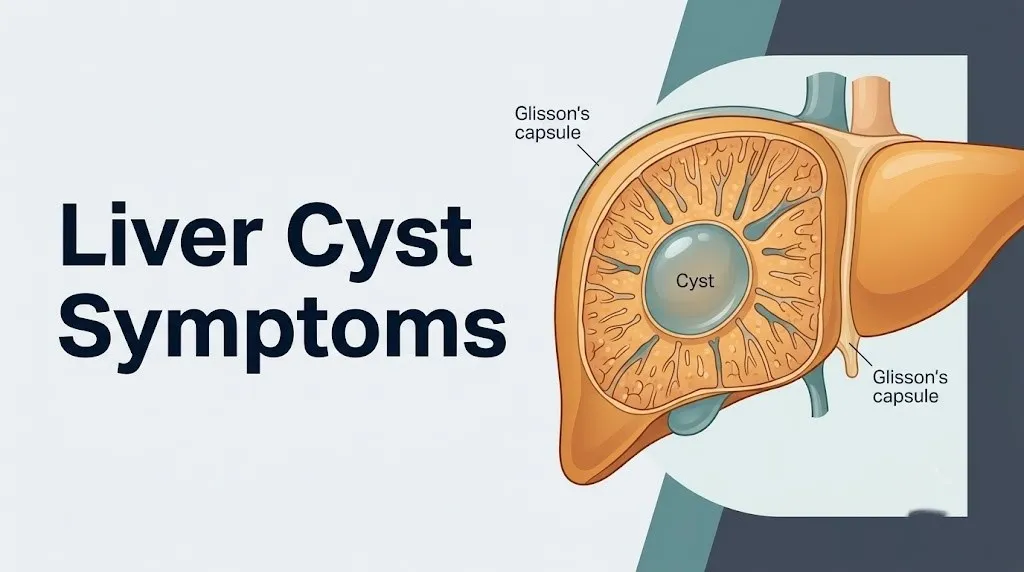
The liver sits in the right upper quadrant of the abdomen, tucked under the right side of the rib cage. It occupies the right lobe (the larger segment), the left lobe, and the smaller caudate lobe. The entire organ is enclosed in Glisson’s capsule, a fibrous outer covering that is richly supplied with nociceptive nerve fibers. This capsule is the reason liver cysts cause pain when they grow: as a cyst expands, it stretches the capsule, activating those nociceptors and producing the characteristic right upper quadrant aching or pressure.
Most liver cysts form from the biliary epithelium, the same type of epithelial cells that line the bile ducts inside the liver. A developmental failure during fetal bile duct formation can leave behind small epithelial-lined pockets that gradually fill with fluid over decades. These are the benign simple hepatic cysts that make up the majority of incidental cyst findings on abdominal imaging.
Not all liver cysts are the same, and not all carry the same risk. The EASL distinguishes several distinct hepatic cyst categories with importantly different clinical behaviors, ranging from entirely benign fluid-filled sacs to parasitic cysts capable of producing life-threatening rupture and anaphylaxis. Understanding which type you have is the first question to ask your gastroenterologist or hepatologist after any cyst is identified on imaging.
People with a personal or family history of polycystic kidney disease should know that hepatic cysts are present in up to 75 to 80 percent of people with autosomal dominant polycystic kidney disease (ADPKD), making their liver involvement far more likely than in the general population.
Liver Cyst Symptoms: The Complete Clinical Picture
Liver cyst symptoms range from complete absence in the majority of cases to a recognizable pattern of right upper abdominal discomfort, bloating, referred shoulder pain, and in serious cases, jaundice, fever, or sudden severe pain from a complication.
The most practically important fact about liver cyst symptoms is this: the presence or absence of symptoms correlates more closely with cyst size and location than with cyst number. A single large cyst (generally above 4 centimeters in diameter, and particularly above 10 centimeters) is far more likely to produce symptoms than multiple small cysts, regardless of how many there are.
| Symptom | Mechanism | Typical Cyst Size | Clinical Significance |
|---|---|---|---|
| Right upper quadrant aching | Glisson’s capsule distension by expanding cyst | Often above 4 cm | Common; warrants monitoring |
| Right shoulder pain | Phrenic nerve irritation via diaphragm contact | Often above 8 cm | Indicates large or superiorly located cyst |
| Bloating and fullness | Gastric fundus compression by large cyst | Often above 8 cm | Moderate; affects eating comfort |
| Early satiety | Mechanical gastric compression | Often above 8 cm | May cause unintentional weight loss |
| Nausea | Gastric displacement and vagal nerve stimulation | Variable | Correlates with meal timing |
| Jaundice | Bile duct compression causing obstructive cholestasis | Any size if bile duct contact | Red flag; requires prompt evaluation |
| Fever and rigors | Cyst infection (secondary bacterial colonization) | Any size | Medical emergency |
| Sudden severe pain | Cyst rupture or intracystic hemorrhage | Any size | Emergency |
Pregnant women represent a population at specific risk for rapid symptom onset from pre-existing liver cysts. Elevated estrogen and progesterone levels during pregnancy may accelerate cyst growth, and the mechanical displacement of abdominal organs by the enlarging uterus can exacerbate compression symptoms even from cysts that were previously asymptomatic.
Right Upper Abdominal Pain From Liver Cysts
Right upper abdominal pain from liver cysts is produced primarily by distension of Glisson’s capsule, the fibrous outer layer of the liver that contains pain-sensing nociceptive nerve fibers supplied by branches of the right lower intercostal nerves and the right phrenic nerve.
Think of Glisson’s capsule like a fitted membrane wrapped snugly around the entire organ. When a cyst grows inside the liver or on its surface, it pushes outward against this membrane from the inside. The capsule has no room to expand freely because it is anchored by the surrounding rib cage and abdominal wall. That outward pressure activates the nociceptors embedded in the capsule, producing the characteristic dull aching, pressure, or heaviness in the right upper abdomen, below the right rib cage, that most people with symptomatic liver cysts describe.
The pain is typically not sharp in nature (sharp, colicky pain suggests gallbladder involvement rather than liver capsule distension). It does not usually change dramatically with movement or body position. It tends to be persistent rather than episodic, though it may worsen after large meals because eating causes the stomach to expand, pressing further against an already-crowded upper abdomen.
According to a clinical review published in Hepatology, right upper quadrant discomfort is the most frequently reported symptom in patients with symptomatic simple hepatic cysts, present in approximately 80 percent of those presenting with symptoms.
People with pre-existing right-sided rib injuries, recent abdominal surgery, or liver conditions such as non-alcoholic fatty liver disease may experience pain that is harder to attribute specifically to a cyst. Any new or worsening right upper abdominal pain warrants evaluation by a primary care physician, who can order a hepatic ultrasound to assess cyst size and character as a first step.
Liver Cyst Shoulder Pain Explained
The right shoulder pain associated with liver cysts is a classic example of referred pain, produced when the phrenic nerve (arising from cervical nerve roots C3, C4, and C5) is irritated by a large hepatic cyst that contacts the inferior surface of the diaphragm.
The phrenic nerve serves two functions: it carries motor signals to the diaphragm muscle, and it carries sensory information from the central diaphragm and the pericardium back to the spinal cord at the C3-C5 level. The C3, C4, and C5 dermatomes also supply the skin over the right shoulder and right-sided neck. When a large hepatic cyst in the right lobe’s superior segment presses against the underside of the diaphragm, the phrenic nerve interprets this mechanical irritation as coming from the shoulder region, because both the diaphragm’s central surface and the shoulder skin share the same spinal cord entry point.
This referred right shoulder pain has no direct musculoskeletal cause. Pressing on the shoulder does not reproduce it. Rotating the shoulder does not worsen it. It does not respond to topical analgesics or physical therapy. These features are what help distinguish it from rotator cuff injury or cervical radiculopathy.
The same phrenic nerve pathway explains why gallbladder attacks, diaphragmatic irritation from blood (as in a ruptured ectopic pregnancy), and subphrenic abscess all produce similar referred right shoulder pain. The anatomy is consistent: any structure that contacts the right hemidiaphragm from below can irritate the C3-C5 phrenic nerve and produce shoulder discomfort.
Quick Tip:
- Right shoulder pain without any shoulder injury, especially combined with right upper abdominal discomfort or bloating, should prompt consideration of hepatic, gallbladder, or subdiaphragmatic causes.
- Tell your doctor the shoulder pain came on around the same time as any abdominal symptoms and that there was no shoulder injury, as this is the specific clinical detail that points toward referred diaphragmatic irritation.
- People over 60 with this shoulder-abdominal pain combination may have their shoulder pain attributed to degenerative joint disease; specifically request abdominal imaging if no shoulder cause is found.
Key Takeaway: Right shoulder pain with right upper abdominal aching and no history of shoulder injury is the phrenic nerve referred pain pattern of a large liver cyst, and it should prompt a hepatic ultrasound, not a shoulder MRI.
Bloating, Nausea, and Early Satiety From Liver Cysts
Bloating, nausea, and early satiety from liver cysts are produced by direct mechanical compression of the stomach (specifically its fundus, the upper dome-shaped portion) and adjacent duodenum by large cysts located in the left lobe of the liver or in right lobe segments positioned near the gastroduodenal junction.
The stomach’s fundus sits directly adjacent to the inferior surface of the liver’s left lobe. When a large left lobe cyst expands downward, it reduces the functional space available for the stomach to expand after eating. The result is the sensation of fullness after consuming only a small amount of food, the clinical phenomenon called early satiety. If the cyst also presses against the vagus nerve branches running alongside the stomach, it may trigger nausea through vagal activation of the brainstem’s vomiting center (the area postrema in the medulla oblongata).
Bloating in liver cyst patients is different from the gas-related bloating of irritable bowel syndrome. The liver cyst bloating is a persistent pressure or fullness in the upper abdomen that does not fluctuate significantly with bowel movements. It does not correlate with specific foods in the way that functional gastrointestinal bloating does. It may gradually worsen over months as the cyst grows, and then remain stable if cyst growth plateaus.
Mayo Clinic notes that large symptomatic hepatic cysts can cause patients to unintentionally limit their food intake due to discomfort, which over months can contribute to weight loss and nutritional deficiency.
Women in their 40s and 50s may attribute these digestive symptoms to perimenopause-related gastrointestinal changes, which can delay recognition of a hepatic cyst as the cause. Any persistent early satiety, upper abdominal fullness, or unexplained nausea lasting more than two to four weeks warrants evaluation by a primary care physician, who should include abdominal ultrasound in the workup rather than assuming a functional gastrointestinal cause.
Asymptomatic Liver Cyst: When No Symptoms Is Still a Concern
An asymptomatic liver cyst is one that produces no pain, discomfort, or digestive symptoms and is identified incidentally during imaging performed for an unrelated reason, such as an abdominal ultrasound for gallstones or a CT scan after a minor abdominal trauma.
The vast majority of incidentally discovered liver cysts are benign simple cysts that will remain stable for years and never require treatment. A 2020 study published in JAMA Internal Medicine evaluating incidental hepatic lesions found on CT imaging reported that the overwhelming majority of simple-appearing cysts in adults without known malignancy were benign on long-term follow-up, regardless of size at initial discovery.
However, “asymptomatic” does not automatically mean “safe to ignore.” The clinical concern with any newly discovered liver cyst is whether it is truly a simple benign cyst or whether it represents a cyst type that carries risk (biliary cystadenoma, echinococcal cyst, or a cystic-appearing hepatic malignancy). This determination cannot be made from symptoms alone.
To describe a newly found liver cyst to your physician effectively:
- Note exactly where you feel any discomfort, specifically whether it is right-sided, upper abdominal, or referred to the shoulder.
- State when the symptoms began relative to the imaging that found the cyst.
- Report any fever, chills, or jaundice you have experienced, even briefly.
- Tell the provider about any travel history to areas where Echinococcus is endemic (parts of South America, Central Asia, Mediterranean countries, sub-Saharan Africa, Middle East).
- Disclose any family history of polycystic kidney disease or polycystic liver disease.
- Bring the actual imaging report (not just the verbal result) to your appointment so the radiologist’s characterization of the cyst can be reviewed.
People who are immunocompromised (organ transplant recipients on immunosuppression, people with HIV, those receiving chemotherapy) should request immediate gastroenterologist evaluation of any newly discovered hepatic cyst rather than waiting for a routine appointment, because infection risk and atypical presentations are more frequent in immunocompromised individuals.
Liver Cyst Types and How Each One Presents Differently
Liver cysts are not a single condition but a category of five clinically distinct entities, each with a different origin, growth pattern, symptom profile, complication risk, and management strategy.
| Cyst Type | Origin | Symptom Pattern | Key Complication | Who Is Most Affected |
|---|---|---|---|---|
| Simple hepatic cyst | Developmental biliary epithelial malformation | Asymptomatic or right upper quadrant pressure if large | Rare; hemorrhage if large | Adults over 40; females more than males |
| Polycystic liver disease (ADPLD) | Genetic mutation (PRKCSH, SEC63, PKD1, PKD2) | Progressive abdominal distension, early satiety, pain | Portal hypertension, malnutrition | Females predominantly; those with ADPKD |
| Biliary cystadenoma | Mucinous biliary epithelium neoplasm | Right upper quadrant pain, palpable mass | Malignant transformation to cystadenocarcinoma | Predominantly middle-aged females |
| Echinococcal (hydatid) cyst | Echinococcus granulosus or E. multilocularis larval stage | Right upper quadrant pain, urticaria with minor leakage | Rupture with anaphylaxis, secondary infection | Persons with livestock exposure in endemic regions |
| Von Meyenburg complex | Biliary hamartoma, congenital ductal plate malformation | Usually asymptomatic | Benign; rarely associated with cholangiocarcinoma | Adults; found incidentally at any age |
The EASL Clinical Practice Guidelines emphasize that distinguishing these cyst types requires imaging characterization, not symptom assessment alone. A simple cyst on ultrasound appears as a thin-walled, anechoic (completely dark), rounded lesion with smooth borders and no internal septations. Any internal septations, solid components, wall thickening, or calcifications should prompt further evaluation with MRI and specialist review.
Older adults above 65 may present with multiple cyst types simultaneously, including simple cysts and von Meyenburg complexes appearing on the same imaging study. Distinguishing these from hepatic metastases in a patient with any cancer history requires specialist hepatology or radiology review.
Key Takeaway: The type of liver cyst determines everything about risk and management: a simple cyst usually needs only monitoring, while a biliary cystadenoma or echinococcal cyst requires specialist evaluation and often intervention regardless of symptoms.
Polycystic Liver Disease Symptoms
Polycystic liver disease (PLD), also called autosomal dominant polycystic liver disease (ADPLD), produces symptoms that are distinct in character and severity from those of simple hepatic cysts, primarily because PLD involves hundreds to thousands of cysts replacing progressively larger portions of the hepatic parenchyma rather than a single or small number of discrete lesions.
The dominant symptom of advanced PLD is massively increased abdominal girth, reflecting a liver that can grow to many times its normal 1.5 kilograms as cysts multiply and enlarge. The liver’s normal volume is approximately 1,500 milliliters. Published case series in Journal of Hepatology have reported PLD livers exceeding 10,000 milliliters in the most severely affected patients, producing an abdomen that resembles late-stage pregnancy in appearance.
Symptoms produced by this massive hepatomegaly include:
- Severe abdominal distension and visible abdominal protrusion
- Profound early satiety and inability to eat a normal-sized meal
- Chronic fatigue from reduced caloric intake and nutritional compromise
- Shortness of breath from upward diaphragmatic displacement reducing lung expansion
- Lower extremity edema from inferior vena cava compression by the enlarged liver
- Back pain from postural compensation for the anterior abdominal weight shift
- Pain and pressure at the costal margins from the enlarged liver pressing against the lower ribs
Despite this dramatic anatomical change, the liver’s metabolic function (producing clotting factors, metabolizing drugs and toxins, synthesizing albumin) is usually preserved in PLD until very late disease stages because the remaining non-cystic hepatic parenchyma compensates. Liver function test results (ALT, AST, albumin, INR) are often normal even in severely affected patients.
Females with PLD who have used oral contraceptives or hormone replacement therapy represent the population at highest risk for rapid PLD progression. The National Institute of Diabetes and Digestive and Kidney Diseases notes that estrogen accelerates cyst growth in PLD, and cessation of exogenous estrogen is often the first clinical recommendation for women with symptomatic PLD.
Echinococcal Liver Cyst Symptoms
Echinococcal liver cysts, also called hydatid cysts, are caused by larval infection with the tapeworm Echinococcus granulosus (producing cystic echinococcosis) or Echinococcus multilocularis (producing alveolar echinococcosis, the more invasive and dangerous form), and their symptom pattern includes features entirely absent from other liver cyst types.
The clinical presentation of echinococcal cysts depends on cyst size, location, and whether the cyst wall has been compromised by leakage or rupture. Slow-growing intact cysts may remain asymptomatic for years, sometimes for a decade or more, because the parasite’s growth rate is gradual and the host’s immune system is initially suppressed by parasite-derived immune-modulatory proteins. When symptoms do develop from an intact cyst, they mirror simple cyst symptoms: right upper quadrant heaviness, pressure, and in large cysts, referred shoulder pain.
What makes echinococcal cysts distinctly dangerous is the consequence of rupture or leakage. The hydatid cyst fluid contains protoscolices (larval tapeworm heads) and parasite antigens, which are profoundly allergenic to the human immune system. Minor leakage produces urticaria (hives), pruritus, and in some cases, bronchospasm. Major rupture can produce life-threatening anaphylaxis with systemic IgE-mediated hypersensitivity response, along with the risk of seeding daughter cysts throughout the peritoneal cavity (peritoneal dissemination of echinococcosis).
According to the Centers for Disease Control and Prevention, echinococcosis in the United States occurs primarily in immigrants from endemic regions, including parts of the Mediterranean, Middle East, Central Asia, sub-Saharan Africa, and South America, and in people who have had close contact with sheep, cattle, or dogs in these areas.
Anyone presenting with liver cyst and a history of livestock exposure or travel to endemic regions should disclose this history explicitly to their physician, since standard imaging cannot always distinguish echinococcal cysts from other liver cysts without serological confirmation (ELISA or Western blot for Echinococcus-specific IgG antibodies).
Liver Cyst Jaundice and Bile Duct Symptoms
Jaundice from a liver cyst occurs when the cyst compresses an intrahepatic bile duct or the common bile duct or common hepatic duct, blocking the normal flow of bile from the liver into the duodenum and causing bilirubin to accumulate in the blood and tissues.
Bile is produced continuously by hepatocytes and drains through a network of progressively larger intrahepatic bile ducts that converge into the common hepatic duct, which joins the cystic duct (from the gallbladder) to form the common bile duct, emptying into the duodenum at the ampulla of Vater. When a liver cyst grows in a position that directly contacts or compresses any segment of this drainage pathway, the back-pressure of obstructed bile causes obstructive cholestasis: bilirubin accumulates in hepatocytes, spills into the bloodstream, and deposits in skin and sclera (the whites of the eyes), producing the yellow discoloration called jaundice.
The symptoms of bile duct involvement from a liver cyst include:
- Yellowing of the skin and whites of the eyes (jaundice)
- Dark urine (from elevated urinary bilirubin excretion, producing a tea-colored or cola-colored appearance)
- Pale or clay-colored stools (from reduced bilirubin delivery to the bowel, which normally gives stool its brown color)
- Itching (pruritus) of the skin, produced by bile salt deposition in cutaneous tissues
- Right upper quadrant pain escalating from the background aching of capsular distension
Jaundice from a liver cyst is a clinically important finding because it narrows the differential diagnosis and raises the possibility of biliary cystadenoma, a mucin-secreting tumor with malignant potential, or of echinococcal cyst with intrabilliary rupture.
According to the American College of Gastroenterology, any liver cyst producing jaundice should be evaluated urgently by a gastroenterologist or hepatologist, with same-day liver function tests (total and direct bilirubin, ALP, GGT, ALT, AST) and cross-sectional imaging (MRI with MRCP is preferred for visualizing bile duct anatomy and cyst-duct relationships).
People with pre-existing cholestatic liver conditions such as primary sclerosing cholangitis or primary biliary cholangitis may develop jaundice from superimposed cyst compression at lower cyst volumes than otherwise healthy individuals, and their abnormal baseline liver function tests make identifying the new contribution of cyst-related obstruction more clinically complex.
Key Takeaway: Jaundice, dark urine, pale stools, or itchy skin in a person known to have a liver cyst is not a routine follow-up finding. It requires same-day evaluation by a gastroenterologist, not a scheduled appointment.
Liver Cyst Complications and Warning Signs
Liver cyst complications include cyst rupture, intracystic hemorrhage, secondary infection, biliary communication, and in the specific case of biliary cystadenoma, malignant transformation into biliary cystadenocarcinoma.
Cyst rupture can be spontaneous (from rapid fluid accumulation exceeding the cyst wall’s tensile strength) or traumatic (from blunt abdominal impact). Rupture of a simple cyst produces acute peritoneal irritation with sudden severe abdominal pain, but is rarely life-threatening because simple cyst fluid is non-toxic. Rupture of an echinococcal cyst is a different category of emergency because of the anaphylaxis risk.
Intracystic hemorrhage occurs when a blood vessel within or adjacent to the cyst wall bleeds into the cyst cavity. This produces sudden onset of severe, acute right upper quadrant pain that is categorically different from the gradual aching of capsular distension. The pain peaks rapidly and may be accompanied by nausea, vomiting, and diaphoresis (sudden cold sweating). On imaging, hemorrhagic cysts show heterogeneous internal contents rather than the uniformly clear fluid of a simple cyst.
Secondary infection (infected hepatic cyst or liver abscess) develops when bacteria colonize the cyst fluid, most commonly via hematogenous spread or from biliary communication. Symptoms include fever above 38.5 degrees Celsius, rigors (shaking chills), and right upper quadrant pain with systemic signs of sepsis (racing heart rate, rapid breathing, confusion). This presentation requires emergency evaluation.
Warning signs that a liver cyst has become complicated:
- Sudden severe abdominal pain replacing or dramatically worsening the prior dull aching
- Fever with chills and right upper quadrant pain together
- Jaundice appearing acutely in someone with a known liver cyst
- Skin rash or hives appearing in the context of known liver cyst (possible echinococcal leakage)
- Rapid abdominal swelling over hours rather than gradual growth over weeks
Mayo Clinic notes that biliary cystadenocarcinoma (malignant transformation from biliary cystadenoma) can present with the same symptom pattern as its benign counterpart, making histological evaluation after surgical removal the only way to definitively confirm or exclude malignancy.
Liver Cyst Symptoms in Women and Hormonal Factors
Women experience symptomatic liver cysts at higher rates than men, and the biological mechanism behind this sex-based difference is better established than most consumer health content acknowledges.
Laboratory and clinical evidence shows that the biliary epithelial cells lining simple hepatic cysts express estrogen receptor alpha (ERalpha) on their cell surfaces. When estrogen binds to these receptors, it stimulates intracystic fluid secretion, promoting cyst expansion. This mechanism, identified in studies published in Hepatology, explains why simple liver cysts are both more prevalent and more frequently symptomatic in premenopausal and perimenopausal women compared to age-matched men.
Women taking exogenous estrogen, whether via combined oral contraceptives or hormone replacement therapy (HRT), have been observed in clinical series to experience more rapid cyst growth compared to women not using these agents. The EASL clinical guidelines note this association and recommend discussing the potential impact of exogenous estrogen on cyst growth with any female patient who has a known liver cyst.
Pregnancy deserves specific attention. Elevated endogenous estrogen and progesterone during pregnancy can accelerate the growth of pre-existing hepatic cysts. Women who were asymptomatic before conception may develop right upper quadrant pain, early satiety, and shoulder pain during the second and third trimesters as previously stable cysts expand under hormonal influence. Hepatic cysts discovered during pregnancy should be evaluated by an obstetrician in consultation with a gastroenterologist or hepatologist to determine whether imaging surveillance or intervention is appropriate.
Postmenopausal women who are not taking HRT tend to have stable or slowly reducing cyst volumes compared to their premenopausal trajectory, which supports the estrogen receptor mechanism. For a postmenopausal woman on HRT who reports new or worsening right upper quadrant symptoms, reviewing HRT continuation with her obstetrician-gynecologist or primary care physician in the context of cyst management is a clinically reasonable and evidence-consistent conversation.
Liver Cyst Symptoms in Older Adults
Liver cysts are substantially more prevalent in older adults, with population imaging studies estimating that simple hepatic cysts are present in 5 to 7 percent of adults over 50 and in a higher proportion still of those over 70, making age one of the strongest predictors of hepatic cyst presence.
The clinical presentation of liver cysts in older adults differs in several clinically meaningful ways. First, the likelihood that a liver cyst in an older adult is an incidental finding on imaging performed for another reason (CT for suspected pulmonary embolism, ultrasound for abdominal aortic aneurysm screening, or CT colonography) is high. Second, the differential diagnosis for a right upper quadrant mass or pain in an older adult is broader and includes hepatocellular carcinoma, hepatic metastases from colorectal cancer, cholangiocarcinoma, and gallbladder carcinoma, all of which must be distinguished from benign hepatic cysts through appropriate imaging characterization.
Third, symptoms that younger adults might attribute immediately to a liver cyst may be attributed incorrectly in older adults to other age-related conditions. Right upper abdominal discomfort may be attributed to musculoskeletal rib or intercostal causes. Early satiety may be attributed to reduced gastric motility with aging (gastroparesis). Referred right shoulder pain may be attributed to degenerative rotator cuff changes or cervical spondylosis, which are genuinely prevalent in this age group.
According to a review published in JAMA Internal Medicine, older adults with incidentally discovered hepatic lesions on CT scanning had a lower rate of malignancy than initially feared in studies from tertiary referral centers, but the rate of malignancy was non-trivial in those with known primary cancers. Any older adult with a cancer history and a new hepatic lesion should be evaluated by a gastroenterologist or hepatologist within two to four weeks, not placed on a routine monitoring schedule without specialist characterization.
People over 65 taking anticoagulant medications (warfarin, apixaban, rivaroxaban) should specifically disclose this to their physician when a liver cyst is identified, since intracystic hemorrhage risk may be elevated and the presenting symptom pattern of sudden severe pain would warrant emergency evaluation in this context.
Liver Cyst Diagnosis: How Doctors Confirm What You Have
Liver cyst diagnosis begins with imaging, and the specific imaging modality, combined with the clinical history, determines whether a cyst can be classified as benign without further workup or whether specialist evaluation is required.
The typical diagnostic pathway from first clinical suspicion to confirmed cyst characterization:
- Primary care evaluation: The physician takes a history of symptoms, travel, occupational exposures, family history of cystic kidney or liver disease, and cancer history. Physical examination may reveal right upper quadrant tenderness or a palpable hepatic margin.
- Hepatic ultrasound (first-line imaging): Ultrasound is the most widely available, radiation-free, and cost-effective first study. A simple cyst on ultrasound appears as a well-defined, anechoic (completely echo-free), thin-walled lesion with posterior acoustic enhancement (a brightening of the tissue behind the cyst, produced by sound waves passing through fluid more easily than through solid tissue). Any ultrasound finding that does not meet all of these criteria requires further imaging.
- CT scan of the abdomen with and without contrast: Provides better anatomical detail, identifies the cyst’s relationship to bile ducts and vessels, and can detect calcifications (common in echinococcal cysts). Contrast-enhanced CT helps identify any solid enhancing components that would suggest a non-benign lesion.
- MRI with MRCP (magnetic resonance cholangiopancreatography): The most detailed modality for characterizing hepatic cysts and their relationship to the biliary tree. MRCP is particularly valuable when bile duct involvement is suspected, as it provides a non-invasive image of the entire biliary anatomy without radiation or contrast injection into bile ducts.
- Laboratory testing: Liver function tests (ALT, AST, ALP, GGT, total and direct bilirubin) assess hepatic function and biliary obstruction. Tumor markers (CA 19-9, AFP) are ordered when biliary cystadenoma or hepatocellular carcinoma is in the differential. Echinococcus serology (ELISA, Western blot) is ordered when the travel and exposure history supports this possibility.
- Specialist review: Any cyst with internal septations, solid components, wall thickening, calcifications, or a clinical history of cancer should be evaluated by a gastroenterologist or hepatologist before a benign diagnosis is assigned.
Women with a newly discovered liver cyst who are currently using oral contraceptives or HRT should discuss this with their primary care physician during the diagnostic workup, as this information is relevant to cyst growth prediction and management planning.
Key Takeaway: A liver cyst cannot be safely classified as benign based on symptoms alone. A hepatic ultrasound showing all four criteria of a simple cyst (anechoic, thin-walled, round, with posterior acoustic enhancement) is the minimum imaging standard for initial benign classification, and any deviation from those four criteria requires MRI and specialist review.
Liver Cyst Prognosis and When Treatment Is Needed
The prognosis for the most common liver cyst type, the simple benign hepatic cyst, is excellent: the vast majority remain stable in size over years of follow-up, produce no serious complications, and require no intervention beyond periodic imaging surveillance.
The EASL clinical guidelines recommend that simple cysts meeting all ultrasound criteria for a benign appearance in a patient without cancer history and without symptoms can be monitored with a repeat ultrasound at six to twelve months after initial discovery to confirm stability, followed by less frequent imaging if the cyst has not grown. Asymptomatic stable simple cysts do not require surgery.
Treatment becomes indicated when:
- A symptomatic simple cyst is large enough (typically above 4 to 10 centimeters, depending on location and symptom severity) to justify intervention for quality of life
- Cyst growth is documented on serial imaging
- Complications (hemorrhage, infection, biliary obstruction) develop
- The cyst type requires intervention regardless of symptoms (echinococcal cysts, biliary cystadenoma)
For symptomatic simple cysts, the procedures available include laparoscopic cyst fenestration (surgical unroofing of the cyst to allow drainage into the peritoneal cavity, the most durable approach), percutaneous aspiration with sclerotherapy (draining the cyst through a needle and injecting a sclerosing agent to collapse the cyst wall, with moderate long-term recurrence rates), and in rare cases, formal hepatic resection.
For echinococcal cysts specifically, the PAIR procedure (puncture, aspiration, injection of a protoscolicide, re-aspiration) is a minimally invasive option for appropriate cyst subtypes, always performed in a center with expertise in echinococcosis management to mitigate anaphylaxis risk.
People with polycystic liver disease who experience severe symptoms may eventually require liver transplantation in the most advanced cases, though this represents a small minority of PLD patients and is only considered after less invasive options have been exhausted and liver function is compromised.
People in their 30s and 40s with a first diagnosis of a simple liver cyst and no symptoms should be reassured by their primary care physician or gastroenterologist that the natural history is favorable, but should schedule follow-up imaging as directed rather than dismissing the finding as permanently irrelevant.
Emergency Symptoms: When to Call 911 or Go to the ER
Certain symptoms associated with liver cysts require immediate emergency evaluation. Do not wait to see if these resolve on their own.
Call 911 or go to the nearest emergency room immediately if you experience:
- Sudden, severe abdominal pain that is categorically worse than any prior right upper quadrant discomfort, especially if it begins abruptly, indicating possible cyst rupture or intracystic hemorrhage
- Fever above 38.5 degrees Celsius (101.3 degrees Fahrenheit) with shaking chills and right upper quadrant pain together, indicating a possible infected hepatic cyst or liver abscess with sepsis risk
- Yellowing of the skin or whites of the eyes (jaundice) appearing acutely or rapidly progressing, combined with any right upper abdominal pain, indicating acute biliary obstruction that may progress to acute cholangitis
- Sudden onset of skin hives (urticaria), throat tightening, difficulty breathing, or swelling of the face or tongue in a person with a known echinococcal liver cyst, indicating possible cyst rupture with anaphylaxis requiring immediate epinephrine and emergency support
- Rapidly worsening abdominal distension with severe pain over hours rather than the gradual distension of polycystic liver disease, indicating acute hepatic decompensation or internal hemorrhage
- Any loss of consciousness, extreme low blood pressure (feeling faint when standing), or cold, clammy skin in a person with a known liver cyst, indicating possible hemorrhagic shock from cyst rupture
These presentations can indicate hepatic abscess with systemic sepsis, biliary obstruction progressing to acute cholangitis, anaphylactic reaction to ruptured echinococcal cyst, or hemorrhagic shock from cyst rupture, all of which require emergency medical assessment, not an urgent care appointment or a phone call.
Frequently Asked Questions About Liver Cyst Symptoms
What does a liver cyst feel like?
A liver cyst typically feels like a dull ache, pressure, or heaviness in the right upper abdomen, below the right rib cage, produced by the cyst stretching Glisson’s capsule, the fibrous outer layer of the liver.
Larger cysts may produce a referred pain in the right shoulder through phrenic nerve irritation when the cyst contacts the underside of the diaphragm.
Many liver cysts produce no sensation at all and are discovered only when abdominal imaging is performed for an unrelated reason.
Can a liver cyst make you feel sick or cause nausea?
A liver cyst can cause nausea when it is large enough to compress the stomach’s fundus or stimulate vagal nerve branches running alongside the stomach, producing both nausea and a sensation of fullness after eating very little.
This pattern of early satiety and upper abdominal fullness after eating is most common with large cysts, generally above 8 centimeters, located in the left lobe of the liver or adjacent to the gastroduodenal junction.
If nausea is severe, persistent, or accompanied by vomiting, jaundice, or fever, evaluation by a primary care physician or gastroenterologist is warranted promptly rather than after a prolonged waiting period.
Should I be worried about an incidentally found liver cyst?
An incidentally found liver cyst is not automatically a cause for concern, as the majority of such cysts in adults without cancer history are benign simple cysts with an excellent long-term prognosis.
The key step after any incidental hepatic cyst discovery is confirming that it meets all imaging criteria for a simple benign cyst, specifically that it is anechoic, thin-walled, round, and shows posterior acoustic enhancement on ultrasound, because any deviation from these features requires further evaluation with MRI and specialist review.
Any person with a cancer history should have an incidentally found liver cyst evaluated by a gastroenterologist or hepatologist within two to four weeks rather than waiting for routine follow-up.
What are the warning signs that a liver cyst is serious?
The warning signs that a liver cyst requires urgent evaluation include sudden severe abdominal pain, fever with chills, jaundice (yellowing of the skin or eyes), dark tea-colored urine, pale stools, and rapidly worsening abdominal distension.
Skin hives or throat tightening in a person with a known liver cyst and a history of livestock exposure or travel to echinococcosis-endemic regions may indicate echinococcal cyst leakage, which is a medical emergency requiring immediate evaluation.
Any of these signs should prompt same-day evaluation by an emergency medicine physician, not a scheduled office appointment.
Can a liver cyst go away on its own?
Simple benign hepatic cysts do not typically resolve or disappear without intervention, but the majority remain stable in size for years and never produce symptoms requiring treatment.
Spontaneous resolution of simple hepatic cysts has been reported in case series but is not the expected clinical course, and no lifestyle modification or dietary change is known to cause cyst reabsorption.
Echinococcal cysts, biliary cystadenomas, and infected or hemorrhagic cysts do not resolve without medical or surgical treatment and require specialist management.
How do doctors tell if a liver cyst is cancerous?
Doctors use a combination of imaging characteristics and laboratory markers to determine whether a liver cyst has malignant features, because symptoms alone cannot distinguish benign from malignant cystic liver lesions.
MRI with MRCP is the preferred imaging modality for characterizing internal cyst architecture, since solid components, internal septations, papillary projections, and wall thickening are imaging features associated with biliary cystadenoma and its malignant counterpart, biliary cystadenocarcinoma.
Tumor markers including CA 19-9 and CA-125 may be elevated with mucinous biliary tumors, but normal levels do not exclude malignancy, and definitive diagnosis requires histological examination of surgically removed cyst tissue.
Closing
The practical reality of liver cysts is that most people who receive this diagnosis will never need treatment. A stable, simple, asymptomatic cyst found on imaging in an adult without cancer history is almost always benign, and the primary clinical action is confirming its imaging characteristics and scheduling a follow-up ultrasound.
What matters is knowing the specific symptoms that should change your response from “wait and watch” to “see someone today.” Right upper quadrant pain combined with fever, jaundice, sudden severe pain, or skin hives in anyone with a known echinococcal exposure history are the triggers that require emergency evaluation, not a next-week appointment.
If you’ve been told you have a liver cyst and you’re experiencing any abdominal discomfort, right shoulder pain, or digestive changes, bring the actual imaging report to your primary care physician or gastroenterologist and ask specifically about the cyst’s imaging classification, its current size, and the recommended surveillance interval. That conversation is the most actionable next step.