What Causes Calluses on Feet and How to Address Them
What causes calluses on feet is repeated mechanical stress, specifically friction and pressure, triggering a specific cellular response in the skin that produces thickened, hardened layers of dead skin cells as a protective adaptation. The cause of any individual callus is not just “wearing bad shoes,” but a combination of the underlying foot anatomy, gait pattern, footwear, and in some cases systemic health conditions that determine exactly where and why the callus forms.
According to the American Podiatric Medical Association (APMA), foot disorders including calluses and corns affect a large proportion of the adult population, with foot pain from thickened skin being among the most frequent complaints seen in podiatric practice. In people with diabetes, a plantar callus is not merely a cosmetic issue: published research in Diabetes Care has found that plantar calluses concentrate subcallus pressure by a factor of three to five compared to surrounding plantar skin, making them a leading precipitant of diabetic foot ulcers and non-traumatic lower limb amputations.
This article covers the specific cellular mechanism by which calluses form, the biomechanical reasons they develop at specific locations on the foot, every major structural and systemic cause, and the clinical thresholds that determine when a callus requires professional evaluation by a podiatrist rather than home management.
What Causes Calluses on Feet: The Cellular Mechanism Explained
Calluses on feet form when repeated mechanical stress activates a protective proliferative response in the skin’s basal layer, producing more skin cells than are shed, and causing the outermost skin layer to progressively thicken.
The skin of the foot’s weight-bearing surfaces has the same layered structure as skin elsewhere on the body: the stratum basale (innermost proliferating layer), the stratum spinosum, the stratum granulosum, and the stratum corneum (the outermost layer of flattened, protein-rich dead cells called corneocytes). The stratum corneum is normally approximately 15 to 20 cell layers thick on non-plantar skin, but on the plantar surface it is already substantially thicker because of the normal loads the foot bears during walking.
When a specific skin site is subjected to repeated friction or pressure beyond what the existing stratum corneum can protect against, keratinocytes in the stratum basale detect the mechanical signal through a process called mechanotransduction. Mechanical loading activates integrin receptors on the keratinocyte surface, which recruit focal adhesion kinase (FAK) and activate downstream signaling through the mitogen-activated protein kinase (MAPK) / ERK1/2 pathway. This cascade tells the keratinocytes to divide faster, producing more cells that migrate upward through the spinosum and granulosum layers and accumulate in the stratum corneum as additional corneocytes.
Simultaneously, the rate at which the stratum corneum sheds its surface corneocytes (desquamation) slows in response to the mechanical environment, because the pressure and friction physically impede the enzymatic processes that normally loosen surface corneocyte attachments. The combined result is a net accumulation of corneocytes in the stratum corneum: the layer thickens, hardens, and takes on the yellowed, waxy appearance of a callus (medically termed tyloma or hyperkeratosis).
According to the American Podiatric Medical Association, this callus formation process is fundamentally a protective physiological response: the thickened skin distributes mechanical load across a larger volume of tissue, reducing peak stress at the focal contact point. The problem arises when the callus itself becomes thick enough to concentrate pressure rather than distribute it, or when the underlying cause (gait problem, foot deformity, wrong shoes) is not addressed.
People with diabetes experience the same callus formation mechanism but with an important additional risk: peripheral neuropathy impairs the protective pain sensation that normally alerts a person to excessive pressure before ulceration occurs, allowing calluses to progress to dangerous subcallus hemorrhage or breakdown without warning.
How Friction and Pressure Cause Foot Calluses
Friction and pressure cause foot calluses through two mechanically distinct processes that both trigger the same keratinocyte proliferation response but at different tissue depths and with slightly different clinical appearances.
Pressure is a compressive force applied perpendicular to the skin surface, such as the force of body weight pressing down through the plantar surface during standing or walking. When this pressure is concentrated on a small area repeatedly, the keratinocytes beneath that area receive a consistent mechanotransduction signal to increase their division rate. The resulting callus is broad-based, diffuse, and relatively flat, with a gradual transition from normal surrounding skin to the thickened central area. Pressure calluses are the most common type on the plantar (bottom) surface of the foot.
Friction is a shear force applied parallel to the skin surface, such as the sliding of a shoe upper against the dorsum of a toe with each step. Shear forces stretch and compress keratinocytes from a different orientation than vertical pressure, but they activate the same integrin-mediated mechanotransduction pathways. Friction-induced calluses tend to be more localized and discrete than pressure calluses, and they often develop on the top or sides of toes rather than the plantar surface.
Think of the callus-forming process like the way a hand develops toughened skin from using a shovel: the first day of digging produces blisters (acute fluid accumulation beneath the epidermis when shear force separates the epidermal layers before the stratum corneum has thickened). With repeated use over weeks, the stratum corneum thickens ahead of future shear force, preventing blister formation. The thick skin on a laborer’s palm is the same biological response as the callus on a runner’s forefoot.
Key differences between pressure and friction calluses:
| Feature | Pressure Callus | Friction Callus |
|---|---|---|
| Force direction | Perpendicular to skin surface | Parallel to skin surface |
| Typical location | Plantar surface: heel, ball of foot | Toe dorsum, sides of toes, heel counter area |
| Appearance | Broad, diffuse, flat, gradual margins | More discrete, localized, sometimes with clear center |
| Primary cause | Body weight concentration at focal point | Shoe-skin sliding contact |
| Most affected population | All; amplified in foot deformity and obesity | Athletes, people in ill-fitting shoes |
Pregnant women in the second and third trimesters experience both pressure and friction callus formation from two simultaneous mechanisms: increased body weight raises plantar pressure across the entire foot, and the loosening of plantar ligaments from relaxin hormone allows the foot arch to splay and flatten, redistributing pressure to new areas that the skin was not previously conditioned to handle.
Plantar Pressure Distribution and Where Calluses Form on the Foot
The location of a callus on the foot is not random. It directly maps to the areas of highest plantar pressure concentration during walking, standing, and running, and the pressure distribution pattern is determined by the foot’s bony architecture, soft tissue padding, muscle function, and gait mechanics.
During normal barefoot walking, peak plantar pressures are distributed across five main anatomical regions in a specific sequence: heel strike first, then lateral midfoot, then fifth metatarsal head, across to first through third metatarsal heads, and finally through the great toe during push-off. A normal foot has an intact heel fat pad and metatarsal fat pad that distribute these forces over a relatively broad area, preventing focal pressure concentration.
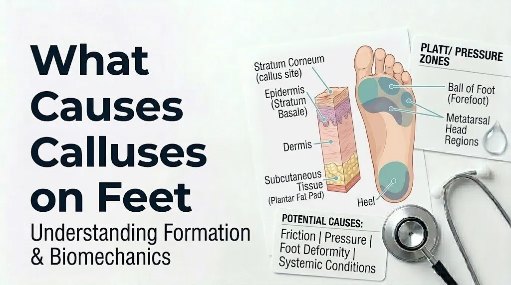
When this distribution is disrupted by any cause, pressure concentrates in a smaller area and callus formation begins at that site.
Plantar pressure zones and typical callus formation:
| Anatomical Zone | Normal Pressure Range | Conditions Increasing Pressure | Callus Pattern |
|---|---|---|---|
| Calcaneal tuberosity (heel) | High during initial contact | Heel fat pad atrophy, pes cavus, tight Achilles | Central or posterior heel callus |
| Lateral midfoot | Low in normal arch | Pes planus (flat foot) increasing lateral contact | Lateral arch callus |
| Fifth metatarsal head | Moderate | Supinated gait, pes cavus, wide forefoot | Lateral ball-of-foot callus |
| Second and third metatarsal heads | Moderate-high | Hallux valgus, first ray insufficiency, Morton’s toe | Central forefoot callus |
| First metatarsal head | High during push-off | Normal; reduced in hallux valgus | First MTP callus or reduced callus when hallux valgus present |
| Great toe tip | Low during normal gait | Hallux rigidus, compensatory gait | Tip-of-toe callus |
According to research published in the Journal of Foot and Ankle Research, pedobarographic studies consistently show that callus location in a patient’s foot corresponds with areas of elevated peak plantar pressure measured during gait analysis, with a correlation coefficient above 0.75 in most published studies. This means a podiatrist who examines the location of a patient’s callus can make a clinically informed prediction about the underlying biomechanical cause before ordering any tests.
Older adults over 65 show altered pressure distribution compared to younger adults because of plantar fat pad atrophy, reduced intrinsic foot muscle strength, and age-related changes in gait, producing higher forefoot pressures and greater callus formation at the central metatarsal heads.
What Causes Heel Calluses: The Calcaneal Fat Pad and Ground Contact Forces
Heel calluses form primarily because the calcaneal tuberosity (the posterior-inferior surface of the heel bone, the calcaneus) is the first point of contact with the ground during walking and absorbs some of the highest impact forces of the entire gait cycle, with peak heel pressures reaching two to three times body weight during the initial contact phase of normal walking at faster speeds.
The primary anatomical protection against this force concentration is the plantar heel fat pad, a specialized adipose structure composed of fibro-elastic fat chambers arranged in a honeycomb pattern that absorbs and dissipates impact energy. This structure is not simple adipose tissue: the fat chambers are enclosed by fibrous septa anchored to the calcaneal periosteum, creating a biomechanical shock absorber uniquely suited to the repetitive compressive loads of walking. When the heel fat pad is intact and well-hydrated, it distributes calcaneal pressure across a broad area, reducing the focal pressure that drives callus formation.
Heel calluses develop when this fat pad protection is insufficient relative to the applied load. The most important causes:
Plantar heel fat pad atrophy occurs progressively with aging and after prolonged corticosteroid injection in the heel region. As the fat chambers become smaller and their fibrous walls less elastic, the pad thins and loses shock-absorbing capacity, concentrating heel pressure on the calcaneal skin directly.
Barefoot walking on hard surfaces eliminates the additional shock absorption provided by footwear midsoles, subjecting the heel fat pad to the full unmitigated impact of each step.
Pes cavus (high-arched foot) reduces the contact area of the heel with the ground by increasing the angle of heel strike, concentrating force on a smaller posterior calcaneal surface.
Dry skin on the heel, common in people with hypothyroidism, diabetes, and in older adults with reduced dermal moisture, makes the heel skin more prone to fissuring around the margins of pressure-induced calluses, creating the painful cracked heel condition clinically associated with deep heel calluses.
According to the Mayo Clinic, heel calluses associated with painful cracking (heel fissures) that extend into the dermis warrant evaluation by a podiatrist, particularly in people with diabetes, because these fissures create open portals for bacterial entry into the deeper plantar tissues.
People with rheumatoid arthritis frequently develop prominent heel calluses because the disease process destroys the synovial lining of the subtalar and ankle joints, altering heel contact mechanics and redistributing ground reaction forces toward the posterior calcaneal skin.
Key Takeaway: Heel calluses form because the plantar heel fat pad, which absorbs impact forces during the initial contact phase of walking, becomes insufficient to distribute those forces broadly when it atrophies with age, when hard surfaces eliminate footwear cushioning, or when foot architecture concentrates heel contact on a smaller area than normal.
What Causes Calluses on the Ball of Foot: Metatarsal Head Pressure
Calluses on the ball of the foot form beneath the metatarsal heads, the five rounded distal ends of the metatarsal bones that bear the full body weight during the push-off phase of gait when the heel has lifted and the entire body weight passes forward through the forefoot.
The first metatarsal head normally bears the greatest load during push-off because the great toe (hallux) provides the primary propulsive lever for forward movement, with the two sesamoid bones beneath the first metatarsal head acting as pulleys for the flexor hallucis brevis tendon and spreading the contact force over a slightly larger area. When first metatarsal function is normal, the central second and third metatarsal heads share the remaining forefoot load.
Calluses on the ball of foot at the second, third, or fourth metatarsal heads typically indicate that these central metatarsals are bearing more load than they should. This pattern, called metatarsalgia (ICD-10: M77.4), has several common underlying causes:
- Hallux valgus (bunion): as the great toe drifts laterally toward the second toe, the first metatarsal head loses its mechanical advantage, and the lesser metatarsal heads bear compensatory increased load
- Morton’s toe: an anatomically longer second metatarsal than first, shifting peak forefoot pressure to the second metatarsal head
- Forefoot equinus: a tight Achilles tendon that keeps the heel elevated during gait, transferring weight-bearing time disproportionately to the forefoot
According to the American College of Foot and Ankle Surgeons (ACFAS), the distribution of callus across the metatarsal heads is one of the most reliable clinical indicators of forefoot loading patterns, and it directly guides the decision about whether custom foot orthoses (with metatarsal padding) or surgical correction of underlying deformity is the appropriate intervention.
People who wear high-heeled footwear regularly develop ball-of-foot calluses through a biomechanically direct mechanism: heel elevation in a high-heeled shoe transfers approximately 75% of body weight to the forefoot (compared to approximately 50% in flat footwear), chronically overloading the metatarsal heads even in the absence of structural foot deformity.
What Causes Calluses on Toes: Dorsal and Tip Pressure Patterns
Calluses on the toes form through friction from shoe contact rather than the compressive plantar pressure mechanisms that drive heel and ball-of-foot calluses, and their location on the toe surface directly reveals which shoe component is causing the problem.
Dorsal toe calluses (on the top surface of toes) form when the toe knuckle contacts the upper of the shoe with each step. This most commonly occurs at the proximal interphalangeal (PIP) joint of a toe that is curled downward (either from a hammertoe deformity or from a shoe too short for the foot that forces the toes to curl to fit inside). With each step, the dorsal skin over the PIP joint slides against the shoe’s lining, generating friction that triggers localized stratum corneum thickening. The callus that develops on a hammertoe PIP joint is often mistaken for a corn (heloma durum), but a callus is diffuse and lacks the defined central nucleus or core that characterizes a corn.
Tip-of-toe calluses form when the toe tip is pressed against the front of the shoe, typically from a shoe too narrow from front to back. The distal phalanx tip bears the friction and compressive force with each step, producing a small, concentrated callus that can be painful when pressed.
Calluses between toes occur less often and typically indicate a soft corn (heloma molle), which is not a true callus but a macerated, white, soft lesion from the pressure of one toe pressing against an adjacent toe’s bony prominence. The moist interdigital environment prevents the normal dry cornification of a typical callus, producing the soft, whitish appearance.
Quick Tip:
- A callus on the top of the second toe that has been present for months suggests either hammertoe deformity or a shoe with insufficient toe box depth. A podiatrist or foot and ankle specialist can assess which is responsible with a weight-bearing examination.
- Calluses at the tip of the big toe most often indicate hallux rigidus (limited first MTP joint motion), compensatory gait patterns from a rigid first toe, or footwear too short for the foot length.
- Children developing toe calluses warrant evaluation by a pediatrician or pediatric orthopedic specialist to exclude juvenile idiopathic arthritis, which can produce toe joint deformities generating secondary callus formation.
According to the American Podiatric Medical Association, any callus on a toe that is painful even at rest (rather than only with shoe contact) should be evaluated by a podiatrist, because resting pain in a toe callus suggests either a deeply embedded heloma or a sub-lesional bursitis, both of which require professional management rather than self-care.
Footwear as a Cause of Calluses on Feet
Footwear causes foot calluses through three distinct mechanisms, each producing a predictable callus location pattern that differs from calluses caused by foot deformity or systemic conditions.
The first mechanism is insufficient toe box width. When the shoe’s toe box is narrower than the foot’s natural width across the metatarsal heads, the shoe compresses the metatarsals medially and laterally, redistributing normal plantar pressure from the first and fifth metatarsal heads inward to the second, third, and fourth heads. Calluses develop at these compressed metatarsal heads in the classic “central metatarsal callus” pattern. Pointed-toe shoes are the most consistent cause of this pattern.
The second mechanism is insufficient length. A shoe shorter than the foot length forces the toes into plantarflexion (curling downward) to fit inside, causing the dorsal PIP joints to contact the shoe upper and the toe tips to press against the front box. This produces simultaneous dorsal toe and toe-tip calluses.
The third mechanism is heel counter friction. A shoe heel counter that is too rigid, too low-cut, or positioned at the wrong height for an individual foot creates a shear force at the posterior superior heel during walking, producing a callus at the specific heel location that the counter contacts. Low-cut heel counters in athletic shoes frequently produce calluses at the Achilles insertion level (the posterior superior calcaneal region) in people with prominent posterior heels.
According to research published in the Journal of Foot and Ankle Research, shoe-related foot disorders are among the most prevalent causes of foot pain in adults, with studies finding that approximately 70 to 80% of adults wear shoes shorter than their foot length at the widest point. This widespread discrepancy between foot width and shoe width directly contributes to the high prevalence of central forefoot calluses in the general population.
Occupational footwear worn in workplaces requiring safety boots, specific uniform footwear, or prolonged hard-surface standing often provides less cushioning and less accurate fit than recreational footwear, making people in these occupations substantially more likely to develop plantar and toe calluses than office workers with footwear freedom.
Key Takeaway: The location of a shoe-related callus directly reveals which footwear design element is causing it: central metatarsal calluses point to a too-narrow toe box, tip-of-toe and dorsal PIP calluses point to a too-short shoe, and posterior heel calluses point to a poorly positioned heel counter.
Gait Abnormalities and Foot Deformities as Causes of Calluses
Gait abnormalities cause calluses on feet by altering the timing, magnitude, and distribution of plantar forces during walking, concentrating pressure at anatomical sites that are not designed to bear sustained high loads.
Normal walking involves a specific sequence: heel strike, loading response, midstance, push-off, and swing phase. Each phase applies specific forces to specific plantar regions in a brief, distributed manner. When gait is abnormal, whether from pain compensation (antalgic gait), neurological conditions, muscle weakness, or joint stiffness, the force distribution changes and some plantar regions are overloaded while others are underloaded. The overloaded regions develop calluses.
Overpronation (excessive inward rolling of the foot during midstance) is one of the most frequent gait-related callus causes. As the foot rolls inward excessively, the arch collapses and the first metatarsal head loads early and heavily, often producing a callus under the first MTP joint and great toe. Simultaneously, the heel’s contact angle shifts medially, sometimes producing a medial heel callus pattern rather than the central posterior heel callus typical of normal gait.
Supination (insufficient inward roll, excessive outward foot position during midstance) concentrates forces on the lateral forefoot and lateral heel, producing calluses at the fifth metatarsal head and lateral heel region. People with high-arched feet frequently supinate and develop this pattern.
Equinus gait (walking with the heel raised due to Achilles tendon tightness or neurological spasticity) dramatically increases forefoot pressure during the entire stance phase, producing severe central forefoot calluses that are disproportionate to footwear or foot structure alone. This pattern is particularly common in people with cerebral palsy or post-stroke spasticity.
A gait analysis performed by a podiatrist or physical therapist, including pedobarographic pressure measurement, is the most objective method for identifying gait-related callus causes and guiding orthotic prescription or physical therapy to normalize force distribution.
Adolescents with rapidly changing foot mechanics during growth spurts may develop new callus patterns at anatomical sites that were previously uncallused, as bone growth outpaces soft tissue adaptation and temporarily alters plantar pressure distribution.
Flat Feet and High Arches as Causes of Foot Calluses
Both flat feet (pes planus) and high-arched feet (pes cavus) cause foot calluses by altering the plantar contact area of the foot with the ground, concentrating pressure in predictable anatomical locations specific to each arch type.
Pes planus (flat foot) reduces or eliminates the normal medial longitudinal arch, increasing the total plantar contact area but disproportionately loading the medial midfoot and first metatarsal head. In a flat foot, the medial longitudinal arch (normally elevated and not in contact with the ground during standing) contacts the ground partially or fully, creating friction and pressure along the medial arch and inner heel border that the skin in these locations is not conditioned to handle. Calluses forming along the medial arch and inner heel border in a person without other obvious causes should prompt assessment for pes planus by a podiatrist with weight-bearing examination and possibly foot X-ray.
Pes cavus (high-arched foot) has the opposite pressure pattern: the arch is elevated above normal, drastically reducing the plantar contact area to essentially two zones: the heel and the ball of the foot (metatarsal heads). With less surface area to distribute body weight, the pressure at these contact points is substantially higher than in a normally arched foot. A high-arched foot during walking concentrates impact at the posterior lateral heel on initial contact and at the lateral and central metatarsal heads during push-off, producing heavy calluses at these specific sites and relative absence of callus at the arch.
According to the American College of Foot and Ankle Surgeons, pes cavus that causes painful plantar calluses warrants evaluation for the neuromuscular conditions that frequently underlie high-arch development in adults, particularly Charcot-Marie-Tooth disease (hereditary motor and sensory neuropathy), which causes progressive foot muscle wasting producing the high-arch, claw-toe pattern. Any adult who develops a progressively higher arch alongside distal leg muscle weakness should be evaluated by a neurologist in addition to a foot and ankle specialist.
People with rheumatoid arthritis frequently develop a specific pes planus pattern from synovitis-driven destruction of the posterior tibial tendon, which is the primary support structure of the medial arch.
Bunions and Hammertoes as Causes of Calluses on Feet
Bunions (hallux valgus) and hammertoes are two of the most consistent structural foot deformities that cause calluses in predictable, diagnostically recognizable patterns, because both deformities alter plantar pressure distribution in well-characterized ways.
Hallux valgus (bunion) is a lateral deviation of the great toe at the first metatarsophalangeal (MTP) joint, with associated medial prominence of the first metatarsal head. The deformity produces calluses through three concurrent mechanisms. First, the prominent medial first metatarsal head generates friction against the medial shoe, producing a callus over the bunion prominence. Second, as the great toe drifts laterally and loses its normal propulsive mechanics, the lesser metatarsal heads (second and third) bear compensatory forefoot load, producing central metatarsal head calluses. Third, the deviated great toe frequently overlaps or underlaps the second toe, creating toe-to-toe pressure that produces interdigital calluses or corns at the first-second web space.
Hammertoe is a flexion contracture at the proximal interphalangeal (PIP) joint of a lesser toe, causing the middle segment of the toe to elevate and press against the shoe upper. The dorsal PIP joint skin is subjected to repetitive friction from above (shoe upper) while the toe tip is pressed against the shoe from below and in front. This produces the classic hammertoe dual callus: a dorsal PIP callus from shoe contact and a distal tip callus from the toe pressing against the shoe front.
According to research published in the Journal of the American Podiatric Medical Association, hallux valgus affects an estimated 23% of adults aged 18 to 65 and up to 35% of adults over 65, with women affected approximately twice as frequently as men, partly attributed to the higher prevalence of narrow, pointed-toe footwear use in females across the lifespan. The associated callus burden in this population is substantial and often motivates the initial podiatric consultation.
Children and adolescents with juvenile bunions, which are structurally different from adult bunions in their underlying cause and management, should be evaluated by a pediatric orthopedic surgeon or pediatric podiatrist rather than managed with adult-pattern orthotics.
Key Takeaway: A callus over a bony prominence on the medial side of the big toe joint indicates hallux valgus (bunion) deforming the first MTP joint mechanics; a callus on the top of a lesser toe’s middle knuckle indicates hammertoe, and both patterns require structural assessment by a podiatrist before any callus removal attempt, because removing the callus without addressing the underlying deformity will simply allow it to recur.
Plantar Fat Pad Atrophy as a Cause of Calluses in Older Adults
Plantar fat pad atrophy is the single most prevalent structural cause of callus formation in adults over 60, producing a pattern of forefoot and heel calluses that is directly linked to the age-related thinning and mechanical failure of the foot’s natural cushioning system.
The plantar fat pad consists of two distinct specialized adipose structures: the heel fat pad beneath the calcaneal tuberosity and the metatarsal fat pad beneath the metatarsal heads. Unlike ordinary subcutaneous fat, these pads are organized into fibro-elastic compartments enclosed by fibrous septa that connect to the plantar fascia and calcaneal periosteum. This architecture gives the pads their shock-absorbing and pressure-distributing function, which ordinary adipose tissue lacks.
With aging, the fat cells within these compartments lose volume and the fibrous septa lose elasticity. The pad thins, its compartmentalized architecture becomes less organized, and its ability to distribute plantar impact forces across a broad area diminishes. The result is higher focal pressure at the heel and metatarsal heads for the same walking load, driving accelerated callus formation at these sites. Heel fat pad thickness decreases by an estimated 30 to 40% between age 20 and age 60, according to sonographic measurement studies cited in the Journal of Foot and Ankle Research.
Corticosteroid injections into the heel (sometimes used for plantar fasciitis) can accelerate local fat pad atrophy by reducing the adipocyte cell volume and fibrous septa integrity at the injection site, producing iatrogenic heel fat pad atrophy that worsens plantar pressure concentration over time.
According to the Mayo Clinic, older adults with painful forefoot calluses from metatarsal fat pad atrophy benefit from metatarsal padding in footwear and custom foot orthoses that offload the affected metatarsal heads, but callus removal without padding or orthoses allows the callus to reform rapidly because the underlying pressure concentration has not been addressed.
People over 65 taking long-term oral corticosteroids (for rheumatoid arthritis, inflammatory bowel disease, or other conditions) are at amplified risk for plantar fat pad atrophy from both age and drug-related mechanisms simultaneously, producing early-onset severe forefoot calluses that warrant podiatric evaluation even at younger ages than typical for fat pad atrophy.
What Causes Calluses on Feet in Athletes and People Who Stand All Day
Athletes and people who stand or walk for prolonged periods occupationally develop foot calluses through the sheer cumulative volume of mechanical stress applied to the plantar skin, which exceeds the skin’s capacity to fully normalize through normal desquamation between loading episodes.
In endurance athletes (marathon runners, triathletes, ultramarathon runners), the callus pattern reflects the specific biomechanics of their gait and their footwear. Runners who strike with their heel first develop central posterior heel calluses. Forefoot strikers develop central metatarsal head calluses. Athletes running high mileage in flexible minimalist shoes develop thicker and more widespread calluses than those in cushioned shoes because less footwear material mediates the skin-ground interface. A well-conditioned runner’s plantar calluses are generally protective and should not be aggressively removed, as doing so exposes the less-conditioned skin beneath to higher injury risk in subsequent training sessions.
Ballistic athletes (basketball players, soccer players, tennis players) who involve frequent direction changes experience high friction forces at the lateral forefoot and toe tips, producing calluses at these sites from shear rather than simply compressive loads.
Occupational standing on hard floors (retail workers, healthcare workers, manufacturing workers, kitchen workers) generates sustained plantar pressure at the metatarsal heads over 8 to 10 hour shifts. Unlike gait, prolonged static standing concentrates load at fixed anatomical sites for extended periods without the pressure relief that occurs during the swing phase of walking. This sustained loading pattern is an efficient driver of callus formation, particularly at the second and third metatarsal heads.
According to the National Institute for Occupational Safety and Health (NIOSH), prolonged occupational standing is associated with foot discomfort in a substantial proportion of workers who stand more than 4 hours per shift, with callus formation and plantar pain being among the most frequently reported musculoskeletal outcomes in this population.
Female athletes are at higher risk for calluses on the lateral forefoot and fifth metatarsal head compared to male athletes because of wider pelvis-to-hip angle (Q angle), which produces a slight biomechanical supination tendency during running that loads the lateral forefoot more than in male athletes with similar training volumes.
Diabetes, Peripheral Neuropathy, and the Dangerous Callus
A foot callus in a person with diabetes mellitus is not the same clinical entity as a callus in a non-diabetic person, and treating it as such is a clinical error that can contribute to limb-threatening outcomes.
The danger begins with the mechanism of neuropathy. Diabetic peripheral neuropathy (ICD-10: E11.40) damages the sensory nerve fibers (primarily Aβ and C fibers) that normally signal pain, pressure, and temperature from the plantar skin. When a callus grows thick enough to concentrate subcallus pressure at dangerous levels, the normal pain signal that would prompt a person to rest, change shoes, or seek care is absent. The person continues walking on a callus that is generating pressure sufficient to produce ischemia and necrosis in the subcallus dermal tissue.
Research published in Diabetes Care has demonstrated that plantar calluses concentrate vertical ground reaction force by a factor of three to five in the subcallus tissue compared to adjacent non-callused plantar skin. This pressure concentration, in the context of the already compromised microvascular circulation of the diabetic foot (from diabetic microvascular disease affecting the plantar capillary network), produces areas of repetitive ischemia-reperfusion injury beneath the callus with each step.
The result is subcallus hemorrhage (a dark brown-black discoloration visible beneath the surface of the callus), followed by progressive tissue necrosis, and ultimately callus breakdown and diabetic foot ulcer (ICD-10: E11.621) formation. According to the American Diabetes Association, more than 60% of non-traumatic lower limb amputations in the United States occur in people with diabetes, and the majority of these amputations are preceded by a foot ulcer. Plantar callus is identified as a primary precipitating factor in a substantial proportion of these ulcers.
Red flags in a diabetic patient’s foot callus requiring same-day podiatric evaluation:
- Any dark discoloration (purple, brown, or black) beneath the callus surface suggesting subcallus hemorrhage
- Any break in the skin at the callus margin or center
- Any surrounding warmth, redness, or swelling
- Any drainage from or near the callus
- Any callus larger than approximately 2 centimeters in diameter
- Any callus present in a person with known peripheral arterial disease alongside diabetes
The American Diabetes Association recommends that people with diabetes have their feet examined by a podiatrist or primary care physician at every diabetes-related appointment, with callus debridement performed by a trained healthcare provider (podiatrist or wound care specialist) rather than at home, because improper self-debridement creates open wounds that are at high risk for infection in the context of diabetic immune impairment.
When Calluses on Feet Signal a Problem Requiring Professional Evaluation
Most foot calluses in otherwise healthy adults are benign and manageable with appropriate footwear and basic self-care. The circumstances that convert a callus from a manageable skin change to a condition requiring professional evaluation are specific and recognizable.
The following situations require evaluation by a podiatrist or primary care physician:
- Any callus in a person with confirmed diabetes mellitus, peripheral neuropathy, or peripheral arterial disease, regardless of size or symptoms
- A callus with any area of dark discoloration beneath it (subcallus hemorrhage)
- A callus that shows any open skin break, even a small one
- A callus accompanied by pain that is present at rest (not just with walking or shoe contact)
- A callus with surrounding redness, warmth, swelling, or any discharge
- A callus that has been present and managed for 4 to 6 weeks without improvement from footwear modification and appropriate emollient care
- A callus in a person with rheumatoid arthritis or other inflammatory joint disease, because callus management interacts with the underlying joint deformity
- Heel calluses with deep fissures that bleed or are painful with weight-bearing
- Any callus accompanied by a change in the surrounding nail, skin color, or hair distribution (which may suggest developing peripheral arterial disease)
For a podiatric evaluation of a foot callus, bring the following:
- Your current shoes (all types: work shoes, athletic shoes, everyday shoes), as the podiatrist will examine them for wear patterns
- Your complete medical history, particularly any diagnosis of diabetes, peripheral neuropathy, peripheral arterial disease, or rheumatoid arthritis
- A description of when and where you feel pain or discomfort, and whether it changes with different shoes
- Any over-the-counter treatments you have tried and how the callus responded
According to the American Podiatric Medical Association, scalpel debridement of plantar calluses by a podiatrist reduces plantar pressure at the debridement site by as much as 29% in the immediately post-debridement period, but this reduction is temporary unless the underlying mechanical cause is simultaneously addressed with orthotics, footwear modification, or structural correction.
Older adults with painful calluses who lack transportation or access to regular podiatric care are particularly vulnerable to callus complications, as functional limitations may prevent adequate footwear assessment and self-care. Primary care physicians caring for this population should incorporate foot examination into routine visits and facilitate podiatric referral when calluses are present alongside diabetes or vascular disease.
When Calluses Reach an Emergency Level
Certain presentations involving calluses on feet require immediate emergency medical evaluation and should not wait for a routine podiatrist appointment.
Go to the nearest emergency room immediately or call 911 if you have a callus or foot wound and experience:
- Spreading redness beyond the callus border onto the foot or ankle, particularly in a person with diabetes, advancing more than 1 to 2 centimeters in a matter of hours, indicating possible spreading cellulitis or necrotizing fasciitis
- Fever above 38.5 degrees C (101.3 degrees F) with foot pain or swelling in a person with diabetes, indicating possible deep space foot infection or bacteremia from a foot wound
- Rapid skin color change to black or dark gray over the callus site or surrounding skin, indicating possible wet gangrene from ischemic tissue necrosis
- Inability to feel any sensation in a foot that has a known open callus wound, combined with systemic signs of infection, because neuropathy may be masking the extent of a severe infection
- Foul-smelling discharge from beneath a callus in a person with diabetes, indicating polymicrobial infection with anaerobic bacterial involvement
These presentations can indicate necrotizing fasciitis, deep space foot infection with sepsis risk, or wet gangrene from peripheral arterial disease combined with callus breakdown, and they require emergency surgical and infectious disease assessment, not a next-day podiatrist appointment.
Callus Versus Corn Versus Plantar Wart: How to Tell Them Apart
Calluses on feet are frequently confused with corns and plantar warts, and the distinction matters because each has a different cause, mechanism, and appropriate management.
A callus is a broad, diffuse area of hyperkeratosis without a defined central core, typically found on weight-bearing plantar surfaces or bony prominences. It is caused by repeated friction and pressure and represents a normal protective skin response. Calluses are generally not painful when the thickened tissue itself is palpated, though they may be uncomfortable under body weight.
A corn (heloma) is a smaller, more discrete lesion with a hard central nucleus or plug of keratin that points downward into the dermis, producing sharp, focal pain when pressed from above. Hard corns (heloma durum) form on the dorsal toe surface; soft corns (heloma molle) form in the interdigital spaces. The central nucleus differentiates a corn from a callus on clinical examination. Both have the same underlying cause (focal pressure or friction), but the corn’s confined geometry concentrates the keratin into a plug-shaped structure rather than a flat plate.
A plantar wart (verruca plantaris) is caused by human papillomavirus (HPV), typically types 1, 2, or 4, infecting the plantar epidermis through a small break in the skin. Plantar warts disrupt normal skin lines (dermatoglyphics), which is visible to the naked eye as an interruption of the skin fingerprint pattern over the wart. Calluses and corns maintain normal skin line architecture running through them. Plantar warts also show pinpoint black dots when the surface is gently pared, representing thrombosed capillaries within the wart’s vascular supply. These black dots are absent in calluses and corns.
Comparison table for differential diagnosis:
| Feature | Callus | Corn (Heloma Durum) | Plantar Wart |
|---|---|---|---|
| Borders | Diffuse, gradual | Discrete, well-defined | Well-defined, disrupted skin lines |
| Central core | Absent | Present (hard keratin plug) | Pinpoint black dots (thrombosed capillaries) |
| Pain pattern | Pressure from weight-bearing | Sharp focal pain with direct pressure | Pain with lateral squeeze (side-to-side pressure) more than direct press |
| Skin lines | Intact | Intact | Interrupted |
| Cause | Friction and pressure | Focal friction and pressure | HPV infection |
| Location | Broad weight-bearing areas | Toe dorsum, interdigital | Any plantar surface; most common at pressure points |
| Self-care appropriate? | Often, in non-diabetic | Sometimes | Generally no; requires treatment by primary care physician or podiatrist |
According to the National Library of Medicine (MedlinePlus), the lateral squeeze test is clinically useful for distinguishing plantar warts from calluses: squeezing the lesion from both sides (rather than pressing directly from above) produces pain in a plantar wart but not in a callus, because the wart’s dermal capillary network is squeezed by lateral compression while a callus has no such vascular component.
Immunocompromised individuals, including people with HIV infection, organ transplant recipients on immunosuppressive therapy, and people receiving chemotherapy, are at substantially higher risk for extensive, treatment-resistant plantar warts that are clinically more difficult to distinguish from calluses because of their atypical growth patterns, and they should be evaluated by a dermatologist or podiatrist rather than attempting self-diagnosis.
Frequently Asked Questions About What Causes Calluses on Feet
What is the most common cause of calluses on the bottom of feet?
Repeated friction and pressure from walking and standing, combined with footwear that concentrates plantar forces on specific metatarsal heads or the heel, is the most common cause of calluses on the bottom of feet in the general adult population.
The specific location of a plantar callus directly maps to the area of highest plantar pressure concentration, which is determined by foot architecture, gait pattern, footwear design, and body weight.
In adults over 60, plantar fat pad atrophy that thins the natural shock-absorbing cushion beneath the heel and metatarsal heads is the primary amplifying factor that drives callus formation even in people with normal foot anatomy and appropriate footwear.
Why do I keep getting calluses on my feet even with good shoes?
Recurring calluses despite appropriate footwear usually indicate an underlying structural foot problem, such as a hammertoe, bunion, high or low arch, or gait abnormality, that is concentrating plantar pressure at a specific site regardless of shoe quality.
The footwear alone can only partially compensate for abnormal pressure distribution from structural causes; addressing the structural cause itself through custom orthoses, physical therapy, or in some cases surgical correction is needed to eliminate the underlying pressure pattern.
A board-certified podiatrist or orthopedic foot and ankle surgeon can perform a pedobarographic gait analysis to identify exactly where the excess pressure is concentrated and why, which guides the appropriate structural intervention.
Are foot calluses dangerous if you have diabetes?
Foot calluses in people with diabetes are a recognized high-risk condition because plantar calluses concentrate subcallus pressure by three to five times compared to surrounding skin, which in the context of diabetic microvascular disease and neuropathy can produce ischemic tissue injury leading to diabetic foot ulcers.
The American Diabetes Association recommends that people with diabetes have calluses professionally debrided by a podiatrist rather than self-treating at home, and that any callus showing dark discoloration beneath it, skin breakdown, or surrounding redness be evaluated the same day by a healthcare provider.
A diabetic foot wound with spreading redness or fever requires immediate emergency room evaluation, not a scheduled podiatrist appointment, because deep space infection from a callus breakdown can progress to limb-threatening sepsis within hours.
What is the difference between a callus and a corn on the foot?
A callus is a broad, diffuse area of thickened skin without a defined center, caused by widespread friction or pressure, while a corn (heloma) is a smaller, discrete lesion with a hard central keratin nucleus or core that points into the dermis and produces sharper focal pain when directly pressed.
Both are caused by the same underlying mechanism of keratinocyte hyperproliferation from repeated mechanical stress, but the corn’s confined geometry concentrates the keratin into a plug rather than a flat plate because the pressure is applied to a very small focal point rather than a broad area.
A podiatrist can distinguish the two during a clinical examination, which guides the appropriate treatment approach, since removing a corn’s central nucleus requires different technique than managing a diffuse callus.
What causes calluses on the heels specifically?
Heel calluses form when the plantar heel fat pad, which normally absorbs and distributes the high impact forces of heel strike during walking, becomes insufficient to protect the calcaneal skin from focal pressure concentration.
The most common causes include plantar heel fat pad atrophy with aging, barefoot walking on hard surfaces that eliminates footwear cushioning, pes cavus (high-arched foot) that reduces heel contact area, and dry skin from conditions such as hypothyroidism or diabetes that predispose the heel skin to callus and fissure formation.
Heel calluses with painful deep fissures that bleed or a heel callus in a person with diabetes warrants evaluation by a podiatrist, because fissures create entry points for bacterial infection into the deeper plantar tissues.
When should I see a doctor for a callus on my foot?
See a podiatrist or primary care physician promptly for any callus if you have diabetes, peripheral neuropathy, or peripheral arterial disease, even if the callus appears minor and is not currently painful, because neuropathy may mask the warning signs of dangerous subcallus tissue breakdown.
In people without these conditions, a callus warrants professional evaluation if it shows any dark discoloration beneath it, any open skin break, surrounding redness or warmth, or has failed to improve after 4 to 6 weeks of appropriate self-care including footwear modification and emollient use.
Go to the emergency room immediately if a foot callus or nearby wound is accompanied by spreading redness on the foot, fever, or foul-smelling discharge, because these signs indicate possible deep space infection with sepsis risk.
The Bottom Line on Your Foot Calluses
Calluses on feet always form for a reason, and that reason is a specific excess of mechanical stress at a specific anatomical location. The question is not whether to manage the callus but whether the underlying cause is footwear, gait mechanics, a structural deformity, or a systemic health condition, because only addressing the cause prevents recurrence.
Start with an honest assessment of your footwear. Bring your most-used shoes to a podiatrist appointment and ask for a weight-bearing examination of your foot mechanics. If you have diabetes, peripheral neuropathy, or peripheral arterial disease, skip self-treatment entirely and schedule a podiatric evaluation before attempting any callus management at home.
Any callus showing dark coloration beneath it, a skin break, spreading redness, or fever requires same-day medical attention. You now have the anatomical and clinical framework to assess your own callus pattern accurately and ask the right questions when you see your provider.