Cracked Tooth Symptoms: What Your Pain Is Telling You
Cracked tooth symptoms typically include sharp pain when biting down or releasing pressure, sensitivity to hot, cold, or sweet foods, and intermittent pain that is difficult to pinpoint to a single tooth. These symptoms occur because structural damage to the tooth allows fluid movement within microscopic channels in the dentin, activating nerve fibers in the dental pulp in a pattern that is distinctly different from ordinary cavity pain or gum disease discomfort.
According to the National Institute of Dental and Craniofacial Research (NIDCR), dental pain is one of the most frequently reported unmet health needs among American adults, with tooth fractures representing a leading cause of tooth loss in people over 40. A cracked tooth does not heal on its own. Without treatment, a hairline fracture can progress from a minor structural irregularity to a pulp-involved fracture requiring root canal therapy, and ultimately to an unsalvageable split tooth requiring extraction.
This article covers every clinically recognized symptom of a cracked tooth, explains the exact physiological mechanism behind each one, differentiates fracture types and their distinct symptom profiles, and gives you clear guidance on when a symptom warrants routine dental care versus when it requires the emergency room that same day.
Cracked Tooth Symptoms: The Core Clinical Picture
Cracked tooth symptoms are a constellation of dental pain patterns produced when structural damage to the tooth disrupts the normally sealed environment of the dentin and dental pulp, exposing pressure-sensitive and temperature-sensitive nerve fibers to external stimuli.
The defining hallmark is pain that comes and goes. Unlike the constant ache of a cavity reaching the nerve, cracked tooth pain tends to appear sharply with a specific trigger and then disappear just as quickly. This intermittent quality is exactly what makes it so difficult to diagnose and so easy to dismiss.
The core symptom set recognized by the American Association of Endodontists (AAE) includes:
- Sharp pain when biting down, particularly on one side or one specific food
- Pain upon release of biting pressure (sometimes sharper than the biting-down pain itself)
- Sensitivity to cold, heat, or sweet stimuli that lingers briefly after the stimulus is removed
- Localized soreness around the gum line of the affected tooth
- Difficulty pinpointing which tooth is actually causing the pain
- Intermittent episodes of spontaneous mild discomfort with pain-free periods between
Older adults with large existing silver amalgam restorations experience this symptom pattern at disproportionately high rates. The AAE notes that posterior teeth (premolars and molars) bearing large restorations are the most common site for clinically significant cracks, because the restoration effectively acts as a wedge that concentrates occlusal forces at the remaining natural tooth walls during chewing.
What Is a Cracked Tooth and How Does It Happen?
A cracked tooth is a tooth with a fracture line that may extend through the enamel (the outer mineralized layer), into the dentin (the inner mineralized layer containing millions of microscopic fluid-filled channels called dentinal tubules), and potentially through to the dental pulp (the neurovascular core of the tooth containing blood vessels, lymphatic tissue, and nerve fibers).
Think of the tooth’s internal structure like a water-filled sponge wrapped in a hard outer shell. The dentinal tubules are microscopic conduits, roughly 1 to 4 micrometers in diameter, running from the pulp outward to the enamel-dentin junction. When a crack breaches the enamel and enters the dentin, that sealed sponge is compromised. External forces, temperatures, and pressure can now move fluid within those tubules in ways the intact tooth never allowed.
Cracks in teeth happen through several routes:
- Sustained heavy biting forces, especially on hard foods like ice, hard candy, popcorn kernels, or unpopped seeds
- Bruxism (involuntary tooth grinding and clenching, occurring during sleep or waking hours), which applies repeated lateral and compressive forces far exceeding normal occlusal loads
- Trauma from a direct blow to the face or jaw
- Large existing restorations (especially amalgam fillings) that expand and contract with temperature changes, creating internal stress on surrounding tooth structure over decades
- Sudden temperature changes (pouring ice water over a hot tooth, for example)
- Teeth that have previously undergone root canal therapy, which removes the pulp tissue and significantly reduces the moisture content of the dentin, making the tooth substantially more brittle
A 2019 study published in the Journal of Endodontics found that teeth with large intracoronal restorations were nearly four times more likely to develop clinically significant fractures than unrestored teeth of the same age group.
People with diagnosed bruxism carry a particularly elevated fracture risk. Sleep bruxism generates occlusal forces estimated at 250 to 1,000 pounds per square inch, compared to normal chewing forces of 13 to 200 pounds per square inch. That cumulative nightly force is how cracks propagate in teeth that appear otherwise healthy.
Types of Cracked Teeth and Fracture Classifications
The American Association of Endodontists (AAE) classifies tooth fractures into five distinct categories, and the type of fracture determines the severity of symptoms, the treatment required, and the likelihood of saving the tooth.
| Fracture Type | Description | Pain Pattern | Typical Outcome |
|---|---|---|---|
| Craze line | Hairline crack in enamel only, does not penetrate dentin | Usually no pain; cosmetic only | Monitoring; no treatment typically required |
| Fractured cusp | A cusp (pointed chewing surface) breaks, usually around a large restoration | Sharp pain when biting in specific spot; relief when released | Crown; often good prognosis |
| Cracked tooth (incomplete) | Fracture from crown toward root; has not split the tooth | Intermittent sharp pain on biting and release; temperature sensitivity | Crown or root canal then crown; good to guarded prognosis |
| Split tooth | Fracture has divided the tooth into two distinct segments | Severe pain; often bleeding at fracture site | Extraction usually required; some roots may be retained |
| Vertical root fracture | Fracture begins at the root and extends toward the crown | Often minimal pain until infection develops; periodontal swelling | Extraction nearly always required |
The distinction between an incomplete cracked tooth and a split tooth is not always visible to the naked eye. The fracture line in an incomplete crack may be microscopic in width but extend deep into the dentin or pulp. This is why a tooth can hurt intensely with no crack visible on routine dental examination or standard periapical X-ray.
Vertical root fractures are particularly dangerous because they often produce few symptoms until a periapical infection is already established. A 2021 study published in the International Endodontic Journal found that vertical root fractures accounted for 11 to 13 percent of tooth extractions in endodontically treated teeth, with delayed diagnosis as the primary driver of poor outcomes.
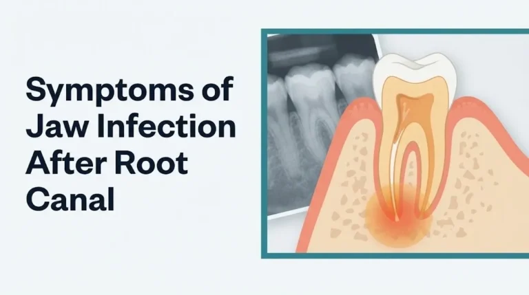
People who have had prior root canal treatment should be specifically aware that their treated teeth are at elevated risk for vertical root fractures precisely because the removal of pulp tissue reduces dentin moisture content by up to 9 percent, according to research published in the Journal of Endodontics, making the tooth structure measurably more brittle.
Symptoms of a Cracked Tooth: Pain When Biting and Releasing
Pain when biting down and, more specifically, pain when releasing bite pressure is the most diagnostically distinctive symptom of a cracked tooth, and the underlying mechanism explains exactly why this occurs the way it does.
When you bite down on a cracked tooth, the two segments of the fracture compress slightly together under load. When you release, the segments spring back apart. This spring-back movement creates a rapid pressure change within the dentinal tubules adjacent to the crack line, pushing and pulling the fluid inside them.
Here is the mechanism the clinical literature supports. According to the hydrodynamic theory of dentin sensitivity, substantiated by Brannstrom’s experimental work and confirmed by subsequent electrophysiological research, rapid fluid movement within dentinal tubules mechanically deforms the nerve endings of A-delta fibers within Raschkow’s plexus (the subodontoblastic nerve network lining the inner pulp wall). A-delta fibers are fast-conducting, myelinated sensory fibers that produce the sharp, brief, localized pain people describe as “electrical” or “shooting.” The pain on release is often sharper than the pain on biting because the rebound decompression movement is faster.
To describe this symptom accurately to your dentist:
- Note exactly which side of your mouth the pain occurs on.
- Identify whether it is the biting-down moment, the releasing moment, or both that triggers pain.
- Rate the pain on a 0-to-10 scale at its peak.
- Note how many seconds the pain lasts after the trigger.
- Identify which specific foods, textures, or biting angles reliably reproduce it.
People with temporomandibular joint disorder (TMD) may find this symptom pattern harder to isolate because their existing jaw joint pain can mask or mimic the crack-specific bite pain. An endodontist or dentist experienced in cracked tooth syndrome diagnosis will use a Tooth Slooth (a small plastic biting device that loads individual cusps selectively) to isolate exactly which cusp of which tooth reproduces the symptom.
Hairline Crack in Tooth Symptoms
A hairline crack in a tooth produces symptoms that are often dismissed as minor sensitivity or attributed to other causes, precisely because the crack itself is invisible on visual examination and frequently absent on standard dental radiographs.
The symptom profile of a hairline crack depends entirely on how deep the fracture has extended. A hairline crack confined to the enamel (a craze line) typically produces no pain at all, or at most a fleeting sensitivity to cold that disappears within a second. A hairline crack that has reached the dentin produces the full hydrodynamic-theory-mediated symptom set: brief sharp pain with biting and temperature changes, localized gum tenderness, and occasional spontaneous twinge sensations.
Clinically significant hairline crack in tooth symptoms include:
- Transient sensitivity to cold water or cold air that resolves in under three seconds (enamel-level crack, generally lower urgency)
- Sensitivity to cold that lingers for four seconds or longer (suggesting dentin or early pulp involvement)
- Pain with sweet foods or beverages (osmotic fluid movement within dentinal tubules, mediated by the same hydrodynamic mechanism)
- Localized gum soreness when pressing with a finger directly over the suspected tooth
- Intermittent spontaneous tingling or aching that appears briefly and then vanishes
The challenge with hairline cracks is that standard periapical radiographs miss most of them. A fracture line must be roughly parallel to the X-ray beam and wide enough to appear as a radiolucency. Most clinically symptomatic cracks are oriented vertically or obliquely and are too thin to appear on two-dimensional X-ray. Research published in the Journal of Endodontics has found that cone-beam computed tomography (CBCT) has substantially higher sensitivity for detecting incomplete fractures, though it is not used as a first-line tool for every tooth complaint due to higher radiation exposure relative to conventional radiography.
Young adults (ages 18 to 35) who grind their teeth at night are an increasingly common demographic presenting with hairline crack symptoms in lower first molars, often on teeth that appear clinically pristine with no visible crack and no radiographic findings.
Key Takeaway: Sharp pain when releasing bite pressure is the most diagnostically specific single symptom of a cracked tooth. If you feel it repeatedly on the same tooth, that tooth needs evaluation by a dentist, not monitoring at home.
Cracked Molar Symptoms
Cracked molar symptoms are typically more pronounced than cracks affecting front teeth, because molars bear the highest occlusal loads in the mouth, receive the most concentrated biting force, and are most likely to contain large restorations that weaken their remaining structure.
The first and second mandibular (lower) molars are the most frequently fractured teeth in clinical endodontic practice. These teeth carry the greatest functional load and are most often restored with large amalgam fillings that, over decades, expand and contract with temperature cycling, widening existing micro-cracks and creating new ones.
Cracked molar symptoms include all the core cracked tooth symptoms described earlier, but with specific characteristics:
- Pain that is lateralized to one side of the lower or upper jaw, often difficult to pinpoint to a specific tooth without clinical testing
- Sensitivity to chewing any texture, not just hard foods, once the crack has reached the dentin
- Gum swelling localized to one root of a multi-rooted molar (a classic sign of a vertical root fracture or crack extending below the gumline)
- A narrow, isolated periodontal pocket (a deep pocket between the tooth and gum detectable only with a periodontal probe) alongside a single root of an otherwise periodontally healthy tooth
The last symptom deserves emphasis. A narrow, isolated pocket deeper than 5 millimeters next to an otherwise periodontally healthy tooth is a red flag for a crack extending below the gumline. The American Association of Endodontists identifies this clinical finding as one of the most reliable indicators of a subgingival crack when combined with patient-reported bite pain.
Older adults, particularly postmenopausal women, face an additional layer of risk. Bone density changes associated with osteoporosis do not directly fracture teeth, but the medications used to treat osteoporosis, specifically bisphosphonates like alendronate (Fosamax), alter bone remodeling in the jaw (bisphosphonate-related osteonecrosis of the jaw, BRONJ). If a cracked molar requires extraction in a patient on long-term bisphosphonate therapy, healing may be severely impaired. These patients need consultation between their dentist, endodontist, and prescribing physician before any extraction.
Cracked Tooth Pain That Comes and Goes
The intermittent nature of cracked tooth pain is not a sign that the problem is resolving. It is a structural feature of how incomplete fractures behave under variable loading conditions.
Pain comes and goes because not every bite, every temperature exposure, or every food texture activates the fractured segments in the same way. When you eat soft foods that don’t load the crack line, you feel nothing. When you happen to bite at the angle that compresses or springs the fracture segments, the sharp pain fires. When you move to a softer bite, it vanishes.
This pattern is clinically distinct from the constant throbbing pain of irreversible pulpitis, the dull ache of a sinus infection (which can mimic upper molar tooth pain), or the pressure-related pain of a tooth abscess. The episodic quality of crack-related pain is produced by the mechanical intermittency of fracture segment movement, not by the continuous chemical inflammatory process that drives most other tooth pain types.
People with cracked tooth pain that comes and goes sometimes wait months or years before seeking evaluation, particularly when pain-free intervals are long. The American Dental Association (ADA) and the AAE both document cases where symptom-free intervals led patients to defer treatment until the crack had propagated to an irreparable extent. A crack that produces intermittent pain today can become a split tooth, a vertical root fracture, or an infected tooth in months without intervention.
The pain-comes-and-goes pattern also varies with food temperature. Cold sensitivity that disappears in under three seconds during a pain-free interval may intensify to a lingering 10- to 15-second ache when the crack has deepened and the pulp has begun inflammatory changes. Tracking this progression (how long cold sensitivity lasts, whether it’s getting longer) gives your dentist clinically useful information about whether the pulp is moving toward irreversible pulpitis.
Cracked Tooth With No Visible Crack or Pain
A cracked tooth can exist without any visible external crack and, in early stages, without any pain at all. This is clinically important and frequently misunderstood by both patients and, in some cases, by general practitioners without specific cracked tooth syndrome training.
Craze lines are hairline fractures confined entirely to the enamel surface. They are visible as fine lines under dental transillumination (a fiberoptic light pressed against the tooth to illuminate internal structures) but produce no symptoms. They require no treatment, only monitoring. Mistaking a craze line for a clinically significant crack is one of the most common sources of patient confusion and unnecessary anxiety.
The more concerning scenario is a crack that extends into the dentin or toward the pulp but has not yet produced consistent symptoms because the crack line is oriented in a position that doesn’t consistently load during normal chewing. These teeth are genuinely asymptomatic between episodes and may show nothing on periapical X-ray.
Signs that a “no-pain” tooth may harbor a significant crack include:
- Intermittent episodes of sharp sensitivity to cold, resolving in under three seconds, that have been present for months
- A history of noticing occasional brief discomfort while chewing that quickly passes
- A large existing restoration in the tooth in question, particularly an old amalgam filling
- History of facial or jaw trauma, even years prior
- Tooth that has previously undergone root canal treatment (making it more brittle)
- Active bruxism, especially with no occlusal guard currently in use
According to the Centers for Disease Control and Prevention (CDC), approximately 26 percent of American adults have untreated dental conditions. Many asymptomatic cracks fall into this category, discovered incidentally during routine dental examination or only when they progress to pain.
Key Takeaway: The absence of constant pain does not rule out a clinically significant crack. Intermittent brief sensitivity to cold, combined with a history of large restorations or bruxism, is enough justification for a focused evaluation by a dentist using transillumination and selective bite testing.
Cracked Tooth Nerve Symptoms and Pulp Involvement
When a crack has extended into the dental pulp, the symptom profile shifts from intermittent and mechanical to persistent and inflammatory, signaling that a fundamentally different nerve fiber population is now activated.
In an intact tooth, pain signals travel primarily through A-delta fibers in Raschkow’s plexus. These fast-conducting fibers produce brief, sharp, well-localized pain in response to mechanical or thermal stimuli. When the crack reaches the pulp chamber and bacteria or bacterial byproducts penetrate the pulpal tissue, a second type of nerve fiber, the C-fiber, becomes dominant. C-fibers are slow-conducting, unmyelinated sensory fibers that produce the throbbing, poorly localized, deep aching pain that people describe as “the worst toothache of my life.”
Pulp involvement symptoms are clinically distinguishable from early crack symptoms:
- Spontaneous toothache that occurs without any biting, eating, or temperature stimulus
- Persistent pain after a cold stimulus is removed, lasting longer than 15 to 30 seconds (a clinically reliable indicator of irreversible pulpitis)
- Throbbing pain that worsens when lying down (due to increased venous pressure in the pulpal vasculature when the head is horizontal)
- Tooth pain that wakes you from sleep
- Visible swelling around the gum line of the tooth, indicating that infection has begun spreading to the periodontal tissue or periapical region
The distinction between reversible pulpitis and irreversible pulpitis is clinically critical. Reversible pulpitis produces sensitivity that resolves quickly after the stimulus is removed and can be treated by eliminating the irritant (in this case, treating the fracture with a crown). Irreversible pulpitis produces lingering, spontaneous, worsening pain and requires root canal therapy (endodontic treatment) to remove the inflamed or necrotic pulp tissue.
People with diabetes mellitus face a specific heightened risk at this stage. Diabetic patients, particularly those with poorly controlled blood glucose, have impaired neutrophil function and compromised microvascular supply to periodontal tissue. What presents as a manageable pulpal infection in a non-diabetic patient can escalate rapidly to a spreading fascial space infection in a person with poorly controlled diabetes. Any diabetic patient experiencing spontaneous toothache or tooth swelling should contact their dentist the same day, not wait for their next scheduled appointment.
Cracked Tooth Infection Symptoms
A cracked tooth that allows bacteria to reach the pulp and periapical tissues can develop into a dental abscess, and the symptoms at this stage represent a genuine clinical escalation that goes beyond dental inconvenience.
Periapical abscess formation occurs when pulp tissue becomes necrotic (dead) and bacteria multiply in the pulp chamber and begin migrating through the root canal system to the periapical tissue (the bone and soft tissue at the root tip). The immune system responds with an acute inflammatory reaction, producing the collection of pus that defines a dental abscess.
Cracked tooth infection symptoms include:
- Persistent, severe throbbing toothache that does not resolve with over-the-counter pain medications at standard doses
- Swelling of the gum tissue adjacent to the tooth, which may feel fluctuant (fluid-filled) to touch
- Fever, typically above 100.4°F (38°C)
- A bad taste in the mouth or foul odor that appears suddenly (pus draining through a sinus tract in the gum, called a “gum boil” or parulis)
- Swollen lymph nodes under the jaw or in the neck
- Facial swelling visible externally, particularly below the eye for upper tooth infections or along the jaw for lower tooth infections
The Journal of Endodontics has published case series documenting the trajectory from periapical abscess to fascial space infection (infection spreading through the natural tissue planes of the head and neck) in patients who did not receive timely treatment. This progression can occur within 24 to 72 hours in immunocompromised individuals or those with diabetes.
Critically, a dental abscess at this stage is not a “wait until morning” situation. Any cracked tooth infection producing visible facial swelling, fever, or swollen neck lymph nodes requires same-day dental evaluation. If a dentist cannot see you that day, go to an emergency room. Antibiotics alone do not resolve a dental abscess. The source of infection (the necrotic pulp) must be treated or the tooth extracted.
Broken Tooth Symptoms vs. Cracked Tooth Symptoms
A broken tooth and a cracked tooth are not interchangeable terms clinically, and the symptom profiles differ in ways that tell you something important about the extent of damage and the urgency of treatment.
A broken tooth involves visible structural loss: a cusp has fractured off, a large piece of the tooth crown has separated, or the tooth has split into identifiable segments. A cracked tooth may have no visible structural loss at all. The fracture is present but the two segments remain in contact.
| Feature | Cracked Tooth | Broken Tooth |
|---|---|---|
| Visible damage | Often invisible without magnification or special light | Visible fracture, missing piece, or sharp edge |
| Pain pattern | Intermittent, triggered by biting or temperature | Often immediate and constant if pulp exposed |
| Bite sensitivity | Sharp pain on biting and releasing | Pain may be severe on any contact |
| Temperature sensitivity | Common; brief sensitivity to cold | Severe if pulp is directly exposed to oral environment |
| Urgency | Urgent (days to week); emergency if infected | Urgent to emergency depending on pulp exposure |
| Self-visible? | Rarely | Usually yes |
A broken tooth that has exposed the dental pulp is a dental emergency. The pulp, now directly exposed to the bacteria-rich oral environment, will become infected without prompt treatment. Pain from an exposed pulp is typically severe, constant, and made worse by both hot and cold.
By contrast, a cracked tooth without pulp exposure can be evaluated at an urgent dental appointment (within days), not necessarily at an emergency room, unless infection symptoms are present.
Children and adolescents who break a tooth during sports or a fall should be evaluated the same day, both to assess pulp status and, in the case of a permanent tooth fragment that has been cleanly separated, because fragments can sometimes be bonded back to the tooth if treatment occurs within hours. The American Academy of Pediatric Dentistry (AAPD) recommends that any dental trauma in a child involving a permanent tooth be evaluated by a dentist or pediatric dentist within 24 hours.
Key Takeaway: A broken tooth with visible structural loss and immediate severe pain is a same-day dental emergency. A cracked tooth producing intermittent bite pain without swelling or fever is urgent but generally not an emergency room situation, unless infection develops.
Symptoms of a Cracked Tooth Under a Crown or Filling
A cracked tooth under an existing crown or large filling is one of the most diagnostically challenging presentations in clinical dentistry, because the restoration itself conceals the fracture and can mask some of the classic pain patterns.
A dental crown covers the entire visible portion of the tooth. A crack that develops under an existing crown can propagate downward toward or below the gumline without producing the typical bite-pain-and-release pattern, because the rigid crown redistributes occlusal forces differently than a natural tooth surface. Patients are often surprised to learn their crowned tooth is cracked because they assumed the crown protected the underlying tooth from fracture.
Symptoms suggesting a crack beneath a crown or filling include:
- Persistent dull aching around a crowned or filled tooth, different in character from any previous sensitivity related to that tooth
- Pain when biting in a specific location even through the crown, suggesting fracture segment movement beneath the restoration
- Gum swelling localized to one area around a crowned tooth, particularly along a single root
- A narrow, deep periodontal pocket (detected by a dentist using a periodontal probe) alongside one root of a crowned tooth that is otherwise periodontally normal
- Recurrent sensitivity to cold in a tooth that previously had no sensation issues
According to the American Association of Endodontists, cracks beneath existing crowns frequently occur in teeth that received large core build-ups or post-and-core restorations after root canal treatment. The post (a metal or fiber pin placed in the root canal to support the crown) can act as a lever during biting forces, concentrating stress at the root and initiating or propagating a vertical root fracture.
People who clench or grind their teeth while wearing a night guard should know that a night guard reduces but does not eliminate parafunctional forces. Teeth can still fracture under a crown in active bruxers, particularly if the night guard is worn or no longer properly fitted. Annual evaluation of the guard’s fit and occlusal balance is part of responsible bruxism management.
Cracked Tooth Causing Ear Pain, Jaw Pain, and Headache
A cracked tooth can produce pain felt in the ear, jaw, temple, or side of the face through a process called referred odontogenic pain, and it is one of the primary reasons cracked tooth syndrome is consistently misdiagnosed for months or longer.
Referred pain occurs because the trigeminal nerve (cranial nerve V), specifically its mandibular branch (V3) and maxillary branch (V2), innervates both the teeth and the surrounding structures of the face, jaw, ear region, and temple. When pulpal or periodontal pain signals from a cracked tooth travel along the trigeminal nerve, the brain can mislocalize the source. Pain arising from a lower molar is frequently felt as ear pain. Pain from an upper molar is frequently felt as sinus pressure or cheek pain. Pain from any posterior tooth can radiate to the temple and be interpreted as a tension headache.
Clinical patterns of referred cracked tooth pain:
- Ear pain with no ear infection findings on otoscopic examination, occurring on the same side as a posterior tooth with bite sensitivity
- Unilateral facial aching or jaw soreness, worse on one side, that intensifies after meals
- Persistent headaches localized to the temple on one side, especially in a known bruxer
- Sinus pressure symptoms affecting only one side, in the absence of nasal congestion, runny nose, or bilateral symptoms
Distinguishing cracked tooth-origin referred pain from temporomandibular joint disorder (TMD), primary ear pathology, or sinusitis requires a clinical evaluation that includes selective bite testing, periodontal probing, transillumination, and sometimes CBCT imaging. A dentist cannot diagnose ear pathology, and an otolaryngologist cannot diagnose dental fractures. If you have ear or jaw pain that persists and your ear examination is normal, request a focused dental evaluation specifically for cracked tooth syndrome.
People with a documented history of bruxism and new-onset unilateral ear pain should consider a dental evaluation before proceeding to an ear, nose, and throat specialist, particularly if the ear pain correlates with meals or specific chewing motions.
Fractured Tooth Symptoms in Older Adults and High-Risk Groups
Fractured tooth symptoms in older adults and people with specific medical conditions often present differently, progress more rapidly, or carry consequences that are more serious than the same fracture in a healthy young adult.
Older adults (65 and older) accumulate fracture risk through several converging mechanisms: decades of occlusal wear, a high prevalence of large existing restorations (many placed 30 or 40 years ago and now structurally compromised), lower salivary flow rates (common with many medications used in older adults, including antihistamines, antidepressants, and antihypertensives), and reduced dentin remineralization capacity. Symptoms in older adults may be muted because aging reduces the density of nerve fibers in the dental pulp, a process called pulpal sclerosis, meaning that the classic sharp bite pain may be less prominent even as fracture severity increases.
High-risk group symptom profiles and clinical considerations:
| Population | Specific Risk | Clinical Impact on Symptoms | Action Required |
|---|---|---|---|
| People with diabetes | Impaired immune response, microvascular disease | Faster abscess progression, higher fascial spread risk | Same-day dental evaluation for any swelling or fever |
| Bruxers (sleep/awake) | Parafunctional forces up to 1,000 psi | Accelerated crack propagation, bilateral fracture risk | Night guard fitting plus urgent evaluation of symptomatic teeth |
| Bisphosphonate users | Altered bone remodeling, BRONJ risk | Extraction complications, altered healing | Multidisciplinary consultation before any extraction |
| Endodontically treated teeth | Dentin dehydration, increased brittleness | Higher vertical root fracture risk, less warning pain | Prompt evaluation of any new symptoms in root canal-treated teeth |
| Immunocompromised patients | Reduced infection containment | Rapid spread of dental abscess to fascial spaces | Same-day or emergency evaluation for any swelling |
| Older adults | Pulpal sclerosis, reduced pain perception | Classic symptoms may be absent despite significant damage | Routine dental evaluation every 6 months mandatory |
A 2022 paper published in the Journal of the American Dental Association (JADA) found that endodontically treated teeth in patients over 55 had a fracture rate approximately 3.5 times higher than vital (non-root-canal-treated) teeth in the same age group, with vertical root fractures accounting for the majority of those cases.
People with active chemotherapy or recent organ transplant who develop any dental pain or swelling should contact both their oncologist or transplant team and their dentist before undergoing any dental procedure, because dental treatment protocols may need modification based on their current immune status and platelet counts.
Key Takeaway: Older adults and people with diabetes, bruxism, large restorations, or prior root canal treatment need to know that their fracture risk is substantially elevated, and that classic pain symptoms may be absent or reduced even when significant damage has occurred.
How a Cracked Tooth Is Diagnosed
Diagnosing a cracked tooth is one of the most technically challenging tasks in clinical dentistry, and it often requires several complementary diagnostic tools rather than a single test.
The diagnostic process typically follows this sequence:
- Detailed symptom history: The dentist asks about bite pain, release pain, temperature sensitivity, duration, location, and which foods or angles reproduce the pain. This narrows the suspect tooth or teeth.
- Visual examination with magnification and transillumination: A fiberoptic light is pressed against each tooth. Intact teeth transmit light evenly. A fracture interrupts light transmission, appearing as a dark line across the tooth. This is often the first definitive visual confirmation of a crack.
- Selective bite test using Tooth Slooth: The patient bites on a small plastic cone placed over individual cusps in sequence. The cusp that reproduces the pain when loaded, or when released, identifies the fracture location within the tooth.
- Periodontal probing: The dentist uses a periodontal probe to measure pocket depths around all roots of the tooth. A single deep, narrow pocket alongside one root in an otherwise healthy mouth strongly suggests a subgingival crack.
- Periapical radiograph: Standard dental X-ray. It may not show the crack itself (most fractures are not visible on standard X-ray), but it can show periapical bone changes indicating infection or abscess, and it shows the size and depth of existing restorations.
- Cone-beam computed tomography (CBCT): Three-dimensional X-ray imaging that provides cross-sectional views of the tooth and surrounding bone. Research published in the Journal of Endodontics confirms CBCT has significantly higher sensitivity than conventional radiography for detecting vertical root fractures and incomplete fractures, though it involves higher radiation exposure and is used selectively rather than routinely.
- Pulpal vitality testing: The dentist applies a cold stimulus (refrigerant spray or ice) or electrical pulp testing to assess whether the pulp is alive, inflamed, or necrotic. This determines whether root canal treatment is needed alongside crack management.
Patients with multiple large posterior restorations and intermittent bite pain should ask their dentist specifically to perform transillumination and selective bite testing, not just a visual examination. These additional steps add minutes to an appointment but make the diagnostic difference between an identified, treatable crack and months of unexplained tooth pain.
Emergency Symptoms: When to Call 911 or Go to the ER
Certain cracked tooth-related complications can progress to life-threatening emergencies. Do not wait to see if these symptoms resolve on their own.
Call 911 or go to the nearest emergency room immediately if you experience:
- Swelling of the floor of the mouth (the area under the tongue) or swelling that is pushing your tongue upward, which can indicate Ludwig’s angina, a rapidly spreading infection of the submandibular and sublingual fascial spaces that can close the airway
- Swelling extending from the jaw or face down into the neck or upper chest
- Difficulty swallowing or pain with swallowing that has appeared alongside facial or jaw swelling from dental infection
- Difficulty breathing, a feeling of throat tightening, or shortness of breath in the context of facial swelling from dental infection
- Trismus: an inability to open your mouth beyond roughly 2 centimeters (about the width of two fingers) due to pain and muscle spasm, combined with facial swelling and fever
- Fever above 103°F (39.4°C) combined with facial or neck swelling from dental origin
- Confusion, neck stiffness, or sensitivity to light in the presence of facial swelling and dental pain (signs suggesting infection may have reached the meningeal spaces)
For an abscess with swelling confined to the gum tissue or cheek without the above signs: Go to a dentist the same day or to an urgent care dental clinic if your dentist cannot accommodate you. If neither is available, go to the emergency room. Antibiotics prescribed in an ER are a bridge measure only. The infected tooth must be treated by a dentist promptly.
Ludwig’s angina and deep fascial space infections of dental origin carry reported mortality rates of up to 40 percent without rapid surgical airway management and aggressive intravenous antibiotic treatment, according to case series published in the Journal of Oral and Maxillofacial Surgery. These presentations can deteriorate from “bad toothache with some swelling” to a complete airway emergency in hours.
These presentations require emergency medical assessment. An urgent care center, dental office, or phone call to a dentist is not sufficient when these signs are present.
Frequently Asked Questions About Cracked Tooth Symptoms
What does a cracked tooth feel like?
A cracked tooth typically feels like a sharp, brief, electrical pain when biting down on a specific food or angle, often followed by an equally sharp or sharper pain when releasing the bite.
Temperature sensitivity to cold, and sometimes heat or sweet foods, is also common, usually resolving within a few seconds in early-stage cracks.
If the fracture has reached the pulp, the sensation shifts from intermittent sharpness to a persistent, throbbing ache that can occur without any chewing trigger.
Can a cracked tooth have no symptoms at all?
Yes. Craze lines, which are fractures confined to the enamel surface, produce no pain and are often found incidentally during routine dental examination.
Cracks that have extended slightly into the dentin may also produce no consistent symptoms, particularly if the fracture orientation does not load during normal chewing patterns.
However, even a symptom-free crack beneath a large restoration or in a root-canal-treated tooth can propagate without warning, which is why regular dental examinations remain clinically necessary even when you feel no pain.
How do I know if my tooth crack has reached the nerve?
A crack that has reached the dental pulp (the nerve chamber) typically produces pain or sensitivity that lingers for more than 15 to 30 seconds after the cold or heat stimulus is removed, rather than resolving within a few seconds.
Spontaneous pain that occurs without biting, eating, or temperature exposure, particularly throbbing pain that worsens when lying down, is a strong clinical indicator of pulpal involvement.
A dentist can confirm pulp status using cold testing or electrical pulp testing at your appointment; these tests take minutes and provide clear diagnostic information about the pulp’s health.
Can a cracked tooth cause ear pain or jaw pain?
Yes. A cracked tooth, particularly a lower molar, can produce pain felt in the ear, jaw, or temple through referred pain carried by the trigeminal nerve (cranial nerve V), which innervates both the teeth and surrounding facial structures.
Unilateral ear pain with a normal ear examination, or jaw pain that worsens after eating, should prompt a dental evaluation specifically for cracked tooth syndrome before assuming the source is the ear or jaw joint.
The American Association of Endodontists identifies referred odontogenic pain as one of the most common reasons cracked tooth syndrome goes undiagnosed for extended periods.
What happens if you leave a cracked tooth untreated?
An untreated cracked tooth can progress from a minor, treatable fracture to pulp infection, periapical abscess, and in some cases a spreading fascial space infection that requires hospitalization and surgical management.
The tooth itself may progress from a repairable crack (treated with a crown) to an irreparable split tooth (requiring extraction), with the timing of that progression unpredictable and often rapid.
Leaving a cracked tooth untreated is never clinically appropriate once symptoms have begun; the treatment required becomes more extensive, more expensive, and more invasive with each stage of progression.
Can a cracked tooth heal on its own?
No. Teeth do not regenerate enamel or dentin, and a fracture line in tooth structure cannot close or repair itself through any biological process.
Symptom reduction during pain-free intervals does not mean the crack has improved; it means the fracture line was not loaded in a way that triggered the hydrodynamic pain mechanism during that period.
Only dental treatment, ranging from a crown for early-stage incomplete fractures to root canal therapy plus crown or extraction for advanced fractures, can address the structural damage.
The Bottom Line on Recognizing a Cracked Tooth
Sharp pain when biting, a jolt of sensitivity when you release chewing pressure, or persistent cold sensitivity on one specific tooth are not symptoms to watch and wait through. These patterns have a precise physiological explanation: fractured tooth structure is moving under load, shifting fluid in your dentin, and firing nerve fibers that are telling you something structural has changed.
The difference between a crown fitted in the early stages of an incomplete fracture and an extraction followed by a bone graft and implant months later often comes down to one thing: how quickly you had the tooth evaluated. Track your symptoms with a specific dentist appointment in mind, not a “when it gets worse” threshold.
If you have swelling, fever, difficulty swallowing, or pain spreading toward your neck alongside dental pain, skip the dental office and go directly to the nearest emergency room. That combination is not a dental inconvenience. It is a medical emergency with an airway at stake