Morton’s Neuroma Symptoms: Full 2026 Clinical Guide
Morton’s neuroma symptoms include a burning, aching, or electric-shock pain in the ball of the foot, most often between the third and fourth toes, accompanied by tingling, numbness, and a persistent sensation of standing on a pebble or folded sock that does not move when you adjust your position. These symptoms are produced by compression and progressive scarring of the common plantar digital nerve in the intermetatarsal space, a process that evolves in stages and explains why early-stage symptoms are intermittent while advanced presentations produce constant discomfort even without walking.
According to clinical observation data reviewed by the American College of Foot and Ankle Surgeons (ACFAS), Morton’s neuroma is one of the most common causes of forefoot pain in adults, with women affected approximately 8 to 10 times more frequently than men, primarily due to footwear habits that narrow the intermetatarsal space and chronically compress the plantar digital nerves. The condition is frequently misattributed to plantar fasciitis, metatarsal stress fractures, or general forefoot pain before the nerve entrapment pattern becomes clinically clear.
This guide covers every recognized symptom with its anatomical and neurological mechanism, explains the full progression from early to late-stage presentation, identifies the populations at highest risk, distinguishes Morton’s neuroma from its most important diagnostic mimics, and tells you exactly when footwear changes stop being enough.
Morton’s Neuroma Symptoms: What This Condition Actually Feels Like
Morton’s neuroma symptoms are a cluster of sensory and pain experiences in the forefoot produced by progressive compression and fibrosis of the common plantar digital nerve, including burning pain, electric or sharp shooting sensations, tingling between specific toes, numbness of the adjacent toe surfaces, and a persistent feeling of a foreign object under the ball of the foot.
The combination of these sensations is distinctive enough that experienced clinicians often recognize the symptom pattern before examination. The burning quality reflects inflammatory nociceptor activation from nerve compression. The electric or shooting quality reflects disrupted saltatory conduction along partially demyelinated nerve fibers. The numbness reflects loss of sensory axon function in advanced cases.
The complete recognized symptom set includes:
- Burning pain in the forefoot, centered on the third and fourth metatarsal heads
- Sharp or electric-shock sensations that radiate into the adjacent toes
- Tingling or “fizzing” sensation in the web space between the third and fourth toes
- Numbness affecting the touching surfaces of the third and fourth toes
- A persistent sensation of a pebble, marble, or folded sock under the ball of the foot
- Pain that worsens with walking, prolonged standing, or wearing tight or narrow shoes
- Temporary relief when the shoe is removed and the foot is rested and massaged
- In advanced stages, pain that persists at rest and during the night
The Mayo Clinic notes that symptoms typically begin gradually and may initially appear only during specific activities, such as running or wearing a particular pair of shoes. The early intermittent pattern is mechanistically distinct from the late-stage constant presentation, which reflects two different phases of nerve injury rather than simply worsening of the same process.
People with diabetes mellitus and pre-existing peripheral neuropathy may experience fewer or less intense early pain symptoms because their baseline sensory function is already reduced. In these individuals, numbness may be the presenting symptom rather than pain, which can delay recognition and diagnosis.
What Is Morton’s Neuroma and Which Nerve Is Affected
Morton’s neuroma is a benign perineural fibrosis of the common plantar digital nerve in the intermetatarsal space, most often the third space between the third and fourth metatarsal heads, caused by repetitive mechanical compression of the nerve against the deep transverse intermetatarsal ligament during forefoot weight-bearing.
Despite its name, Morton’s neuroma is not technically a tumor or true neuroma: it contains no neoplastic cells. The correct anatomical term is interdigital neuroma or intermetatarsal neuroma. The pathological process is one of mechanical injury, chronic inflammation, and progressive fibrosis of the nerve sheath rather than abnormal cellular growth. The name “neuroma” reflects the thickened, firm, nodular feel of the affected nerve segment when examined or surgically excised.
The third intermetatarsal space is affected in approximately 80 percent of all clinical cases. The second interspace is the next most common site, accounting for most remaining cases. The first and fourth interspaces are rarely affected. The anatomical reason for the third space’s vulnerability is specific and important: the third common plantar digital nerve in this location regularly receives a communicating branch from the medial plantar nerve, producing a nerve trunk with greater cross-sectional diameter than those in other interspaces. This larger nerve is more susceptible to compression within the fixed anatomical channel formed between the metatarsal heads and beneath the deep transverse intermetatarsal ligament.
The deep transverse intermetatarsal ligament spans the metatarsal heads and forms the roof of the intermetatarsal tunnel through which the digital nerve runs. When the forefoot is loaded during walking or running, the metatarsal heads spread slightly, narrowing the intermetatarsal channels from the sides, while the ground reaction force pushes the nerve upward against the unyielding ligament from below. This produces a pinching compression with every step.
People with rheumatoid arthritis face elevated risk because synovitis of the metatarsophalangeal joints narrows the intermetatarsal space further and adds inflammatory mediators to the local environment, accelerating perineural inflammation and fibrosis progression.
What Does Morton’s Neuroma Feel Like Day to Day
Day to day, Morton’s neuroma feels like walking on a small, hard lump that cannot be found when you remove your shoe, combined with burning or tingling in specific toes that intensifies with activity in closed footwear and temporarily improves when the shoe is removed and the forefoot is massaged.
Think of the affected nerve like a garden hose being stepped on repeatedly, hundreds of times per day. Each step presses the hose against a hard surface. The water (nerve signal) still moves through, but not efficiently, and the hose itself is being slowly damaged by the repeated compression. That damage produces a peculiar mix of pain (from injured nerve tissue releasing inflammatory mediators), abnormal sensation (from disrupted nerve conduction), and eventually, numbness (from axonal loss).
The daily experience changes predictably based on footwear. Narrow or pointed shoes that compress the forefoot laterally narrow the intermetatarsal space from both sides, worsening nerve compression dramatically. High heels shift the forefoot into a plantarflexed position that loads the metatarsal heads with a higher proportion of body weight, increasing the ground-upward compression force on the nerve against the ligament. Removing a constricting shoe and manually massaging the forefoot, which spreads the metatarsal heads apart and temporarily decompresses the intermetatarsal space, provides the characteristic short-term relief that patients consistently report.
Walking barefoot on a firm surface often causes less discomfort than walking in shoes, because the toes can splay naturally and the intermetatarsal space is not artificially narrowed. Walking barefoot on soft, unstable surfaces like sand or thick carpet may worsen symptoms by requiring more active toe flexor engagement, which increases intrinsic forefoot pressure.
For people with hallux valgus (bunion deformity of the first metatarsophalangeal joint), the lateral deviation of the great toe pushes the second toe laterally, which in turn narrows the second and third intermetatarsal spaces and compounds the nerve compression from two directions.
Morton’s Neuroma Pain Location and Distribution
Morton’s neuroma pain is located in the forefoot at the level of the metatarsal heads, most precisely in the third intermetatarsal space between the third and fourth toes, and it radiates distally into the adjacent toe surfaces, most often producing pain, burning, or altered sensation in the third and fourth toes.
The pain’s location distinguishes it from several other forefoot conditions. Unlike plantar fasciitis, which produces heel and arch pain on the medial plantar surface, Morton’s neuroma pain is anterior, positioned in the ball of the foot rather than the heel or mid-arch. Unlike a metatarsal stress fracture, which produces localized tenderness directly over the metatarsal bone shaft, Morton’s neuroma pain is centered in the space between metatarsal heads, not over the bone itself.
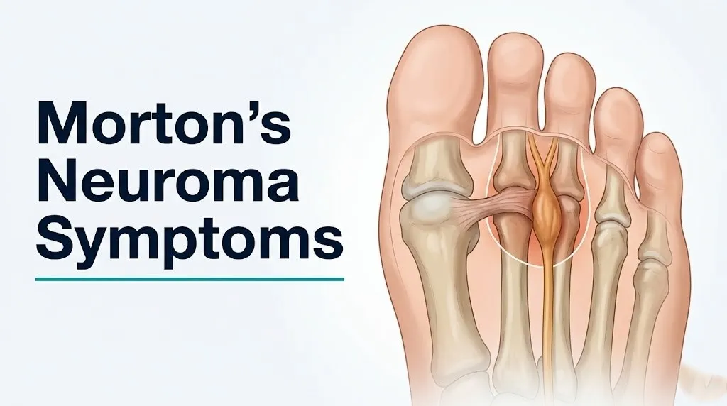
The radiation pattern is diagnostically useful. The burning and tingling from Morton’s neuroma travel distally toward the tips of the third and fourth toes, following the sensory distribution of the third common plantar digital nerve. Proximal radiation toward the ankle or heel does not occur in Morton’s neuroma and should prompt consideration of a more proximal nerve entrapment such as tarsal tunnel syndrome, in which the tibial nerve is compressed at the medial ankle and produces symptoms that can refer throughout the plantar foot and toes.
| Pain Feature | Morton’s Neuroma | Metatarsal Stress Fracture | Tarsal Tunnel Syndrome |
|---|---|---|---|
| Primary location | Third or second intermetatarsal space | Metatarsal shaft (bone) | Medial ankle, diffuse plantar foot |
| Radiation direction | Distally into third and fourth toes | Localized, minimal radiation | Throughout plantar foot, may include toes |
| Worse with | Tight shoes, walking, forefoot compression | Activity, weight-bearing | Prolonged standing, night (often) |
| Better with | Shoe removal, foot massage, rest | Complete rest and offloading | Ankle position change, elevation |
| Palpation finding | Tender interspace with possible click | Direct bone tenderness | Tinel’s sign at medial ankle |
Bilateral Morton’s neuroma (affecting both feet) is less common than unilateral presentation but does occur. According to clinical data reviewed in the Journal of Foot and Ankle Research, bilateral cases occur in approximately 15 percent of patients with Morton’s neuroma, and bilateral presentation should prompt review of systemic risk factors including rheumatoid arthritis and footwear habits affecting both feet equally.
Burning and Electric Shock Sensations in Morton’s Neuroma
The burning and electric shock sensations in Morton’s neuroma are produced by two distinct pathological processes: inflammatory nociceptor activation from prostaglandin and substance P release at the compression site, and disrupted saltatory conduction along partially demyelinated nerve fibers that produces spontaneous abnormal electrical discharges perceived as electric jolts or sharp radiating pain.
The burning quality of Morton’s neuroma pain is not arbitrary. Burning pain is the hallmark of nerve injury at a specific stage: when the nerve is still conducting signals but doing so through tissue that is inflamed, swollen, and irritated. Substance P, released from compressed sensory nerve endings, activates and sensitizes adjacent nociceptive C-fibers and A-delta fibers, lowering their firing threshold so that stimuli that would not normally produce pain (light touch of the shoe upper, the gentle pressure of standing) now trigger a burning pain response.
The electric-shock or shooting quality is mechanistically distinct. It occurs when compression and early demyelination disrupt the organized saltatory conduction of the nerve impulse along the myelin-sheathed axons. Instead of a clean signal traveling efficiently from node to node along the myelin sheath, the signal becomes erratic, jumps between adjacent nerve fibers (ephaptic transmission), and triggers abnormal spontaneous discharges. The patient experiences these discharges as sudden, brief electric jolts or stabs that shoot toward the toes, often triggered by a specific step or shoe pressure.
According to a clinical review published in Foot and Ankle International, the burning and electric-shock quality of forefoot pain, specifically combined with the interspace location and partial toe numbness, is one of the most diagnostically specific symptom combinations for Morton’s neuroma compared to other forefoot conditions.
People with multiple sclerosis or other demyelinating disorders already affecting the peripheral or central nervous system may find that Morton’s neuroma symptoms are harder to attribute accurately to a single cause, because pre-existing demyelination can produce overlapping sensory symptoms. A board-certified neurologist and a board-certified podiatrist working collaboratively may be needed to evaluate these cases.
Numbness and Tingling Symptoms of Morton’s Neuroma
Numbness and tingling in Morton’s neuroma affect the skin surfaces of the third and fourth toes that face each other in the web space, and they reflect progressive dysfunction of the sensory axons within the compressed common plantar digital nerve, ranging from mild intermittent tingling in early stages to persistent complete numbness in advanced cases.
Tingling, clinically described as paresthesia, is an early neurological symptom. It occurs when nerve conduction is disrupted but the nerve fibers are still largely intact. The sensation is often described as pins-and-needles, fizzing, or vibrating in the web space and adjacent toe surfaces. It may last only seconds during the provoking activity and resolve promptly when the foot is rested and unloaded.
Numbness, clinically described as hypoesthesia (reduced sensation) or in severe cases anesthesia (absent sensation), appears later in the progression. It indicates that the sensory axons within the nerve are no longer conducting reliably, either because perineural fibrosis has physically compressed the axons below their functional threshold or because demyelination has progressed to the point where normal signal propagation cannot occur. The specific skin territory affected is the adjacent surfaces of the third and fourth toes: touching these surfaces feels muted, reduced, or absent compared to the equivalent skin on the unaffected side.
The pattern of numbness is anatomically specific. If numbness extends to the entire sole of the foot, the whole foot, or the ankle and leg, Morton’s neuroma is not the explanation, and a more proximal nerve entrapment or systemic neurological condition should be investigated.
Quick Tip:
- To self-assess the numbness pattern: lightly brush a fingertip across the adjacent surfaces of the third and fourth toes on the affected foot and compare the sensation to the same test on the opposite foot. Reduced or absent sensation in this specific territory is consistent with the sensory distribution of the third common plantar digital nerve.
- Any numbness that extends beyond the third and fourth toe web space warrants evaluation by a board-certified podiatrist or board-certified orthopedic foot and ankle surgeon to rule out more proximal nerve involvement.
- People with type 2 diabetes mellitus and pre-existing sensory neuropathy may already have reduced plantar sensation that masks the toe numbness that would otherwise signal Morton’s neuroma, making the pain and burning symptoms more important diagnostic indicators in this population.
Key Takeaway: The combination of burning forefoot pain, electric-shock sensations radiating into the third and fourth toes, and numbness specifically affecting the adjacent surfaces of those two toes is the most diagnostically specific symptom cluster for Morton’s neuroma and should prompt evaluation by a foot specialist within 2 to 4 weeks.
Early Stage Morton’s Neuroma Symptoms
Early stage Morton’s neuroma symptoms are intermittent and activity-dependent: a mild burning or aching in the forefoot that appears after prolonged walking or wearing narrow shoes, resolves within minutes of removing the shoe and resting the foot, and does not yet produce persistent numbness or rest pain.
At this stage, the pathological process is primarily edema within the perineurium and endoneurium of the compressed nerve, accompanied by local inflammatory mediator release. The nerve architecture is still largely intact. The edema is pressure-dependent: it develops during activity when forefoot loading compresses the intermetatarsal space, and it partially resolves with rest when the compression is removed and local tissue fluid pressure normalizes.
Early symptoms are easy to dismiss. A burning sensation after a long day in dress shoes or running shoes that disappears completely within 10 to 15 minutes of rest does not feel like a medical problem. Many people attribute it to fatigue, ill-fitting footwear, or aging feet. This is clinically significant because the early stage is when simple footwear modification and metatarsal pad use have their best evidence base for symptom resolution. A 2019 systematic review published in the Journal of Foot and Ankle Research found that conservative measures including footwear modification and orthotic support show their highest success rates when applied to smaller neuromas at earlier stages of pathological change.
The Mulder’s sign (discussed in detail in a later section) is typically absent or inconsistently present in early-stage Morton’s neuroma because the nerve has not yet developed enough perineural fibrosis to create a firm, palpable, displaceable nodule. Clinical diagnosis at this stage relies more on the characteristic symptom history and response to footwear change than on physical examination findings.
Pregnant women in the second and third trimester may experience earlier-than-expected progression through the early stage due to increased fluid retention (edema affecting the endoneural capillary beds) and ligamentous laxity that allows greater metatarsal head spreading during loading, compressing the intermetatarsal space more dynamically with each step.
Late Stage and Chronic Morton’s Neuroma Symptoms
Late stage Morton’s neuroma symptoms include constant forefoot burning that persists regardless of footwear or activity, prominent numbness in the third and fourth toes that does not resolve with rest, and a palpable or reproducible pain response to gentle interspace compression even without walking.
Late-stage Morton’s neuroma reflects advanced perineural fibrosis with progressive demyelination of the affected nerve axons. By this stage, the epineurium and perineurium have been substantially replaced by dense collagen, the nerve trunk has thickened to a firm, fibrotic nodule, and the demyelinated axons are firing spontaneously and erratically without the need for mechanical provocation.
The constant nature of late-stage pain is mechanistically distinct from the early intermittent pattern. The fibrotic nerve no longer requires compression to produce symptoms: the demyelinated, sensitized nerve fibers discharge spontaneously, and the lowered threshold of surrounding nociceptors means that even light contact with footwear, bedsheets, or air pressure produces a painful response. This spontaneous activity is the same process that produces the constant burning pain of diabetic peripheral neuropathy, post-herpetic neuralgia, and other chronic neuropathic pain conditions, though in Morton’s neuroma it is localized to the specific nerve segment rather than generalized.
Late-stage Morton’s neuroma that has persisted untreated for more than 12 to 18 months is less likely to respond to conservative measures. According to clinical outcome data reviewed by the American College of Foot and Ankle Surgeons, neuromas with extensive perineural fibrosis and established chronic symptoms typically require more aggressive intervention such as corticosteroid injection, sclerosing alcohol injection series, or surgical neurectomy for symptom control.
Older adults over 65 may present at a later stage than younger patients because they are more likely to have attributed forefoot pain to general age-related foot changes and delayed seeking evaluation. Pre-existing sensory changes from age-related peripheral nerve function decline can also mask the early tingling that would otherwise serve as an earlier symptom signal.
Morton’s Neuroma Symptoms at Rest and at Night
Morton’s neuroma symptoms at rest and at night indicate that the condition has progressed beyond the early pressure-dependent edema stage into a phase of established perineural fibrosis and axonal demyelination, where the nerve fires spontaneously without requiring mechanical compression.
Rest pain is a clinically important symptom marker. Early-stage Morton’s neuroma produces symptoms only when the foot is loaded and the intermetatarsal space is compressed. When a patient reports that the burning or tingling persists for more than 30 minutes after all footwear is removed, or that symptoms wake them from sleep, the condition has transitioned to a stage where conservative footwear-only management is no longer an adequate treatment approach.
Nocturnal symptoms from Morton’s neuroma are less pronounced than those seen in tarsal tunnel syndrome or carpal tunnel syndrome, where nighttime positioning consistently worsens nerve compression. In Morton’s neuroma, night pain arises from the spontaneous neural discharge of a chronically irritated, partially demyelinated nerve rather than from a positional compression effect. This distinction is diagnostically useful: tarsal tunnel syndrome produces predictable positional changes in nocturnal symptoms, while Morton’s neuroma night pain is relatively position-independent.
The patient experience of rest pain from Morton’s neuroma is sometimes described as a persistent “buzzing,” “glowing,” or “simmering” sensation in the ball of the foot that does not feel like the sharp mechanical pain of walking on it but is nonetheless disruptive and prevents comfortable sleep.
People with anxiety disorders may find that the hypervigilance component of their condition amplifies their perception of the spontaneous neural discharge associated with rest pain in Morton’s neuroma. This does not make the pain psychosomatic: the underlying nerve pathology is real. However, it does mean that the reported severity of rest symptoms may not always correlate linearly with the degree of objective perineural fibrosis visible on imaging, and this population may benefit from evaluation that includes both a board-certified podiatrist and a pain management specialist for comprehensive symptom management.
Key Takeaway: Symptoms that persist at rest for more than 30 minutes after shoe removal, or that wake you from sleep, mean your Morton’s neuroma has advanced beyond the early conservative treatment window, and evaluation by a board-certified podiatrist or board-certified orthopedic foot and ankle surgeon is warranted promptly.
Morton’s Neuroma Symptoms in Women and With High Heels
Women develop Morton’s neuroma approximately 8 to 10 times more often than men, primarily because narrow-toed and high-heeled footwear mechanically compresses the intermetatarsal space from the sides and loads the metatarsal heads with a higher proportion of body weight, directly increasing nerve compression against the deep transverse intermetatarsal ligament.
The biomechanics are specific. A two-inch heel shifts approximately 57 percent of forefoot body weight onto the metatarsal heads compared to flat footwear, according to biomechanical data reviewed in clinical foot and ankle research. A three-inch heel shifts an even higher proportion. This increased plantar loading force presses the plantar digital nerves upward against the transverse intermetatarsal ligament with each step. Simultaneously, the narrow pointed toe box typical of dress heels compresses the metatarsal heads medially and laterally, squeezing the intermetatarsal space from the sides and leaving the nerve with less available cross-sectional space.
Women who wear high-heeled or pointed-toe shoes for prolonged occupational or social periods accumulate this compression load daily, often for years before the perineural fibrosis reaches the threshold of producing consistent symptoms. This explains why the typical age at diagnosis in women is the fourth to sixth decade: the cumulative mechanical exposure has had decades to produce progressive nerve pathology.
The symptom pattern in women whose Morton’s neuroma is directly footwear-driven is often highly predictable: symptoms appear within 30 to 60 minutes of wearing the offending footwear and resolve within similar time after removal. When this pattern is present and consistent, a trial of wide-toe-box, flat footwear is a reasonable first step before imaging or specialist evaluation, provided the symptoms are not yet constant or present at rest.
For women in occupations requiring formal footwear (legal, financial, medical administration, hospitality), rotating between wide-toe-box professional footwear options throughout the week may reduce cumulative nerve compression enough to allow early-stage symptom resolution. This occupational context should be discussed specifically with a board-certified podiatrist when seeking evaluation.
Morton’s Neuroma Symptoms in Runners and Athletes
Runners and athletes develop Morton’s neuroma from the high-frequency repetitive impact loading of the forefoot during running, jumping, and cutting movements, which generates repeated compression of the plantar digital nerve against the transverse intermetatarsal ligament at rates far exceeding those of normal walking.
A recreational runner covering 30 miles per week takes approximately 50,000 forefoot contact cycles per week. Each cycle compresses the intermetatarsal space. In runners with a forefoot or midfoot strike pattern, this loading is concentrated on the metatarsal heads during push-off, generating peak plantar pressures in the forefoot that are substantially higher than walking pressures. The result is an accelerated accumulation of the same compressive nerve injury that produces Morton’s neuroma in non-athletic populations, sometimes appearing in runners with good footwear choices simply from load volume.
Runners with Morton’s neuroma typically report that symptoms develop during the later miles of a run and persist for 30 to 60 minutes after stopping. Early-stage athletic presentations may be entirely absent during the first 20 to 30 minutes of running, appearing only after the forefoot has been repeatedly loaded for a sufficient time to produce interspace edema and nerve irritation.
Athletes in specific sports face additional anatomical risks. Ballet dancers who spend extended time in pointe position load the metatarsal heads in extreme plantarflexion, producing forefoot compression analogous to but more severe than high heels. Court sports athletes (tennis, basketball, squash) who perform frequent lateral cutting movements generate high transient forefoot loads during direction changes.
According to clinical case data published in the British Journal of Sports Medicine, runners with a narrower foot width relative to shoe width are at lower risk than runners whose foot is wider than their shoe’s toe box, which forces toe compression during push-off and narrows the intermetatarsal spaces from the sides during the most high-load phase of the running gait cycle.
The evaluation for a runner with suspected Morton’s neuroma should include biomechanical gait assessment by a physical therapist specializing in running biomechanics, in addition to clinical evaluation by a board-certified podiatrist.
Morton’s Neuroma vs Metatarsal Stress Fracture Symptoms
The key clinical distinction between Morton’s neuroma and a metatarsal stress fracture is that stress fracture pain is localized to direct palpation over the metatarsal bone shaft, while Morton’s neuroma pain is localized to compression of the intermetatarsal space between metatarsal heads, and stress fractures produce no tingling, numbness, or electric-shock sensations.
Getting this distinction right matters because the treatments are completely different. A metatarsal stress fracture requires offloading, modified weight-bearing, and potentially immobilization. Morton’s neuroma requires footwear modification, nerve decompression strategies, and possible injection. Attempting footwear-only management of an undiagnosed stress fracture can allow the fracture to progress to complete cortical disruption.
The palpation test is the single most clinically useful self-assessment differentiator:
- Find the ball of your foot where the pain is centered.
- Press firmly downward directly over the painful metatarsal bone, about 1 to 2 centimeters proximal to the toe webbing.
- Note whether the sharp pain is directly over the hard bony shaft (stress fracture pattern) or in the soft tissue space between the bones (Morton’s neuroma pattern).
- Then use the opposite thumb and index finger to gently squeeze the intermetatarsal space from the sides (dorsal and plantar surfaces). Pain with this interspace compression is Morton’s neuroma pattern.
- Sensory changes (tingling, numbness, or electric sensation) triggered by either test point strongly toward Morton’s neuroma rather than stress fracture.
Any suspicion of stress fracture, specifically midfoot pain in a runner or active person that worsens consistently with activity and is relieved only by complete rest, warrants imaging. An X-ray may miss early stress fractures. MRI of the foot is the most sensitive imaging tool for detecting early cortical stress reactions and can simultaneously assess the intermetatarsal spaces for neuroma, making it the preferred single imaging test when the differential is genuinely uncertain.
People with osteoporosis or those on long-term corticosteroid therapy are at substantially higher risk of metatarsal stress fracture at lower activity levels than healthy bone would require, and any forefoot pain in these individuals warrants imaging before attributing it to a soft tissue cause.
Morton’s Neuroma vs Plantar Fasciitis and Other Mimics
Morton’s neuroma is most reliably distinguished from plantar fasciitis by pain location: plantar fasciitis produces medial heel and proximal arch pain that is worst with the first steps of the morning, while Morton’s neuroma produces forefoot interspace pain between the third and fourth metatarsal heads that worsens with closed-toe shoes and walking.
The morning start-up pain of plantar fasciitis, produced by overnight contraction of the plantar fascia at its calcaneal insertion and the resulting tensile stress on the first steps of weight-bearing, does not occur in Morton’s neuroma. Morton’s neuroma pain typically begins after a period of walking or standing in footwear, not immediately upon rising. This temporal distinction is clinically reliable.
Other important mimics to distinguish clinically:
- Intermetatarsal bursitis: Inflammation of the intermetatarsal bursa adjacent to the plantar digital nerve produces forefoot pain and swelling similar to Morton’s neuroma, and the two conditions frequently coexist. Pure bursitis produces more aching and less electric or burning pain. It responds better to rest and anti-inflammatory treatment than to footwear modification alone. Ultrasound can distinguish a bursitis-dominant presentation from a neuroma-dominant one.
- Freiberg’s infraction: This is avascular necrosis of the metatarsal head, most often the second metatarsal, producing localized metatarsophalangeal joint pain and stiffness. The pain is located directly at the joint rather than in the interspace, and joint range of motion is restricted and painful in Freiberg’s infraction but not in Morton’s neuroma. X-ray typically shows metatarsal head flattening or collapse in established cases.
- Plantar plate tear: The plantar plate is a fibrocartilaginous structure on the plantar surface of the metatarsophalangeal joint. A plantar plate tear produces pain at the joint itself, often accompanied by toe deformity (hammertoe beginning to develop), and is reproduced by dorsal instability testing of the toe rather than by interspace compression.
According to guidance from the American Academy of Orthopaedic Surgeons, any forefoot pain that does not respond to four to six weeks of appropriate conservative measures warrants clinical evaluation specifically to exclude these diagnostic alternatives before committing to a treatment pathway designed for Morton’s neuroma.
Key Takeaway: If your forefoot pain includes burning, tingling, and electric sensations between specific toes that worsen with tight shoes and improve when you remove them, the clinical picture is much more consistent with Morton’s neuroma than with plantar fasciitis, stress fracture, or most other forefoot conditions, but clinical and imaging confirmation remains the standard before treatment.
Mulder’s Sign and Clinical Examination Findings
Mulder’s sign is a clinical examination finding in Morton’s neuroma in which the examiner simultaneously compresses the metatarsal heads from the sides with one hand while applying upward pressure to the affected intermetatarsal space with the other hand, producing a palpable and often audible “click” along with reproduction of the patient’s familiar pain and sensory symptoms.
The click is produced by the thickened, fibrotic nerve being displaced through the narrowed intermetatarsal space by the combined compression and pressure. It is not a joint click or a tendon-snapping sound. It is the firm nodule of perineural fibrosis being forced through the tight interspace channel. When Mulder’s sign is positive, it is highly specific for Morton’s neuroma: the combination of click and symptom reproduction at the interspace is difficult to attribute to any other forefoot structure.
Mulder’s sign is most reliably positive in moderate-to-advanced cases where sufficient perineural fibrosis has occurred to create a palpable nodule. In early-stage Morton’s neuroma, the nerve is edematous but not yet firmly fibrotic, and the sign may be absent or inconsistently positive. The clinical examination also includes:
- Forefoot squeeze test: Lateral compression of the metatarsal heads reproducing interspace pain (positive in Morton’s neuroma)
- Web space palpation: Direct pressure on the plantar surface of the affected interspace, looking for a tender, firm mass
- Sensory testing: Light touch comparison between the adjacent toe surfaces of the affected and unaffected feet
- Tinel’s sign at the ankle: Tapping over the medial ankle reproducing plantar foot tingling (positive in tarsal tunnel syndrome, not Morton’s neuroma)
Research published in the Journal of Bone and Joint Surgery has found that clinical examination by an experienced foot and ankle specialist, combining Mulder’s sign with the forefoot squeeze test and symptom history, achieves diagnostic accuracy comparable to ultrasound in moderate-to-severe cases. In early or atypical cases, imaging adds meaningful diagnostic precision.
Senior patients over 70 may have reduced forefoot tissue compliance from age-related changes in plantar fat pad thickness and skin elasticity, which can alter the force required to produce Mulder’s sign and may require a more experienced examiner to elicit reliably.
When to See a Podiatrist or Orthopedic Surgeon for Morton’s Neuroma
See a board-certified podiatrist or board-certified orthopedic foot and ankle surgeon for Morton’s neuroma symptoms if forefoot burning, tingling, or numbness has not improved within 4 to 6 weeks of appropriate footwear modification, if symptoms are constant or present at rest, if numbness rather than tingling is the dominant sensory symptom, or if you cannot confidently rule out a metatarsal stress fracture as the cause of your forefoot pain.
The decision framework for professional evaluation is:
| Symptom Pattern | Appropriate Action |
|---|---|
| Intermittent burning with tight shoes only; resolves fully with shoe removal | Footwear modification trial for 4 to 6 weeks |
| Burning persists beyond 30 minutes after shoe removal | Evaluation by board-certified podiatrist within 2 weeks |
| Numbness (not just tingling) in web space or toe surfaces | Evaluation within 1 to 2 weeks |
| Pain present at rest or waking from sleep | Evaluation within 1 week |
| Bony tenderness directly over a metatarsal shaft | Same-day or next-day evaluation; imaging needed to rule out stress fracture |
| Symptoms in a person with diabetes, rheumatoid arthritis, or osteoporosis | Evaluation within 1 week regardless of symptom severity |
| No improvement after 6 weeks of correct conservative management | Specialist evaluation for injection or surgical assessment |
When preparing for a specialist appointment, bring the following information:
- A description of exactly where the pain is and which toes are affected.
- The specific type of footwear that worsens symptoms and what type helps.
- How long symptoms have been present and whether they have changed over time.
- Whether symptoms are present at rest, at night, or only with activity.
- Any previous treatments attempted, including footwear changes, insoles, or over-the-counter pain relief.
- A complete medication list, including any anti-inflammatory medications and any medications for neuropathy or nerve pain.
- Any relevant medical history: diabetes, rheumatoid arthritis, osteoporosis, history of foot fractures.
How Morton’s Neuroma Is Diagnosed With Imaging
Morton’s neuroma is diagnosed with imaging using musculoskeletal ultrasound as the first-line tool for confirming a suspected neuroma and characterizing its size, or MRI of the foot when the diagnosis is uncertain, when multiple interspaces are involved, or when a serious mimic such as a metatarsal stress fracture or joint pathology needs to be excluded.
Musculoskeletal ultrasound for Morton’s neuroma shows the thickened, hypoechoic (dark-appearing) nerve nodule in the affected intermetatarsal space with high accuracy. The examination is performed with the probe positioned on the dorsal (top) surface of the foot, imaging the intermetatarsal space at the level of the metatarsal heads. Dynamic ultrasound, where the examiner applies the equivalent of Mulder’s maneuver while watching the ultrasound image in real time, can visualize the nerve nodule displacing through the interspace during compression, confirming the click that is felt clinically.
A 2020 systematic review published in the Journal of Foot and Ankle Research found that musculoskeletal ultrasound achieves sensitivity of approximately 79 to 91 percent and specificity of approximately 76 to 100 percent for Morton’s neuroma diagnosis when performed by an experienced musculoskeletal sonographer, making it a clinically reliable first-line imaging choice that avoids radiation and is significantly less expensive than MRI.
MRI of the foot provides superior soft tissue contrast and can simultaneously assess the metatarsophalangeal joints, plantar plate, metatarsal bones, and all four intermetatarsal spaces in a single examination. It is preferred when the diagnosis is genuinely uncertain, when a plantar plate tear or Freiberg’s infraction needs to be excluded, or when a stress fracture is suspected and X-ray has been negative.
X-ray of the foot is not diagnostic for Morton’s neuroma because the affected structure is a soft tissue nerve, not bone. X-ray is useful for ruling out metatarsal stress fracture (though early stress reactions may not be visible), Freiberg’s infraction (visible as metatarsal head changes in established cases), and accessory ossicles or arthritis at the metatarsophalangeal joints.
People with implanted metallic hardware in the foot or ankle from previous surgery, or with claustrophobia limiting MRI tolerance, should discuss alternative imaging options with a board-certified podiatrist or board-certified orthopedic foot and ankle surgeon before assuming MRI is the only path to diagnosis.
Frequently Asked Questions About Morton’s Neuroma Symptoms
What does Morton’s neuroma feel like in the early stages?
In the early stages, Morton’s neuroma feels like a burning or mild aching in the ball of the foot that appears after walking in closed-toe shoes and resolves within minutes of removing the shoe and resting.
There may be faint tingling between the third and fourth toes during or after activity, but no persistent numbness at this stage.
The symptoms are intermittent and directly tied to footwear compression of the forefoot, which is why they can be easy to attribute to ill-fitting shoes rather than a nerve problem.
Where exactly does Morton’s neuroma hurt on the foot?
Morton’s neuroma hurts in the forefoot at the level of the metatarsal heads, most often in the interspace between the third and fourth metatarsal heads, corresponding to the web space between the third and fourth toes.
Pain and sensory symptoms radiate distally into the adjacent surfaces of the third and fourth toes, producing the characteristic toe tingling, numbness, and electric sensations.
The second intermetatarsal space (between the second and third toes) is the second most common location; other interspaces are rarely affected.
Can Morton’s neuroma symptoms come and go on their own?
Early-stage Morton’s neuroma symptoms frequently come and go, typically worsening with narrow or high-heeled footwear and resolving with shoe removal, rest, and forefoot massage.
This intermittent pattern reflects pressure-dependent perineurial edema rather than fixed nerve damage, which means footwear modification during this phase may genuinely reduce or resolve symptoms.
However, symptoms that have become constant, are present at rest, or include persistent numbness have progressed beyond the reversible early phase and are unlikely to resolve without professional intervention.
Is Morton’s neuroma pain worse when walking or when resting?
Morton’s neuroma pain is typically much worse with walking, prolonged standing, and wearing closed-toe shoes, all of which compress the intermetatarsal space and load the metatarsal heads.
Rest pain in Morton’s neuroma indicates advanced perineural fibrosis and demyelination, where the damaged nerve fires spontaneously without mechanical provocation.
Rest pain is not characteristic of early-stage Morton’s neuroma and its presence should be communicated specifically to a board-certified podiatrist because it changes the treatment approach significantly.
How do I know if I have Morton’s neuroma or a stress fracture?
Morton’s neuroma pain is centered in the soft tissue interspace between metatarsal heads and includes electric, burning, or tingling sensations, while metatarsal stress fracture pain is localized to direct pressure over the metatarsal bone shaft and does not produce electric or tingling sensations.
Pressing directly over the metatarsal bone shaft (not the interspace) and producing sharp, localized pain suggests stress fracture rather than neuroma.
Any uncertainty between these two diagnoses warrants imaging, as MRI can evaluate both the intermetatarsal spaces for neuroma and the metatarsal bones for stress reaction in a single examination.
When should I stop managing Morton’s neuroma at home and see a foot specialist?
Stop home management and seek evaluation by a board-certified podiatrist if forefoot burning, tingling, or numbness has not improved after 4 to 6 weeks of wearing wide-toe-box, low-heeled footwear with a metatarsal pad.
Seek evaluation within 1 to 2 weeks if numbness rather than tingling is the dominant sensory symptom, if symptoms are present at rest or waking you at night, or if you have diabetes, rheumatoid arthritis, or osteoporosis.
Seek evaluation within 24 hours if you have direct bony tenderness over a metatarsal shaft, as this pattern requires imaging to rule out a stress fracture before any treatment is attempted.
Closing
Morton’s neuroma has a distinctive enough symptom signature that most people who have it will recognize the burning forefoot pain, the electric sensations into specific toes, and the predictable shoe-removal relief in this article. That recognition is worth something. It tells you that what you are experiencing has a specific anatomical explanation rooted in nerve compression, that it follows a predictable progression, and that early-stage management with footwear modification is a legitimate and evidence-supported first step.
What recognition cannot do is replace clinical examination and imaging when the symptoms have progressed beyond intermittent and activity-dependent. Rest pain, persistent numbness, and symptoms that have not improved in 4 to 6 weeks of correct footwear changes are the signals that the condition has moved past what conservative self-management can address alone.
A board-certified podiatrist or board-certified orthopedic foot and ankle surgeon can confirm the diagnosis with clinical examination and targeted ultrasound, and give you specific options matched to where your condition actually is on the progression spectrum. That conversation is worth having before the symptoms that are currently manageable become the ones that wake you at night.