Left Lower Back Pain in Females: Causes and Warning Signs
What causes left lower back pain in females ranges from strained back muscles and herniated lumbar discs to endometriosis, ovarian cysts, kidney infections, and left-sided colon problems, and the specific cause matters because some of these require same-day evaluation while others are emergencies. The left-sided location narrows the diagnostic picture considerably, because specific anatomical structures (the left kidney, left ovary, left ureter, sigmoid colon, and the left-side lumbar nerve roots) each produce characteristically left-lateralized pain through distinct physiological mechanisms.
According to the National Institute of Arthritis and Musculoskeletal and Skin Diseases (NIAMS), lower back pain affects approximately 80 percent of adults at some point in their lives, but the causes differ meaningfully between females and males because of female-specific reproductive anatomy, hormonal physiology, and the unique position of the left ovary and uterosacral ligament relative to the lumbar spine. Women are also more likely than men to experience back pain with a gynecological or hormonal source that is systematically underdiagnosed.
This article covers every clinically recognized cause of left lower back pain in females with the specific anatomical and physiological explanation for why each one produces pain in exactly that location. It also covers which causes are manageable at home, which require a physician’s evaluation within a few days, and which are genuine emergencies requiring immediate action.
What Causes Left Lower Back Pain in Females
Left lower back pain in females is caused by a combination of musculoskeletal, reproductive, urological, and gastrointestinal conditions that are uniquely concentrated on the left side of the body due to the anatomical positions of specific organs and the neurological pathways by which pain from those organs is perceived as back pain.
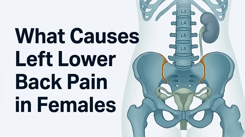
The left lower back refers to the region below the left 12th rib and above the left gluteal fold, corresponding roughly to the area overlying the L3 through S1 vertebral levels and the left iliac crest. Within and near this zone, several anatomically distinct structures can generate pain: the L4-L5 and L5-S1 intervertebral discs, the left sacroiliac joint, the left quadratus lumborum muscle, the left psoas major muscle, the left kidney (which sits between the T12 and L3 vertebrae in the retroperitoneal space), the left ureter, the left ovary, the left uterosacral ligament, and the sigmoid colon.
What makes this clinically complex is that pain from several of these structures is not felt where the structure itself sits. It is felt in the lower back because of a neurological process called viscero-somatic convergence, where sensory fibers from internal organs and sensory fibers from the back muscles share the same relay points in the spinal cord. The brain cannot reliably distinguish which source triggered the signal.
Key causes of left lower back pain in females, ranked by frequency of clinical presentation:
- Left-sided lumbar muscle strain or myofascial pain (most frequent overall cause)
- Left-sided lumbar disc herniation at L4-L5 or L5-S1
- Left sacroiliac joint dysfunction
- Left-sided sciatica (compression of the left L4, L5, or S1 nerve root)
- Dysmenorrhea (menstrual-cycle-related referred lumbar pain)
- Endometriosis affecting left adnexal structures or left uterosacral ligament
- Left ovarian cyst
- Left kidney infection (pyelonephritis) or left ureteral calculus
- Uterine fibroids (leiomyomata)
- Pelvic inflammatory disease
- Left colon pathology (diverticulitis, irritable bowel syndrome)
- Pregnancy-related sacroiliac joint laxity
Older women (over 60) face a different probability distribution across these causes, with degenerative disc disease at L4-L5 and L5-S1 more likely to drive left lower back pain than reproductive causes, while postmenopausal ovarian pathology becomes a clinically important consideration distinct from hormonal cycle-related causes.
Why Left Lower Back Pain Is Different in Females
Left lower back pain presents differently in females than in males because females have a distinct reproductive organ system in the pelvis whose nerve supply overlaps with the lumbar and sacral nerve pathways that serve the lower back.
The hypogastric plexus is a network of autonomic nerve fibers that innervates the uterus, ovaries, fallopian tubes, and upper vagina. Its sympathetic components travel with spinal cord levels T10 through L1, while its parasympathetic components arise from sacral cord levels S2 through S4. These are the same spinal cord levels that receive sensory signals from the left lower back, left hip, and left leg. When any pelvic organ in a female becomes inflamed, distended, or structurally disrupted, the visceral pain signals traveling through the hypogastric plexus converge at the dorsal horn of the spinal cord onto the same interneurons that handle somatic signals from the lumbar musculature. This is the anatomical basis of referred pain in females: the brain receives overlapping signals and interprets some of the pelvic organ pain as originating in the lower back.
Males do not have uterine or ovarian structures. Their pelvic organ innervation pattern (prostate, seminal vesicles, vas deferens) overlaps less extensively with the L1-L2 lumbar dermatomes and produces a different referred pain geography. This difference is not minor. The American College of Obstetricians and Gynecologists (ACOG) notes that gynecological conditions are frequently under-recognized as causes of chronic pelvic and lower back pain in women, contributing to diagnostic delays averaging 7 to 10 years for conditions like endometriosis.
The left side matters specifically because: the left ovary and its ligamentous attachments (the left infundibulopelvic ligament and left round ligament) are positioned closer to the left lumbar musculature than the right ovary is to the right lumbar musculature in most women, due to sigmoid colon positioning in the left lower quadrant. The sigmoid colon itself also sits entirely on the left side of the pelvis, adding another left-specific pain source absent on the right.
| Cause Category | Left-Side-Specific Anatomy | Pain Mechanism |
|---|---|---|
| Reproductive | Left ovary, left fallopian tube, left uterosacral ligament | Viscero-somatic convergence via hypogastric plexus T10-L1 |
| Urological | Left kidney, left ureter along left psoas major | Renal plexus T10-L1, ureteral colic referred to L1-L2 |
| Gastrointestinal | Sigmoid colon, descending colon | Colonic afferents via superior hypogastric plexus T10-L2 |
| Musculoskeletal | Left sacroiliac joint, left L4-L5/L5-S1 disc, left quadratus lumborum | Direct somatic pain via left lumbar and sacral nerve roots |
Left Lower Back Muscle Strain and Herniated Disc in Women
The most frequent single cause of left lower back pain in females is musculoskeletal injury, either left-sided lumbar muscle strain involving the quadratus lumborum or erector spinae muscle groups, or a posterolateral herniation of the L4-L5 or L5-S1 intervertebral disc compressing the left L4, L5, or S1 nerve root.
Think of the intervertebral disc like a water-filled cushion between each pair of vertebral bones. Its outer ring, the annulus fibrosus, is a tough collagen structure that holds the inner gel, the nucleus pulposus, under pressure. When the annulus develops a tear from repetitive loading, bending, or a single forceful event, the nucleus can push through. If it pushes posterolaterally to the left (the most anatomically common direction), it compresses whichever nerve root is exiting the spinal canal on the left at that level. Compression of the left L5 nerve root produces sharp or burning pain radiating from the left lower back down the left lateral leg to the left foot. Compression of the left S1 root produces pain along the left posterior thigh and into the left heel.
Pure muscular strain of the left quadratus lumborum (a flat muscle running from the 12th rib and the L1-L4 transverse processes down to the left iliac crest) produces a localized, achy, non-radiating pain in the left lower back that worsens with lateral bending toward the right or with twisting. Distinguishing this from a disc herniation matters clinically because disc herniation with nerve root compression requires different management.
The American Academy of Family Physicians (AAFP) states that acute low back pain from musculoskeletal sources in adults without red flags is typically self-limiting over 4 to 6 weeks in many patients, though this applies specifically to adults without nerve compression symptoms. Women who experience left lower back pain with concurrent left leg pain, numbness, or weakness in the left foot are not in the self-limiting category and require evaluation by a primary care physician or a board-certified spine specialist.
Women with scoliosis (lateral curvature of the lumbar spine) are at higher risk for asymmetric disc loading that predisposes to left-sided disc herniation if the curvature places greater compressive force on the left side of the L4-L5 or L5-S1 disc. A primary care physician evaluating left-sided back pain with radiculopathy in a woman should ask about prior scoliosis diagnosis and any prior spinal imaging.
Quick Tip:
- Left lower back pain from a herniated disc typically includes a radiating component (pain traveling into the left buttock, hip, or leg) that pure muscle strain does not produce
- Pain that worsens with sitting and improves with walking is more consistent with lumbar disc herniation than with pure muscle strain, which typically worsens with movement
- Women who are breastfeeding face restrictions on some imaging modalities and pain medications; a primary care physician should be informed of breastfeeding status before any diagnostic workup or treatment is initiated
Sacroiliac Joint Dysfunction and Sciatica on the Left Side in Women
Left-sided sacroiliac (SI) joint dysfunction is a disproportionately common cause of left lower back pain in females compared with males, driven by the wider female pelvis, greater laxity of pelvic ligaments throughout the menstrual cycle, and the repeated ligamentous stretching that occurs during pregnancy and the postpartum period.
The left sacroiliac joint connects the left sacrum (the triangular bone at the base of the spine) to the left ilium (the upper blade of the pelvis). It is stabilized by the iliolumbar ligament, the anterior sacroiliac ligament, and the posterior sacroiliac ligament, all of which undergo cyclic hormonal influence in females. When these ligaments become lax, inflamed, or mechanically stressed, the joint produces a deep, aching pain in the left lower back, left buttock, and sometimes the left posterior thigh, typically worsening with prolonged standing, stair climbing, or rolling over in bed.
Sciatica on the left side occurs when the left sciatic nerve (formed by the L4, L5, S1, S2, and S3 nerve roots exiting the left side of the sacral plexus) is compressed or irritated. In most cases, this compression occurs at the level of the lumbar disc, but in a subset of women it occurs at the level of the piriformis muscle in the left gluteal region. The piriformis muscle originates from the anterior sacrum and inserts on the greater trochanter of the femur, and the sciatic nerve passes either beneath or through it. Inflammation or spasm of the left piriformis compresses the sciatic nerve, producing left lower back and buttock pain with radiation down the left posterior thigh.
The FABER test (Flexion, ABduction, and External Rotation of the hip) and the FADIR test (Flexion, ADduction, and Internal Rotation) are physical examination maneuvers a clinician uses to differentiate sacroiliac joint pain from hip joint pathology and lumbar disc-related sciatica. A positive FABER test (pain in the left sacroiliac joint region during this maneuver) points toward SI joint dysfunction rather than a disc herniation, which is more reliably identified by the straight leg raise test.
Women who have delivered a baby within the previous 12 months have a measurably higher rate of left-sided or bilateral SI joint dysfunction, as documented in research published in the Spine journal, because of the residual ligamentous laxity from the relaxin-driven changes of pregnancy. A woman in the first postpartum year who presents with persistent left lower back pain that is worse with position changes and bears the distribution pattern described above should be evaluated for postpartum SI joint dysfunction by a primary care physician or a board-certified physical medicine and rehabilitation specialist, and referred to a pelvic floor physical therapist with obstetric specialization.
Key Takeaway: Left-sided sacroiliac joint dysfunction is more common in females than males and is driven by hormonally influenced ligament laxity; it produces left lower back and buttock pain that worsens with position changes, not with forward bending, which distinguishes it from lumbar disc herniation.
Menstrual Cycle and Left Lower Back Pain
Left lower back pain during or just before menstruation is caused by prostaglandin F2-alpha (PGF2-alpha) and prostaglandin E2 (PGE2) released by endometrial cells as the uterine lining begins to shed, triggering uterine smooth muscle contractions and referred pain in the lumbar region via the spinal dorsal horn.
The prostaglandin mechanism works like this: as progesterone levels drop at the end of the luteal phase, endometrial stromal cells activate the cyclooxygenase-1 and cyclooxygenase-2 (COX-1 and COX-2) enzyme pathways, which convert arachidonic acid into prostaglandins. PGF2-alpha binds to receptors on uterine smooth muscle, causing forceful, rhythmic contractions that temporarily reduce uterine blood flow. The resulting uterine ischemia (inadequate blood supply) activates pain-sensing nerve fibers (nociceptors) in the uterine wall. These nociceptive signals travel through the hypogastric plexus (T10-L1 sympathetic pathway) to the dorsal horn of the spinal cord at the T10-L1 levels, where they converge with somatic signals from the lumbar muscles and lower back. PGE2 simultaneously sensitizes pain receptors at the spinal level, amplifying the total pain experience.
The pain is perceived in the lower back because the dorsal horn neurons that receive these visceral signals also receive input from the skin and muscles of the lower back and lower abdomen. This is the same mechanism that causes referred pain in other visceral conditions, such as shoulder tip pain from diaphragmatic irritation.
Primary dysmenorrhea refers to menstrual pain without an identifiable pelvic pathology, driven purely by this prostaglandin mechanism. According to ACOG, primary dysmenorrhea affects 50 to 90 percent of menstruating people, making it one of the most prevalent causes of recurrent lower back and pelvic pain in females of reproductive age. Left lower back pain that appears consistently 12 to 24 hours before menstrual flow begins and resolves within 2 to 3 days of onset, with no associated fever, abnormal discharge, or pain outside the menstrual window, fits the pattern of primary dysmenorrhea-related referred lumbar pain.
Secondary dysmenorrhea refers to menstrual pain caused by an underlying pelvic pathology such as endometriosis, adenomyosis, or fibroids. Pain that is worsening cycle over cycle, that begins earlier than the onset of flow, or that persists beyond the period itself warrants evaluation by an obstetrician-gynecologist to rule out secondary causes.
Adolescents with primary dysmenorrhea often experience left lower back pain as a prominent symptom alongside lower abdominal cramping, because young women frequently have higher prostaglandin production during menstruation compared with older women, according to research published in Obstetrics and Gynecology. This population should be evaluated for both primary and secondary causes if pain consistently disrupts school or daily activities.
Endometriosis Causing Left Lower Back Pain
Endometriosis causes left lower back pain when endometrial-like tissue implants on the left uterosacral ligament, left ovary, left pelvic peritoneum, or in rare cases the left ureter or left colon, triggering a cycle of inflammation, fibrosis, and nerve sensitization that produces chronic pelvic and lumbar pain.
Endometriosis (ICD-10 N80) occurs when tissue that resembles the endometrial lining of the uterus grows outside the uterine cavity. The most widely supported mechanistic theory is retrograde menstruation: during menstruation, some endometrial cells flow backward through the fallopian tubes into the pelvic cavity rather than out through the cervix. In women with endometriosis, these cells implant on pelvic surfaces and continue to respond to monthly hormonal fluctuations, proliferating during the estrogen-dominant follicular phase and breaking down during the progesterone-withdrawal menstrual phase. This monthly breakdown causes local bleeding, inflammatory cytokine release (interleukin-1 beta, interleukin-6, tumor necrosis factor alpha), and progressive fibrotic scarring.
The left uterosacral ligament, which runs from the posterior cervix to the left sacrum, is one of the most clinically important sites of endometriotic implantation. When endometrial tissue implants here, it sensitizes the sacral nerve plexus and the inferior hypogastric plexus, producing deep left-sided pelvic pain and left lower back pain that characteristically worsens in the second half of the menstrual cycle and during menstruation itself, and may be provoked by deep penetration during sexual intercourse (dyspareunia).
The Endometriosis Foundation of America reports that endometriosis affects an estimated 10 percent of females of reproductive age globally, approximately 190 million people, and that the average diagnostic delay from symptom onset to confirmed diagnosis is 7 to 10 years. Left lower back pain is one of the most frequently underattributed symptoms of endometriosis, particularly because it can persist between menstrual cycles in women with fibrotic or deep infiltrating endometriosis affecting the left uterosacral ligament or left sacral nerve plexus.
A woman experiencing left lower back pain that cycles with her menstrual pattern, worsens with menstruation, causes pain with sex, and has been present for more than six months without a clear musculoskeletal explanation should ask her obstetrician-gynecologist about evaluation for endometriosis, including a pelvic examination, transvaginal ultrasound (for endometriomas), and discussion of diagnostic laparoscopy, which remains the definitive diagnostic tool for peritoneal and ligamentous endometriotic implants.
Ovarian Cyst and Left Lower Back Pain in Women
Left lower back pain caused by a left ovarian cyst occurs when the cyst stretches the left ovarian capsule, compresses adjacent pelvic structures, or, in the case of cyst rupture or torsion, triggers an acute inflammatory response that activates pain fibers running through the left ovarian plexus (sympathetic fibers from T10 to L1).
An ovarian cyst (ICD-10 N83) is a fluid-filled sac on or within the left ovary. Functional cysts, the most common type, include follicular cysts (from an ovarian follicle that fails to release an egg and continues to enlarge) and corpus luteum cysts (from the follicle that releases the egg, which then fills with fluid rather than resolving normally). Most functional cysts are smaller than 5 centimeters and resolve spontaneously within one to three menstrual cycles without causing persistent pain.
Cysts that produce left lower back pain are typically those that have grown large enough to compress surrounding pelvic structures, that have developed internal complications (hemorrhage into the cyst), or that are causing ovarian torsion, a surgical emergency in which the ovary twists on its ligamentous stalk, cutting off its blood supply. The pain of ovarian torsion is characteristically sudden, severe, and one-sided, often accompanied by nausea and vomiting.
The T10-L1 origin of the ovarian plexus explains why left ovarian pathology produces pain in the left lower back and left flank rather than purely in the left lower abdomen. The brain receives the pain signal from the T10-L1 dorsal horn and maps it to the broad dermatome zone served by these levels, which includes the left lower back, left hip, and left inguinal region.
The American College of Obstetricians and Gynecologists states that ovarian cysts detected incidentally on imaging in premenopausal women are present in up to 8 percent of examinations, the majority of which are functional and resolve without intervention. However, cysts in postmenopausal women carry a different clinical significance and require evaluation by a gynecologist, as the risk of ovarian malignancy increases substantially after menopause.
Women who experience sudden, severe left lower back or left pelvic pain with nausea should be evaluated in an emergency setting, as this presentation pattern is consistent with ovarian torsion, which requires surgical intervention within hours to preserve ovarian function.
Key Takeaway: Left ovarian cyst pain in the lower back is explained by the ovarian plexus arising from T10-L1, the same spinal levels that receive lower back sensory input; cyst-related left lower back pain that is sudden and severe with nausea requires emergency evaluation for ovarian torsion.
Uterine Fibroids and Pelvic Inflammatory Disease as Left Back Pain Causes
Uterine fibroids (leiomyomata, ICD-10 D25) and pelvic inflammatory disease (PID, ICD-10 N73) both cause left lower back pain through distinct but related mechanisms involving pelvic organ distension, inflammatory nerve sensitization, and mass effect on adjacent pelvic and lumbar structures.
Uterine fibroids are benign smooth muscle tumors of the myometrium (the muscular wall of the uterus). They grow under the influence of estrogen and progesterone, which is why they expand during reproductive years, often peak in the perimenopausal decade, and typically shrink after menopause when ovarian estrogen production declines. Subserosal fibroids (those growing on the outer surface of the uterus) and posterior fibroids (those embedded in the posterior uterine wall or broad ligament) are the most likely to produce left lower back pain because of direct mechanical pressure on the uterosacral ligaments, the sacral nerve plexus, and the posterior pelvic peritoneum. A large posterior fibroid can also compress the left ureter, producing secondary left-sided hydronephrosis and left flank pain.
Pelvic inflammatory disease is an ascending polymicrobial infection of the upper female reproductive tract, typically involving the uterus, fallopian tubes, and ovarian surfaces. The most frequently implicated organisms include Neisseria gonorrhoeae and Chlamydia trachomatis, though non-sexually transmitted bacteria including Bacteroides species and Escherichia coli are also recovered. PID produces left lower back pain through two mechanisms: direct inflammatory sensitization of the pelvic peritoneum adjacent to the left adnexa, and referred pain via the hypogastric plexus T10-L1 pathway.
According to the Centers for Disease Control and Prevention (CDC), more than 1 million cases of pelvic inflammatory disease are diagnosed annually in the United States, with the highest incidence in women aged 15 to 24. PID that involves a left-sided tubo-ovarian abscess (a collection of pus around the left fallopian tube and ovary) produces severe left lower back and pelvic pain accompanied by fever above 38.3 degrees Celsius (101 F), abnormal vaginal discharge, and pain with cervical motion on pelvic examination. This presentation requires same-day evaluation by a gynecologist or emergency medicine physician, as untreated tubo-ovarian abscess can progress to rupture and sepsis.
Women taking combined oral contraceptives experience substantially lower rates of pelvic inflammatory disease because the progestin-thickened cervical mucus reduces ascending bacterial infection, as documented in a long-term study published in the British Journal of Obstetrics and Gynaecology. However, hormonal contraceptive use does not eliminate PID risk entirely, particularly in women with intrauterine devices (IUDs) in the weeks immediately following insertion.
Left Kidney and Ureter Causes of Left Lower Back Pain
Left lower back pain from the left kidney or left ureter is caused by inflammatory distension of the renal capsule (in pyelonephritis or hydronephrosis) or by the forceful ureteral smooth muscle contractions triggered by a calculus obstructing the left ureter, with pain transmitted through the left renal plexus (T10-L1 sympathetic fibers) to the left flank and lower back.
The left kidney sits in the retroperitoneal space between the T12 and L3 vertebral levels, immediately posterior to the left quadratus lumborum muscle and lateral to the left psoas major. Its anatomical position means that renal capsular distension directly presses against the posterior abdominal wall musculature and the overlying thoracolumbar fascia, producing a deep, aching pain in the left costovertebral angle (the angle between the left 12th rib and the lateral border of the erector spinae muscles) that radiates downward toward the left lower back.
Left pyelonephritis (kidney infection) typically produces a constant, dull-to-severe left costovertebral angle and left lower back pain accompanied by fever, chills, nausea, and urinary symptoms (dysuria, frequency, urgency). This distinguishes renal pain from pure musculoskeletal left lower back pain, which is not accompanied by fever or urinary changes. The costovertebral angle (CVA) tenderness test, performed by percussing gently over the left CVA angle, reproduces or worsens renal pain and is a standard physical examination finding in pyelonephritis.
Left ureteral calculi (kidney stones lodged in the left ureter) produce a different pain character: colicky, meaning it comes in waves of severe intensity, radiating from the left flank downward toward the left lower back, left groin, and left labia in females. This referred pain pathway follows the path of the left ureter as it descends along the left psoas major, crosses the left iliac vessels, and enters the posterior bladder wall. According to the National Institute of Diabetes and Digestive and Kidney Diseases (NIDDK), the lifetime risk of kidney stones is approximately 11 percent in males and 9 percent in females in the United States, with recurrence rates of approximately 50 percent within 10 years after a first stone event.
Women who are pregnant face additional risk: right-sided hydronephrosis is more common in pregnancy due to uterine pressure on the right ureter, but left-sided ureteral dilation and left lower back pain from urinary stasis can also occur. Urinary tract infections are more common in pregnancy due to the dilated, stasis-prone renal collecting system, and pyelonephritis in pregnancy carries an increased risk of preterm labor, requiring prompt antibiotic treatment after consultation with an obstetrician.
| Kidney or Ureter Condition | Characteristic Pain Pattern | Associated Symptoms | Diagnostic Test |
|---|---|---|---|
| Left pyelonephritis | Constant, left CVA and lower back, worsened by CVA percussion | Fever, chills, dysuria, frequency | Urinalysis, urine culture |
| Left ureteral calculus | Colicky waves, left flank to left groin and labia | Nausea, blood in urine (hematuria) | CT scan abdomen/pelvis without contrast |
| Left hydronephrosis | Dull persistent left flank and lower back | May have no urinary symptoms initially | Renal ultrasound, CT scan |
| Left renal abscess | Severe, unrelenting left lower back and flank | High fever, rigors, unwell appearance | CT scan with contrast, blood cultures |
Key Takeaway: Left kidney pain is distinguished from left lower back muscle pain by the presence of fever, nausea, or urinary symptoms, while left ureteral stone pain is colicky and travels from the flank toward the groin, not staying localized to the back like muscle pain does.
Left Colon Causes of Left Lower Back Pain in Females
Left lower back pain caused by left-sided colonic pathology (diverticulitis, irritable bowel syndrome, constipation, or inflammatory bowel disease) occurs because the descending colon and sigmoid colon occupy the left side of the abdominal and pelvic cavity, and their visceral afferent nerve supply travels through the superior hypogastric plexus (T10-L2 sympathetic pathway) to the same spinal cord segments that receive somatic input from the left lower back.
Sigmoid diverticulitis is one of the most clinically important left-sided colonic causes of left lower back pain in females, particularly in women over 40. Diverticula are small pouches that form in weak spots in the muscular wall of the sigmoid colon. When one of these pouches becomes obstructed by fecal matter and subsequently infected, the resulting inflammation (diverticulitis) produces left lower abdominal pain, left lower back pain, fever, and a change in bowel habits (constipation or diarrhea). The pain of sigmoid diverticulitis is typically constant rather than colicky, located in the left lower quadrant and referred to the left lower back, and accompanied by tenderness on palpation of the left lower abdomen.
Irritable bowel syndrome (IBS) produces left lower back pain in a subset of affected women through two mechanisms: gaseous distension of the descending and sigmoid colon (activating stretch receptors whose afferents converge with the left lumbar somatic pathway), and central sensitization of pain processing that amplifies both visceral and somatic signals from the lower body. Research published in the Journal of Pain has found that women with IBS are three times more likely than men to report concurrent lower back pain, reflecting the combined contribution of viscero-somatic referred pain and sex-based differences in central pain sensitization.
Severe constipation with a large fecal mass in the descending colon can produce left lower back pressure and aching that resolves after defecation, which is a helpful diagnostic clue. Pain that consistently improves after a bowel movement and worsens with abdominal distension strongly suggests a colonic rather than a musculoskeletal or reproductive source.
Women with inflammatory bowel disease (IBD), specifically Crohn’s disease or ulcerative colitis, may also experience left lower back pain from two distinct mechanisms: direct colonic inflammation with referred visceral pain, and IBD-associated sacroiliitis (inflammation of the sacroiliac joint), which occurs as an extra-intestinal manifestation in approximately 10 to 20 percent of IBD patients, according to research published in the American Journal of Gastroenterology. Women with IBD and left lower back pain that has a constant, morning-stiff quality that improves with movement warrant rheumatological evaluation for IBD-associated spondyloarthropathy.
Left Lower Back Pain During Pregnancy
Left lower back pain during pregnancy occurs through multiple overlapping mechanisms, including relaxin-mediated sacroiliac joint laxity, mechanical load redistribution as the gravid uterus shifts the body’s center of gravity forward, compression of the left lumbar structures by the enlarging uterus, and increased left ureteral compression producing left-sided hydronephrosis and renal back pain.
Relaxin, a peptide hormone produced initially by the corpus luteum and later by the placenta and decidua, rises sharply in the first trimester and remains elevated throughout pregnancy. Relaxin acts on receptors in the pubic symphysis, sacroiliac joint capsule, and pelvic ligament fibroblasts, stimulating collagenase enzymes that reduce collagen cross-linking and increase ligamentous compliance. This physiological loosening of the pelvic ligaments allows the pelvis to expand during delivery but simultaneously reduces sacroiliac joint stability during the entire pregnancy.
The left sacroiliac joint often bears asymmetric load because of the natural tendency of the pregnant uterus to tilt slightly to the right (dextrorotation), placing greater mechanical demand on the left posterior pelvic structures. Left lower back and buttock pain that worsens with walking, stair climbing, or turning over in bed and is localized to the left posterior iliac crest region fits the pattern of pregnancy-related pelvic girdle pain (PRPGP), which a systematic review in the Spine journal found affects approximately 50 percent of pregnant women in some form during the second and third trimesters.
Left lower back pain in pregnancy that is accompanied by fever, rigors, nausea, vomiting, or urinary symptoms must not be attributed to musculoskeletal causes without evaluation. Pyelonephritis is one of the most serious infections during pregnancy and is more likely to involve the left kidney in the second trimester when uterine growth begins to compress pelvic structures. Any pregnant woman with fever and left lower back or left flank pain should contact her obstetrician immediately.
Pregnant women experiencing left lower back pain in the third trimester who notice a sudden change in pain character (from dull and positional to constant and severe), particularly if accompanied by lower abdominal tightening, should be evaluated for preterm labor by their obstetrician, as uterine contraction pain can be perceived as lower back pain in some presentations.
Left Lower Back Pain in Perimenopause and Postmenopause
Left lower back pain becomes more prevalent and more structurally driven in women during perimenopause (the 2 to 10 years preceding the final menstrual period) and postmenopause, because declining estrogen directly affects the structural integrity of the lumbar intervertebral discs, the trabecular bone of the lumbar vertebral bodies, and the cartilaginous surfaces of the left sacroiliac joint.
Estrogen receptors are present in the cells of the nucleus pulposus (the gel-filled inner core of the intervertebral disc) and in chondrocytes lining articular surfaces. Estrogen promotes proteoglycan synthesis in the nucleus pulposus, helping maintain disc hydration and height. As estrogen levels decline during perimenopause, proteoglycan content in the L4-L5 and L5-S1 discs decreases, the discs lose water content and height, and the annulus fibrosus becomes more susceptible to annular tears. This accelerated disc degeneration is one reason why new-onset degenerative disc disease-related left lower back pain frequently begins or worsens in women during their mid-40s and 50s.
The North American Menopause Society (NAMS) notes that postmenopausal women have a substantially higher rate of vertebral compression fractures compared with premenopausal women, driven by estrogen deficiency-related osteoporosis (accelerated bone resorption by osteoclasts that were previously suppressed by estrogen). A compression fracture of the L3 or L4 vertebral body on the left side can produce acute or insidious left lower back pain in a postmenopausal woman who may not have experienced a traumatic fall, as vertebral fragility fractures frequently occur with minimal force.
Left lower back pain in a postmenopausal woman that is new in onset, constant, and not related to movement or position warrants prompt evaluation. Unlike musculoskeletal pain, which typically varies with activity, vertebral compression fracture pain can be persistently present at rest and worsened only minimally by position change. A primary care physician should order a dual-energy X-ray absorptiometry (DEXA) scan to assess bone mineral density in women over 65 who have not yet been screened, or earlier in women with risk factors including low body weight, prior fracture, smoking history, or long-term glucocorticoid use.
Left ovarian pathology also remains clinically relevant in postmenopausal women. New-onset left lower back pain in a postmenopausal woman accompanied by a pelvic mass detected on examination warrants urgent referral to a gynecologist-oncologist for evaluation of ovarian malignancy, as postmenopausal ovarian cysts with solid components or elevated CA-125 serum levels carry a measurably higher risk of epithelial ovarian cancer than premenopausal ovarian cysts.
Key Takeaway: In perimenopausal and postmenopausal women, declining estrogen accelerates lumbar disc degeneration and increases vertebral fracture risk, making new-onset left lower back pain in this life stage a reason for prompt evaluation by a primary care physician rather than self-management.
Hormonal Mechanisms Behind Left Lower Back Pain in Females
Several distinct hormonal pathways act on lumbar structures, pelvic organs, and pain-processing systems in ways that have no equivalent in males, making left lower back pain in females a partially hormone-driven phenomenon across multiple life stages.
Prostaglandin F2-alpha (from endometrial cells via COX-1 and COX-2) drives the uterine smooth muscle contractions and ischemia that produce menstrual-cycle-related referred lumbar pain, as described in the menstrual cycle section. Beyond the menstrual cycle, prostaglandin upregulation in endometriosis lesions sustains chronic pelvic and lumbar neuroinflammation outside the menstrual window.
Relaxin acts not only during pregnancy but also at lower concentrations during the luteal phase of the menstrual cycle (the two weeks after ovulation). Research published in the Journal of Musculoskeletal Medicine has found that sacroiliac joint laxity measurably increases during the luteal phase in women, particularly those with pre-existing pelvic girdle instability, explaining why some women experience worse left lower back pain in the second half of their menstrual cycle even when not pregnant.
Cortisol, the primary glucocorticoid produced by the adrenal cortex via the hypothalamic-pituitary-adrenal (HPA) axis, amplifies central pain sensitization when chronically elevated. Women with hypothalamic-pituitary-adrenal axis dysregulation from chronic stress, sleep disruption, or conditions like polycystic ovary syndrome (PCOS) may have altered cortisol diurnal rhythms that lower pain thresholds and make both musculoskeletal and visceral left lower back pain more intense and harder to manage with standard analgesics. The National Institute of Mental Health (NIMH) reports that women are approximately twice as likely as men to develop anxiety and stress-related conditions that dysregulate the HPA axis, which has measurable downstream effects on musculoskeletal pain processing.
The interaction between these hormonal pathways means that left lower back pain in females is frequently not a single-source problem. A woman with both lumbar disc disease and endometriosis may experience her disc-related pain amplified during menstruation by prostaglandin-driven central sensitization, even though the two conditions are anatomically unrelated.
Quick Tip:
- Women who track their menstrual cycle alongside their left lower back pain onset and severity can provide a clinician with genuinely useful diagnostic information about whether a hormonal or reproductive pattern is contributing
- Pain that is consistently worse in the 3 to 5 days before menstruation and the first 2 days of flow, then substantially improves, points toward a prostaglandin-mediated or endometriosis-related cause rather than a fixed structural one
- Women with PCOS who also report left lower back pain should mention this connection to their primary care physician or obstetrician-gynecologist, as HPA axis dysregulation in PCOS may be contributing to pain sensitization independent of pelvic structural pathology
Red Flags for Left Lower Back Pain in Women
Certain left lower back pain presentations in females are clinical red flags: symptom features that signal a potentially serious, time-sensitive, or anatomically dangerous underlying cause that requires prompt medical evaluation rather than home management or watchful waiting.
Red flag symptoms that require evaluation by a primary care physician or obstetrician-gynecologist within 24 to 48 hours:
- Left lower back pain with fever above 38 degrees Celsius (100.4 F) without a clear respiratory or viral illness explanation (raises concern for pyelonephritis, pelvic inflammatory disease, or tubo-ovarian abscess)
- Left lower back pain that begins or worsens in the second half of the menstrual cycle and is accompanied by pain with sexual intercourse (raises concern for endometriosis or left adnexal pathology)
- New left lower back pain in a postmenopausal woman with no prior back pain history (raises concern for vertebral compression fracture, ovarian malignancy, or metastatic disease)
- Left lower back pain accompanied by abnormal vaginal bleeding outside the expected menstrual cycle
- Left lower back pain with left leg swelling and warmth (raises concern for deep vein thrombosis, particularly in women using combined oral contraceptives or during the postpartum period)
Red flag symptoms requiring same-day evaluation by a primary care physician or emergency medicine physician:
- Left lower back pain with high fever (above 39 degrees Celsius / 102.2 F), shaking chills, and nausea or vomiting (raises concern for pyelonephritis with sepsis features)
- Sudden onset severe left lower back and left pelvic pain with nausea, not relieved by position change (raises concern for ovarian torsion, requiring surgical evaluation within hours)
- Left lower back or flank pain with blood in the urine (hematuria) and waves of severe pain (typical presentation of left ureteral calculus)
- Left lower back pain with new left leg weakness, left foot drop, or difficulty walking (raises concern for significant left lumbar nerve root compression requiring urgent imaging)
The American Academy of Family Physicians clinical recommendations for lower back pain include bilateral leg weakness, saddle anesthesia (numbness in the perineum, inner thighs, and buttocks), and loss of bladder or bowel control as absolute emergency red flags for cauda equina syndrome, a surgical emergency caused by compression of the cauda equina nerve roots within the lumbar spinal canal below L1-L2.
Women who are pregnant and experience any new or worsening left lower back pain accompanied by uterine tightening, regular cramping, or a change in fetal movement should contact their obstetrician immediately rather than waiting for a scheduled appointment.
Emergency Symptoms: When to Call 911 or Go to the ER
Certain presentations of left lower back pain in females require immediate emergency evaluation. Do not wait to see if these resolve on their own.
Call 911 or go to the nearest emergency room immediately if you experience:
- Sudden, severe left lower back and left pelvic pain with dizziness, lightheadedness, shoulder tip pain, or fainting, particularly if you may be pregnant (possible ectopic pregnancy rupture, which causes life-threatening internal hemorrhage)
- Severe left lower back pain with bilateral leg weakness, numbness in the inner thighs, perineum, or buttocks, or sudden loss of bladder or bowel control (possible cauda equina syndrome, a spinal surgical emergency)
- Left lower back or flank pain with high fever, shaking chills, rapid heart rate, confusion, or altered mental status (possible septic pyelonephritis or sepsis from a left-sided tubo-ovarian abscess)
- Sudden tearing or ripping left-sided back pain in a woman over 50 with a history of high blood pressure or known aortic disease (possible aortic aneurysm or aortic dissection, a life-threatening vascular emergency)
- Severe left lower back or left pelvic pain with sudden nausea and vomiting in a woman with a known ovarian cyst or in the absence of other explanation (possible ovarian torsion, which can cause irreversible ovarian necrosis within 4 to 6 hours without surgical intervention)
- Left lower back pain in a pregnant woman accompanied by heavy vaginal bleeding, severe abdominal pain, or sudden decrease in fetal movement
These presentations can indicate rupture of an ectopic pregnancy, cauda equina syndrome, sepsis, aortic emergency, or ovarian torsion, all of which require emergency medical assessment, not an urgent care appointment or a phone call.
How Left Lower Back Pain in Females Is Diagnosed
The diagnosis of left lower back pain in females begins with a detailed clinical history and physical examination and proceeds to targeted diagnostic testing based on the most likely causes identified from the history, with the specific tests ordered depending on whether the presentation points toward a musculoskeletal, reproductive, urological, or gastrointestinal source.
To describe your left lower back pain effectively to a primary care physician or obstetrician-gynecologist at your appointment:
- Describe the exact location precisely: is it in the left lower back itself, the left flank (between the left 12th rib and left iliac crest), the left buttock, or the left groin? Each location points toward different anatomical structures.
- Characterize the pain quality: is it dull and constant, sharp and stabbing, colicky (coming in waves of severe intensity), or crampy?
- Note the timing: does it correlate with your menstrual cycle? Does it worsen before, during, or after your period? Does it fluctuate or stay constant?
- Identify associated symptoms: fever, chills, urinary changes (burning, frequency, blood in urine), bowel changes, abnormal vaginal bleeding, pain with sex, leg pain, or numbness.
- Describe what worsens and what improves the pain: bending, sitting, walking, lying flat, urination, bowel movements.
- List relevant history: prior endometriosis, ovarian cysts, fibroids, kidney stones, inflammatory bowel disease, spinal problems, prior surgeries, current medications, contraceptive use, and pregnancy status.
| Diagnostic Test | What It Evaluates | When It Is Ordered |
|---|---|---|
| Urinalysis and urine culture | Left kidney infection, ureteral stone, blood in urine | Fever with back pain, urinary symptoms, suspected stone |
| Pelvic ultrasound (transabdominal) | Ovarian cysts, fibroids, uterine abnormalities | Suspected gynecological cause |
| Transvaginal ultrasound | Endometriomas, ovarian cysts, uterine structure detail | Suspected endometriosis, ovarian pathology |
| CT scan abdomen/pelvis without contrast | Ureteral calculus, renal anatomy | Suspected kidney stone (hematuria plus colicky pain) |
| CT scan abdomen/pelvis with contrast | Diverticulitis, abscess, pyelonephritis complications | Fever plus localized left lower quadrant pain |
| MRI lumbar spine | Disc herniation, nerve root compression, sacral pathology | Radiculopathy, leg weakness, neurological symptoms |
| FABER and FADIR tests (physical examination) | Sacroiliac joint dysfunction versus hip pathology | Left SI joint-pattern pain |
| Straight leg raise test | Left lumbar nerve root compression (L4-L5, L5-S1) | Left leg radiculopathy |
| DEXA scan | Bone mineral density for osteoporosis | Postmenopausal women, vertebral fracture risk |
| Laparoscopy | Definitive diagnosis of endometriosis, pelvic adhesions | Suspected endometriosis after imaging is inconclusive |
Diagnostic workup in pregnant women must account for radiation exposure. The American College of Obstetricians and Gynecologists states that MRI (without gadolinium contrast in the first trimester) and renal ultrasound are the preferred imaging modalities for evaluating left lower back pain in pregnancy when musculoskeletal causes cannot explain the clinical picture, reserving CT scanning for situations where the clinical benefit outweighs the radiation risk.
Frequently Asked Questions About Left Lower Back Pain in Females
What organ could cause left lower back pain in a woman?
The organs most likely to cause left lower back pain in a woman are the left kidney, left ovary, left fallopian tube, uterus (via referred pain), and the sigmoid colon.
Each of these organs has nerve supply that travels through spinal cord levels T10 to L1 or S2 to S4, which overlap with the somatic nerve supply to the left lower back, causing organ pain to be perceived as back pain through a process called viscero-somatic convergence.
New left lower back pain without a clear musculoskeletal cause, particularly if accompanied by fever, urinary symptoms, menstrual pattern changes, or pelvic symptoms, warrants evaluation by a primary care physician to determine which organ is involved.
Can endometriosis cause left lower back pain?
Yes, endometriosis can cause left lower back pain when endometrial-like tissue implants on the left uterosacral ligament, left pelvic peritoneum, or left ovary, sensitizing the sacral nerve plexus and inferior hypogastric plexus.
The Endometriosis Foundation of America reports that the average diagnostic delay for endometriosis is 7 to 10 years, and left lower back pain that cycles with the menstrual period, worsens during menstruation, and is accompanied by painful sex is one of the most frequently underattributed symptom patterns for this condition.
A woman with this symptom pattern should request a gynecological evaluation including transvaginal ultrasound and discussion of diagnostic laparoscopy, which remains the definitive diagnostic test.
How do I know if my left lower back pain is kidney or muscle?
Left lower back pain from the kidney is typically accompanied by fever, nausea, urinary symptoms such as burning or frequency, or blood in the urine, while left lower back muscle pain is worsened by specific movements, not accompanied by fever or urinary changes.
The costovertebral angle (CVA) tenderness test, in which percussion over the left CVA angle (below the left 12th rib, lateral to the spine) worsens pain, is a classic clinical sign of left kidney inflammation and helps distinguish renal pain from musculoskeletal pain that reproduces with muscle palpation.
Any left lower back pain accompanied by fever should be evaluated by a primary care physician on the same day rather than self-managed.
What causes left lower back pain before or during a period?
Left lower back pain before and during a period is caused by prostaglandin F2-alpha and prostaglandin E2 released from endometrial cells as they break down, triggering uterine muscle contractions and referred lumbar pain through the hypogastric plexus T10-L1 pathway.
This is the mechanism of primary dysmenorrhea, which according to the American College of Obstetricians and Gynecologists affects 50 to 90 percent of menstruating people.
Pain that is worsening cycle over cycle, begins before the period rather than with it, or persists beyond the first 2 to 3 days of flow warrants evaluation by an obstetrician-gynecologist to rule out endometriosis or other secondary causes.
Should I go to the ER for left lower back pain as a woman?
Go to the emergency room immediately if your left lower back pain is sudden and severe, accompanied by dizziness or fainting (possible ectopic pregnancy rupture), involves bilateral leg weakness or loss of bladder control (possible cauda equina syndrome), or is accompanied by high fever and confusion (possible sepsis).
Left lower back pain with nausea and vomiting in a woman with a known ovarian cyst also warrants emergency evaluation for ovarian torsion, which can cause irreversible ovarian damage within 4 to 6 hours without surgery.
Left lower back pain that is dull, positional, and unaccompanied by fever, neurological changes, or systemic illness can be evaluated by a primary care physician within 24 to 48 hours rather than requiring an emergency visit.
What causes left lower back pain in females during pregnancy?
Left lower back pain during pregnancy is most commonly caused by relaxin-mediated sacroiliac joint laxity, mechanical load redistribution as the uterus enlarges and shifts the body’s center of gravity forward, and left ureteral compression producing mild hydronephrosis.
Relaxin, produced by the corpus luteum and placenta, loosens the ligaments of the sacroiliac joint and pubic symphysis to prepare the pelvis for delivery, but this laxity reduces joint stability throughout pregnancy and produces left lower back and buttock pain that worsens with walking and position changes.
A pregnant woman with left lower back pain accompanied by fever, urinary symptoms, or regular cramping should contact her obstetrician immediately rather than attributing the pain to normal pregnancy-related changes.
What to Do Next
Left lower back pain in females has a specific set of causes that differ from lower back pain in the general population, and identifying the right category (musculoskeletal, reproductive, urological, or gastrointestinal) is the most practical first step toward effective evaluation. The symptom pattern surrounding the pain, its relationship to the menstrual cycle, the presence or absence of fever, and any associated urinary, bowel, or pelvic symptoms narrow the possible causes in ways that can be identified before any testing is done.
If your left lower back pain has lasted more than 2 weeks without a clear injury explanation, cycles with your period, is accompanied by any symptom from the red flags section, or is new in a postmenopausal context, contact a primary care physician. Bring a written description of when the pain started, how it relates to your cycle, what worsens and improves it, and any associated symptoms.
If any symptom from the emergency section applies to you right now, stop reading and act immediately. The window for surgical intervention in ovarian torsion, ectopic pregnancy rupture, and cauda equina syndrome is narrow, and the difference between acting in the first hour and waiting four hours can determine outcomes. You now have the clinical picture to recognize what you are dealing with.