What Causes Back Spasms: Muscles, Nerves & Red Flags
Back spasms are caused by involuntary, sustained contractions of the skeletal muscles running along the spine, and the reasons range from simple muscular strain to structural spinal problems, electrolyte deficits, nerve compression, and even visceral organ dysfunction. Understanding what causes back spasms in your specific case matters because the underlying mechanism determines how serious the spasm is, how long it will last, and whether self-management is appropriate or professional evaluation is overdue.
According to the National Institute of Arthritis and Musculoskeletal and Skin Diseases (NIAMS), back pain, including spasm, is one of the most common reasons Americans miss work and seek medical care, affecting approximately 80 percent of adults at some point in their lives. The American College of Physicians (ACP) published clinical practice guidelines in 2017, updated with supporting evidence through the mid-2020s, distinguishing acute from chronic low back pain and recommending that clinicians identify the specific underlying cause before managing the presentation.
This article covers every major and clinically relevant cause of back spasms, from the cellular mechanics of why a muscle locks up to the structural spinal problems that trigger protective guarding, the stress hormones that lower your spasm threshold, and the specific symptoms that mean the back pain you are feeling is not a muscle problem at all.
What Causes Back Spasms
Back spasms are caused when a combination of physical load, chemical disruption, structural irritation, or neurological stimulation forces the muscles supporting the spine into an involuntary, sustained contraction they cannot release on their own.
The simplest version: the muscle contracts, the signal telling it to relax either fails to arrive or is overridden, and the muscle locks up. That lockup can last seconds or days depending on what triggered it and whether the underlying cause resolves. The four broad categories of cause are muscular, structural, neurological, and systemic, and most people have more than one contributing factor at any given time.
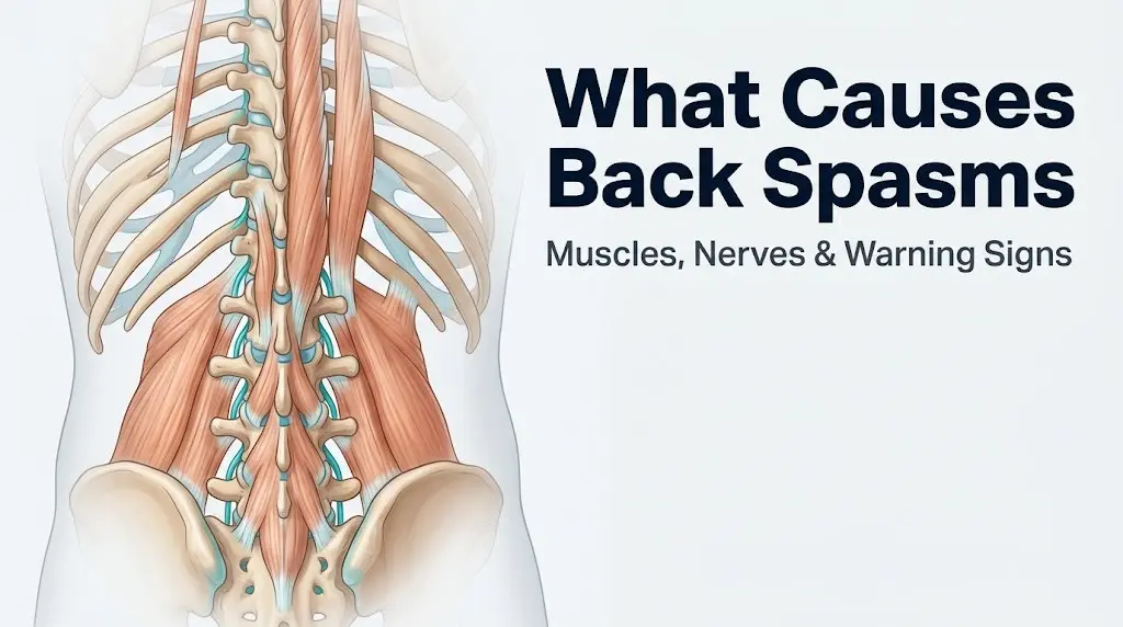
Muscular causes are the most common. Overloading or injuring the erector spinae muscle group (the long columns of muscle running on either side of the spine), the multifidus (a deep segmental stabilizer that runs along the vertebrae), or the quadratus lumborum (a thick rectangular muscle connecting the lower ribs to the iliac crest) produces local inflammation, nociceptor sensitization, and a protective spasm response. The muscle goes into spasm partly as a reflex to prevent further injury.
Structural causes involve the intervertebral discs, facet joints, or spinal canal. When a disc bulges or herniates, or when the spinal canal narrows due to spinal stenosis, nearby nerve roots are irritated. The surrounding muscles respond by contracting protectively around the irritated segment, producing spasm not from direct muscle injury but from neurological input.
Systemic causes include electrolyte deficits, dehydration, and, less commonly, inflammatory conditions or visceral organ referred pain. In these cases, the muscle itself is not injured. Its chemical environment or the nervous system signals it receives have been altered enough to trigger abnormal firing.
What Is a Back Spasm and How Does It Work
A back spasm is the involuntary, sustained contraction of one or more skeletal muscle groups in the back, produced when the normal cycle of muscle contraction and relaxation is disrupted at the cellular level.
Inside a normally functioning muscle cell, contraction begins when a motor neuron fires and releases acetylcholine at the neuromuscular junction. This triggers the release of calcium ions from the sarcoplasmic reticulum (the muscle cell’s internal calcium storage organelle), which allows myosin heads on the thick filaments to bind actin on the thin filaments, pulling the fibers shorter. Relaxation requires the sarcoplasmic reticulum to pump calcium back in, allowing the myosin-actin bond to release.
A spasm occurs when this release cycle fails. The sarcoplasmic reticulum cannot adequately resequester calcium, myosin stays bound to actin, and the muscle cannot shorten further but also cannot lengthen. Think of it like a zipper that has been pulled closed but the pull tab breaks off: the teeth are locked together and there is no mechanism to separate them without outside force. Simultaneously, adenosine triphosphate (ATP) depletion, whether from overuse, ischemia (reduced blood flow), or electrolyte disturbance, removes the energy needed for the myosin release step.
Local injury adds an inflammatory layer. Damaged muscle tissue releases prostaglandins, substance P, and interleukin-6, all of which sensitize local pain receptors and stimulate the gamma motor neuron reflex arc, amplifying motor neuron firing and making the spasm harder to break.
For older adults over 65, this cascade happens at lower thresholds because sarcopenia (age-related loss of muscle mass) reduces the ratio of contractile protein available to buffer stress, while reduced blood flow to paraspinal muscles means ischemic spasm risk is higher during even moderate activity.
What Causes Muscle Spasms in the Back
The most common cause of muscle spasms in the back is acute muscular strain, meaning a physical overload of the paraspinal muscles that tears myofibrils, triggers inflammation, and produces a protective spasm reflex.
Muscular strain happens in predictable scenarios: lifting a heavy object with poor spinal alignment (particularly flexion-rotation, which places asymmetric shear force on the multifidus and erector spinae), sudden awkward movement like twisting while carrying weight, or extended periods of static posture that fatigue muscles without giving them the circulation-driven recovery window they need. A 2020 study published in Spine found that combined lumbar flexion and rotation is the single highest-risk movement pattern for acute paraspinal muscle injury among working-age adults.
Dehydration is the second most common muscular cause. Skeletal muscle is approximately 75 percent water by weight. When total body water decreases by even two to three percent, intracellular fluid volume in muscle cells drops enough to concentrate intracellular electrolytes and disrupt calcium cycling in the sarcoplasmic reticulum. The result is a lowered threshold for spontaneous motor neuron firing and a higher risk of spasm during activity that would otherwise be well-tolerated.
Electrolyte imbalances matter separately from dehydration. Magnesium deficiency reduces the inhibitory tone on motor neuron excitability. Potassium deficiency disrupts the resting membrane potential of muscle cells, making them more likely to fire spontaneously. Sodium deficiency (hyponatremia) destabilizes the electrical gradient across the muscle cell membrane. Any of these deficits, alone or in combination, can produce spontaneous spasm in otherwise healthy muscle.
Key muscular causes of back spasms:
- Acute paraspinal muscle strain from overload or improper lifting mechanics
- Static posture fatigue from prolonged sitting or standing without postural variation
- Dehydration reducing intracellular fluid and disrupting calcium cycling
- Magnesium deficiency lowering motor neuron inhibitory tone
- Potassium deficiency disrupting resting membrane potential
- Post-exercise delayed onset muscle soreness triggering secondary protective spasm
- Athletic overtraining without adequate recovery, common in runners and weightlifters
Women who are postmenopausal should know that decreased estrogen levels are associated with reduced skeletal muscle mass and reduced bone density, both of which shift load patterns onto the paraspinal muscles and increase susceptibility to strain-triggered spasm at lower activity thresholds than younger women.
What Causes Lower Back Spasms
Lower back spasms are caused most often by strain or irritation of the muscles and structures within the lumbar spine region, specifically the quadratus lumborum, the lower segments of the erector spinae, the multifidus at the L4-L5 and L5-S1 levels, and the iliopsoas, a deep hip flexor that shares its lumbar attachment points with the lumbar vertebrae.
The lumbar spine carries the majority of the body’s axial load. Every step you take, every time you sit, every lift you perform, the L1 through L5 vertebral bodies and the discs between them absorb force. When the muscles supporting this region fatigue or are overloaded, the lumbar spine becomes mechanically unstable at a segmental level, and the paraspinal muscles respond with protective spasm to prevent dangerous motion at the affected segment.
The sacroiliac joint, where the sacrum connects to the iliac bones of the pelvis, is a frequently overlooked source of lower back spasm triggers. When the sacroiliac joint is inflamed or hypermobile, it alters load transfer from the legs to the spine, placing eccentric demands on the quadratus lumborum and lower erector spinae that often trigger spasm on one or both sides.
| Cause | Anatomical Structure Involved | Spasm Location | Clinical Distinguishing Feature |
|---|---|---|---|
| Muscular strain | Quadratus lumborum, erector spinae | Bilateral or unilateral lumbar | Worsens with movement, improves with rest initially |
| Disc herniation (L4-L5, L5-S1) | Herniated nucleus pulposus, lumbar nerve root | Unilateral lumbar, may radiate | Associated leg pain or numbness |
| Sacroiliac joint dysfunction | Sacroiliac joint, gluteal muscles | Unilateral, near sacrum | Worsens with single-leg stance, sits-to-stands |
| Iliopsoas tightness | Iliopsoas, lumbar attachments | Deep lumbar, anterior hip | Worsens with prolonged sitting, hip extension stretches |
| Lumbar spinal stenosis | Spinal canal, nerve roots | Bilateral lumbar, radiates to legs | Worsens walking, relieved by bending forward |
Adolescents experiencing lower back spasms should be evaluated for spondylolysis (a stress fracture of the vertebral arch, most common at L5), particularly if they are involved in extension-dominant sports like gymnastics, football, or wrestling, as the American Academy of Orthopaedic Surgeons (AAOS) identifies this group as at elevated risk.
Key Takeaway: Most lower back spasms originate from overloaded or fatigued paraspinal muscles at the lumbar level, but spasms accompanied by leg pain, numbness, or weakness point to a disc or nerve root cause that warrants evaluation by a primary care physician or board-certified physiatrist.
What Causes Back Spasms on Right Side
Right-sided back spasms specifically can reflect several causes that do not apply equally to the left side, including asymmetric loading of the right quadratus lumborum in right-hand-dominant individuals, proximity of the right kidney and ureter, the position of the ascending colon, and referred pain pathways from the liver or gallbladder.
From a purely muscular standpoint, right-dominant individuals (approximately 90 percent of the population) habitually load their right side more heavily during daily tasks. The right quadratus lumborum and right lower erector spinae compensate for this asymmetric loading pattern chronically, making them more prone to fatigue-triggered spasm under the same total load that would not affect the left side.
The right kidney sits in the right posterior retroperitoneal space, directly adjacent to the paraspinal muscles at approximately the T12-L2 level. A right kidney stone moving through the right ureter, a right kidney infection (pyelonephritis), or a right renal cyst can all produce pain that is experienced as right-sided lower back spasm. The distinction matters: true musculoskeletal spasm is typically worsened by spinal movement and relieved by positional changes, while renal-referred pain is constant, may radiate into the right groin or right flank, and is often accompanied by fever, urinary symptoms, or nausea.
Right-sided spasm in the mid and upper lumbar region, particularly when the onset is sudden without a clear mechanical trigger, should also raise the possibility of appendiceal inflammation referred upward, although appendiceal pain more typically presents in the lower right abdomen.
To distinguish right-sided muscular spasm from right-sided referred visceral pain:
- Apply gentle pressure to the right paraspinal muscles, approximately 2 to 3 inches lateral to the lumbar spinous processes.
- Perform a slow, controlled trunk rotation to the right and left. True muscular spasm worsens with movement.
- Note whether pain is present at complete rest in a position with no spinal load. Visceral referred pain often persists regardless of position.
- Check for accompanying fever, chills, nausea, vomiting, urinary urgency, frequency, or blood in the urine. Any of these with right-sided back pain requires same-day medical evaluation.
- Consider the timing: spasm that began during or immediately after physical activity has a clear mechanical trigger; pain that began spontaneously during rest strongly suggests a non-muscular cause.
What Causes Upper Back Spasms
Upper back spasms are caused most often by postural strain, overuse of the rhomboids (which connect the thoracic vertebrae to the medial scapular border), excessive tension in the trapezius (the large diamond-shaped muscle spanning the upper back, neck, and shoulders), and chronic forward head posture that loads the thoracic paraspinal muscles eccentrically.
The thoracic spine, spanning T1 through T12, is the most stable region of the vertebral column because the rib cage provides structural bracing. This stability means thoracic discs rarely herniate in isolation, and spinal stenosis at the thoracic level is uncommon compared to the lumbar region. Upper back spasms are therefore more often muscular in origin than lower back spasms.
The exception is referred pain from the heart, which can produce interscapular pressure, aching, or spasm-like discomfort between the shoulder blades. A 2022 study published in JAMA Internal Medicine noted that up to 25 percent of women presenting with acute myocardial infarction described back or interscapular pain as a primary or sole complaint, rather than the classic anterior chest pressure more commonly associated with heart attack in men. This is a clinically critical distinction: interscapular or upper back “spasm” with associated shortness of breath, jaw pain, left arm discomfort, nausea, or diaphoresis in any adult requires immediate emergency evaluation.
Desk workers who maintain a forward-flexed thoracic posture for six or more hours daily develop chronic eccentric overload of the rhomboids and middle trapezius, producing a pattern of recurrent upper back spasms that typically begin as intermittent tightness between the shoulder blades and progress to frank spasm with even minor postural triggers.
Key causes of upper back spasms:
- Rhomboid strain from prolonged forward-reaching posture (desk work, driving, smartphone use)
- Trapezius overload from carrying shoulder bags, asymmetric loads, or chronic neck tension
- Thoracic facet joint irritation, particularly in people with kyphotic posture
- Referred pain from cardiac ischemia (requires immediate evaluation if accompanied by other cardiac symptoms)
- Referred pain from esophageal disorders or gastroesophageal reflux in the lower thoracic region
- Costovertebral joint strain from coughing, sneezing, or deep breathing after a viral illness
What Causes Back Muscle Spasms From Structural Spine Problems
Structural spinal pathology produces back muscle spasms through a mechanism called protective muscle guarding: the surrounding paraspinal muscles contract involuntarily to splint and stabilize a spinal segment that the nervous system has identified as injured, unstable, or mechanically compromised.
The most common structural causes are intervertebral disc herniation and degenerative disc disease. When the nucleus pulposus (the gel-like inner core of the intervertebral disc) bulges or extrudes through a tear in the annulus fibrosus (the tough outer ring), it can press on a lumbar nerve root or directly irritate the posterior longitudinal ligament. Both events trigger an afferent pain signal that the spinal cord processes as a threat to spinal integrity, producing a motor reflex that contracts the paraspinal muscles around that segment.
Think of a herniated disc pressing on a nerve root the way a pebble under a tent stake irritates everything around it. The tent stake is the nerve root. The surrounding ropes (paraspinal muscles) tighten to try to stabilize the pole. The tightening is involuntary, protective, and often persists long after the initial mechanical event.
Spinal stenosis, the narrowing of the spinal canal due to ligamentum flavum hypertrophy, osteophyte formation (bone spurs), or disc bulging, produces a pattern of spasm that is typically bilateral and worsens with lumbar extension. Facet joint arthropathy, where degenerative changes in the small synovial joints between adjacent vertebrae cause localized inflammation, produces spasm that is often unilateral and worsens with rotation toward the affected side. Spondylolisthesis, the forward slipping of one vertebra on the one below it (most commonly L4 on L5 or L5 on S1), produces shear-driven instability that chronically overloads the multifidus and quadratus lumborum on both sides.
| Structural Cause | Spasm Pattern | Associated Symptoms | Most Affected Age Group |
|---|---|---|---|
| Disc herniation (L4-L5, L5-S1) | Unilateral, lumbar | Leg pain, foot numbness, weakness | 30 to 50 years |
| Spinal stenosis | Bilateral, lumbar | Leg heaviness, neurogenic claudication | 55 years and older |
| Facet arthropathy | Unilateral, lumbar or thoracic | Worsens with rotation, extension | 45 years and older |
| Spondylolisthesis | Bilateral, lumbar | Buttock pain, step-off sign on palpation | 15 to 25 years (isthmic); 50+ (degenerative) |
| Vertebral compression fracture | Acute, localized, severe | Sudden onset, may have kyphosis | Postmenopausal women, older adults |
Postmenopausal women should be aware that osteoporosis-related vertebral compression fractures can present as sudden severe back spasm without a significant traumatic event. The National Institutes of Health estimates that osteoporosis affects approximately 10 million Americans, and vertebral fracture is the most common skeletal complication.
Key Takeaway: Structural causes like disc herniation, spinal stenosis, and spondylolisthesis produce back spasms through a protective nerve-driven reflex, not from direct muscle injury, which is why rest alone rarely resolves structural-cause spasms and imaging is typically needed to identify the underlying pathology.
Neurological Causes of Back Spasms
Neurological causes of back spasms occur when direct irritation or compression of nerve roots or the spinal cord itself produces abnormal motor signals that drive the surrounding muscles into sustained contraction.
The distinction from structural causes is subtle but clinically meaningful: in structural causes, the disc or joint problem is primary and the nerve irritation is secondary. In purely neurological causes, the nerve itself (or the spinal cord) is the primary site of dysfunction. Lumbar radiculopathy, the irritation or compression of a specific lumbar nerve root (most commonly L4, L5, or S1), produces both the referred leg pain people call sciatica and the paraspinal muscle spasm at the level of the compressed root. The spasm in this case is driven directly by the compressed nerve’s abnormal motor output.
Spinal cord disorders such as multiple sclerosis can produce back muscle spasticity and spasm when demyelination disrupts descending inhibitory signals from the brain to the anterior horn cells of the spinal cord. Without adequate inhibition, motor neurons fire at inappropriately high rates, producing tonic muscle contraction. This is distinct from the acute protective spasm of a muscle strain and typically presents as persistent, symmetrical stiffness rather than sudden episodic locking.
Less commonly, spinal epidural abscess (a bacterial infection in the epidural space around the spinal cord) presents with severe localized back spasm, fever, and point tenderness over the affected vertebral level. According to the Mayo Clinic, spinal epidural abscess is a rare but potentially life-threatening condition that is easily missed because the initial presentation resembles a severe mechanical back problem.
According to the National Institute of Arthritis and Musculoskeletal and Skin Diseases (NIAMS), lumbar nerve root compression accounts for approximately five to ten percent of all acute low back pain presentations, with the L4-L5 and L5-S1 levels responsible for the majority of cases.
Signs that a back spasm has a neurological rather than purely muscular origin:
- The spasm is accompanied by shooting pain, burning, or electrical sensation traveling down one or both legs
- Numbness or tingling in the foot, calf, or thigh on the same side as the spasm
- Weakness in the affected leg: difficulty dorsiflexing the foot (L4-L5), extending the big toe (L5), or standing on the tiptoe of the affected side (S1)
- Spasm that occurs bilaterally and symmetrically without a clear strain event
- Spasm accompanied by fever and localized vertebral tenderness (possible epidural abscess)
Can Stress Cause Back Spasms
Psychological stress causes back spasms through a physiologically specific mechanism involving the hypothalamic-pituitary-adrenal (HPA) axis and the sympathetic nervous system, not through vague “tension” that health content rarely explains further.
When the brain perceives a threat (real, anticipated, or psychological), the hypothalamus activates the HPA axis, driving the adrenal glands to release cortisol, and simultaneously activates the sympathetic nervous system, which releases epinephrine and norepinephrine from both the adrenal medulla and sympathetic nerve terminals throughout the body. Among the effects of sustained sympathetic activation is an increase in baseline skeletal muscle tone, particularly in the paraspinal muscles, which are part of the postural musculature that the body prepares for fight-or-flight response.
When cortisol remains chronically elevated due to persistent psychological stress, this elevated baseline paraspinal muscle tone becomes a chronic state. The muscles never fully return to their resting length and tension between stressors. The result is a muscle group that is already operating closer to its contraction limit before any physical load is applied. Minor provocations that would not cause spasm in a relaxed muscle (a slight forward bend, a moment of awkward posture) now cross the threshold and trigger a full spasm.
A 2022 study published in JAMA Internal Medicine found a statistically significant association between clinically elevated psychological distress scores and both the frequency and severity of musculoskeletal pain including back spasm in working-age adults, even after controlling for physical workload and pre-existing structural spine pathology.
People with anxiety disorders or post-traumatic stress disorder (PTSD) are at elevated risk for this mechanism because their HPA axis and sympathetic nervous system maintain a higher baseline activation level. A physiatrist or a pain management specialist can address the neuromuscular component directly through myofascial techniques and specific exercise prescription, while a psychologist or psychiatrist managing the underlying anxiety or trauma response addresses the hormonal driver upstream.
Quick Tip:
- Diaphragmatic breathing exercises activate the parasympathetic nervous system and reduce sympathetic tone in paraspinal muscles; five minutes of slow, deep abdominal breathing can measurably reduce muscle tension in the erector spinae within minutes
- Progressive muscle relaxation targeting the paraspinal muscles specifically (not generic full-body relaxation) is supported by observational evidence as a complementary approach for stress-related back spasm
- People taking beta-blockers for anxiety or cardiovascular conditions should know that these medications blunt the sympathetic response but do not fully eliminate HPA axis cortisol-driven muscle tension
Key Takeaway: Chronic psychological stress raises baseline paraspinal muscle tone through the HPA axis and sympathetic nervous system, meaning your back spasms at lower physical load thresholds and with less provocation than it would in someone with a well-regulated stress response.
What Causes Back Spasms at Night
Back spasms at night are caused by a specific combination of factors that converge during sleep: dehydration accumulated through the day, prolonged static posture that reduces blood flow to paraspinal muscles, the lowest point of cortisol secretion (which reduces natural anti-inflammatory activity), and sleeping positions that place sustained mechanical load on compressed spinal structures.
Cortisol follows a circadian rhythm, peaking around 8 AM and reaching its daily low between midnight and 4 AM. Because cortisol has anti-inflammatory properties, this nocturnal low creates a window during which inflammatory processes in injured or irritated spinal structures are least suppressed. People with disc herniation, facet arthropathy, or ankylosing spondylitis often experience their worst stiffness and spasm in the early morning hours for exactly this reason.
Sleeping position creates sustained mechanical load on specific spinal structures. Side-sleeping without a pillow between the knees allows the top hip to rotate inward, which loads the lower lumbar paraspinal muscles on the side of the top leg asymmetrically throughout the night. Stomach sleeping forces the lumbar spine into extension and the cervical spine into rotation, placing sustained compressive load on the lumbar facet joints and lower erector spinae. Both patterns can trigger spasm that the sleeper feels upon waking or, in severe cases, during sleep itself.
Dehydration is a compounding factor at night. Most adults lose fluid through respiration and minor perspiration during eight hours of sleep without replenishment. If the day’s intake was already borderline, morning dehydration can reach the level (two to three percent body weight loss in water) at which muscle cell calcium cycling is meaningfully disrupted, producing the “morning back spasm” pattern many people experience when first getting out of bed.
People with ankylosing spondylitis (a form of inflammatory arthritis that primarily affects the sacroiliac joints and spine) characteristically experience back spasm and stiffness that is worst at night and in the early morning and improves with movement, which is the opposite of the mechanical back pain pattern (worsens with movement) and is an important distinguishing clinical feature that warrants evaluation by a rheumatologist.
What Causes Back Spasms During Pregnancy
Back spasms during pregnancy are caused by a combination of the hormone relaxin, progressive changes in lumbar lordosis (the inward curve of the lower back), anterior displacement of the body’s center of gravity, and direct pressure from the growing uterus on the iliolumbar ligaments and lower paraspinal muscles.
Relaxin, produced primarily by the corpus luteum and the placenta, increases joint laxity throughout the body in preparation for delivery. While this ligament softening is physiologically necessary, it reduces the passive stability of the sacroiliac joint and lumbar facet joints, forcing the paraspinal muscles to compensate for stability that the ligaments would normally provide. Chronically overloaded stabilizing muscles fatigue faster and spasm at lower load thresholds. This mechanism begins as early as the first trimester, before the uterus is large enough to contribute mechanical pressure.
By the second and third trimesters, the enlarging uterus shifts the body’s center of gravity forward and downward. The lumbar spine responds by increasing its lordotic curve to keep the body balanced over the feet. This exaggerated lordosis places sustained compressive load on the lumbar facet joints and shortens the resting length of the erector spinae at the lumbar level, making these muscles more susceptible to spasm during the flexion movements required for daily tasks.
According to the Mayo Clinic, approximately 50 to 80 percent of pregnant women experience low back pain at some point during their pregnancy, with spasm being one of the most commonly reported presentations in the second and third trimesters.
Pregnant women experiencing back spasms should note the following red flags that require evaluation by an obstetrician-gynecologist or maternal-fetal medicine specialist rather than self-management:
- Rhythmic cramping or tightening in the lower back that comes and goes at regular intervals (possible preterm contractions)
- Back pain accompanied by vaginal bleeding, fluid leakage, or decreased fetal movement
- Fever with back pain during pregnancy (possible pyelonephritis, which is more common in pregnancy due to anatomical changes in the urinary tract)
- Severe, sudden back pain onset without a mechanical trigger at any gestational age
What Causes Severe Back Spasms
Severe back spasms, defined as spasm that prevents movement, produces a locking sensation in the spine, or rates seven or above on the Visual Analog Scale for pain, are caused by the same categories of trigger as mild spasms, but with either a higher magnitude of tissue injury, a greater degree of structural pathology, or an acute neurological event driving the muscular response.
The single most common cause of sudden, severe back spasm in adults aged 30 to 55 is acute disc herniation at the L4-L5 or L5-S1 level. The nucleus pulposus extruding through the annulus fibrosus produces two simultaneous events: direct mechanical irritation of the nearby nerve root, and a dramatic protective guarding response in the surrounding lumbar paraspinal muscles. Both events produce intense pain. The muscle spasm component can be severe enough to produce visible deformity of the lumbar spine, with the person leaning sharply to one side (a presentation called antalgic lean or sciatic scoliosis) as the muscles contract maximally on the side of the herniation.
Sudden severe back spasm following minor activity in a postmenopausal woman or in any older adult with known or suspected osteoporosis should raise immediate concern for a vertebral compression fracture. These fractures can occur with forces as minor as coughing, sneezing, or bending forward to pick up a light object. The pain is typically sudden, severe, well-localized to the level of the fracture, and worsens with any weight-bearing. This presentation warrants urgent evaluation, including plain spinal radiographs at minimum.
Severe back spasm with fever, chills, and localized spinal tenderness in any person with a history of intravenous drug use, a recent spinal injection, a compromised immune system, or a concurrent skin or urinary infection requires emergency evaluation to rule out spinal epidural abscess or vertebral osteomyelitis, both of which can produce catastrophic neurological injury if treatment is delayed.
What Causes Chronic Back Spasms
Chronic back spasms (defined as spasm that recurs regularly over three or more months) are caused by persistent underlying structural pathology, centrally sensitized pain processing, sustained HPA axis dysregulation, or a cycle of incomplete recovery from acute muscle injury followed by re-injury.
Central sensitization is a physiological state in which the central nervous system (the brain and spinal cord) becomes hypersensitive to pain signals after prolonged or repeated pain input. When a person has repeated back spasms over months, the neurons in the dorsal horn of the spinal cord that process incoming pain signals lower their firing threshold, meaning inputs that previously would not register as pain now produce pain signals, and inputs that would have produced mild pain now produce severe pain. The muscle itself may be relatively healthy, but the nervous system is amplifying every signal it receives from it.
Research published in the Annals of Internal Medicine found that people with chronic low back pain show measurable changes in brain gray matter volume and functional connectivity in regions associated with pain processing, decision-making, and emotional regulation, consistent with the neuroplastic changes expected in central sensitization.
Incomplete rehabilitation after acute back injury is a major driver of chronic spasm. When a muscle strain is treated with rest alone without restoring normal strength, flexibility, and neuromuscular coordination, the injured muscle heals with a higher proportion of scar tissue and a reduced ability to tolerate load. The next minor strain produces spasm sooner and more severely. Over months, this cycle produces a chronically reactive muscle with no structural injury visible on imaging, which contributes to the frustrating and common clinical finding of severe, recurrent back spasms in a person whose MRI looks unremarkable.
People with fibromyalgia experience chronic widespread musculoskeletal pain that includes recurrent back spasm, driven by a combination of central sensitization, sleep disruption, and autonomic nervous system dysregulation. Management requires a rheumatologist and typically a multidisciplinary team including a physical therapist and a behavioral health clinician addressing sleep and HPA axis regulation.
Key Takeaway: Chronic back spasms are rarely explained by ongoing tissue injury alone; central sensitization in the spinal cord and brain amplifies pain signals over time, which is why imaging that looks normal does not mean the spasm is not real, and why treatment focused only on the muscle often fails.
Risk Factors That Make Back Spasms More Likely
Several biological, behavioral, occupational, and medical factors meaningfully increase the probability of both a first back spasm episode and recurrent spasm, according to evidence from the American Academy of Orthopaedic Surgeons (AAOS) and clinical epidemiological data.
Physical deconditioning is one of the most consistently identified modifiable risk factors. Weak core musculature (specifically weak multifidus, transversus abdominis, and pelvic floor muscles) reduces the dynamic stability of the lumbar spine, forcing the larger superficial paraspinal muscles (erector spinae, quadratus lumborum) to compensate for stability functions they are not designed to sustain. These overloaded muscles reach spasm thresholds more quickly.
Occupational factors play a substantial role. The Centers for Disease Control and Prevention (CDC) reports that musculoskeletal disorders, including back injury and spasm, account for the largest category of work-related injury in the United States. Jobs requiring repetitive trunk flexion and rotation (nursing, construction, warehouse work), sustained static posture (desk work, driving), or whole-body vibration exposure (truck driving, operating heavy machinery) are associated with substantially elevated rates of back spasm.
| Risk Factor | Mechanism | Most Affected Population |
|---|---|---|
| Physical deconditioning | Reduced core stability shifts load to superficial paraspinals | Sedentary adults, post-surgical patients |
| Obesity (BMI above 30) | Increased axial load on lumbar discs and facets | Adults 35 to 65 years |
| Smoking | Reduced disc nutrition from vascular effects; impairs healing | Adults who smoke regularly |
| Diuretic medication use | Increased electrolyte and fluid loss | Older adults, cardiovascular patients |
| Statin medication use | Associated myopathy lowering spasm threshold | Adults on long-term statin therapy |
| Psychological distress | HPA axis activation raising baseline paraspinal tone | Adults with anxiety, depression, PTSD |
| Prior back injury | Scar tissue, reduced muscle elasticity, incomplete rehab | Athletes, manual workers |
| Sedentary work posture | Static loading of lumbar structures | Desk workers, drivers |
People taking diuretic medications for cardiovascular conditions or hypertension should be aware that these drugs accelerate renal excretion of magnesium and potassium, both of which are electrolytes critical for normal muscle function. Chronic electrolyte depletion from diuretic use is a clinically underrecognized cause of recurrent back spasm in this population, and their primary care physician should periodically monitor serum magnesium and potassium levels.
What Organs Can Cause Back Spasms
Several visceral organs, through the mechanism of referred pain, can produce pain and muscle guarding in the back that is clinically indistinguishable from muscular back spasm without additional investigation.
Referred pain occurs because sensory nerve fibers from visceral organs and sensory fibers from the somatic (body wall and muscle) structures share the same entry points in the dorsal horn of the spinal cord. When a visceral organ generates strong enough pain signals, the brain sometimes misinterprets the source as the somatic structure sharing that spinal entry level. The back muscles respond with protective spasm even though they are not injured.
The kidneys, sitting in the posterior retroperitoneal space at approximately T12-L2 on both sides, are among the most common visceral sources of back spasm. Kidney stones (nephrolithiasis), kidney infections (pyelonephritis), and renal cysts can all produce severe flank and back pain with secondary paraspinal muscle guarding. Kidney stone pain is typically colicky (comes in waves), may radiate to the groin or inner thigh, and is often accompanied by nausea and urinary symptoms. Kidney infection pain is more constant and accompanied by fever, chills, and urinary symptoms.
The abdominal aorta runs along the anterior surface of the lumbar spine. An abdominal aortic aneurysm (AAA) or acute aortic dissection can produce severe back pain with or without muscle guarding. Back pain from aortic pathology is often described as tearing or ripping, is typically constant rather than positional, may be accompanied by abdominal pain, and can be accompanied by signs of cardiovascular instability. This presentation is a medical emergency.
Additional visceral sources of referred back spasm:
- Pancreatic disease (pancreatitis or pancreatic cancer): mid-back and left-sided pain, often boring in character, worse lying flat
- Gallbladder or biliary disease: right upper back or right shoulder referral, often after fatty meals
- Endometriosis (in women): posterior pelvic and lower back pain, cyclically worsening with menstruation
- Prostate disease (in men): deep pelvic and lower back aching, associated urinary symptoms
- Colon disorders including inflammatory bowel disease: variable back referral based on affected segment
A woman between 20 and 45 who experiences lower back spasm that reliably worsens during menstruation and does not respond to typical muscular strain management should discuss endometriosis with her gynecologist, as this is a frequently delayed diagnosis where the referred pain pattern to the lower back is often the most prominent symptom.
Key Takeaway: Back spasm that arrives without a physical trigger, does not change with position, is accompanied by fever, urinary symptoms, nausea, or abdominal pain, or occurs in someone with cardiovascular disease is not necessarily a muscle problem and requires medical evaluation to identify the visceral organ source.
How Are Back Spasm Causes Diagnosed
Diagnosing the specific cause of back spasms begins with a structured clinical history and physical examination, followed by targeted diagnostic testing based on the clinical picture, with the goal of distinguishing between the four broad categories: muscular, structural, neurological, and systemic or visceral.
The clinical history is the most informative diagnostic step. A primary care physician or physiatrist will ask about the onset pattern (sudden during activity vs. gradual vs. spontaneous at rest), the quality and location of pain (localized vs. radiating), associated symptoms (leg pain, numbness, weakness, bladder or bowel changes, fever), relevant medical history (cancer, osteoporosis, inflammatory conditions), and what worsens or relieves the pain.
Physical examination includes palpation of the paraspinal muscles to identify tenderness, spasm, and trigger points; assessment of spinal range of motion; neurological screening (testing sensation, reflexes, and muscle strength in the legs); and provocative tests such as the straight leg raise test (a positive result, meaning reproduction of leg pain with the leg raised between 30 and 70 degrees, suggests lumbar nerve root compression at L4, L5, or S1).
Diagnostic testing is targeted, not routine. The American College of Physicians (ACP) clinical practice guidelines recommend against routine imaging for acute nonspecific low back pain in the absence of red flag symptoms, because the majority of acute back spasm episodes resolve within four to six weeks regardless of imaging findings, and early imaging does not improve outcomes in uncomplicated cases.
| Test | What It Identifies | When It Is Ordered |
|---|---|---|
| Plain spinal X-ray | Vertebral alignment, fractures, significant degeneration | Trauma, suspected fracture, significant degeneration |
| MRI (lumbar or thoracic spine) | Disc herniation, nerve root compression, spinal stenosis, soft tissue pathology | Red flag symptoms, neurological deficits, failure to improve after 4 to 6 weeks |
| CT scan (spine) | Bony detail, fracture assessment, hardware complications | Suspected fracture, post-surgical evaluation, MRI contraindicated |
| EMG and NCV testing | Motor neuron and peripheral nerve function | Suspected radiculopathy, unclear neurological deficit |
| Urinalysis | Kidney infection, kidney stones, blood in urine | Right or bilateral back pain with urinary symptoms or fever |
| CBC and CRP/ESR | Infection, inflammatory condition (ankylosing spondylitis) | Fever, systemic symptoms, morning-predominant spasm |
| Bone density scan (DEXA) | Osteoporosis contributing to vertebral fracture risk | Postmenopausal women, older adults with sudden severe spasm |
People with a history of cancer should know that the American Academy of Orthopaedic Surgeons (AAOS) identifies prior malignancy as one of the highest-priority red flags for expedited spinal imaging, because vertebral metastases can present as back spasm and pain before other signs of cancer recurrence appear.
Emergency Symptoms: When to Call 911 or Go to the ER
Certain back spasm presentations require immediate emergency evaluation. Do not wait to see if these resolve on their own.
Call 911 or go to the nearest emergency room immediately if you experience:
- Loss of bowel or bladder control combined with back pain and leg weakness: this combination suggests cauda equina syndrome, a compression of the nerve roots at the base of the spinal cord requiring emergency surgical decompression. Delays of even hours can result in permanent neurological injury.
- Sudden severe back pain with a tearing or ripping quality that radiates to the abdomen or chest: this pattern suggests aortic dissection or abdominal aortic aneurysm rupture, both of which are immediately life-threatening vascular emergencies.
- Back pain following significant trauma (motor vehicle accident, fall from height, sports collision): possible spinal fracture with or without spinal cord injury; do not move the person and call 911 immediately.
- Back pain with fever above 101 degrees Fahrenheit (38.3 degrees Celsius), chills, and localized vertebral tenderness: this triad suggests spinal epidural abscess or vertebral osteomyelitis, requiring emergency imaging and intravenous antibiotic treatment.
- Progressive weakness in both legs or inability to walk developing over hours: suggests spinal cord compression from any cause; this is a neurological emergency.
- Interscapular or upper back pain with shortness of breath, jaw pain, left arm pain, nausea, or sweating in any adult: requires immediate evaluation to rule out acute myocardial infarction, particularly in women where back or interscapular pain may be the primary cardiac symptom.
- Back pain with blood in the urine and fever in a pregnant woman: possible pyelonephritis, which can progress rapidly to sepsis during pregnancy and requires emergency obstetric evaluation.
These presentations can indicate immediately life-threatening conditions, including aortic rupture, spinal cord compression, cardiac infarction, or sepsis, and require emergency medical assessment, not an urgent care appointment or a phone call.
Frequently Asked Questions About Back Spasms
What causes back spasms suddenly for no reason?
Back spasms that appear without an obvious trigger are typically caused by accumulated muscle fatigue, subclinical dehydration, electrolyte imbalances, or existing structural spinal pathology that reaches a tipping point from a minor unnoticed movement.
A disc herniation can occur during a completely routine motion like bending to pick up a pen, producing sudden severe spasm that feels spontaneous but is the result of pre-existing disc degeneration.
If sudden-onset severe back spasm is accompanied by any neurological symptom (leg weakness, numbness, loss of bladder or bowel control) or occurs in a person with osteoporosis or cancer history, same-day evaluation by a primary care physician or emergency medicine physician is appropriate.
Can dehydration cause back spasms?
Dehydration causes back spasms by reducing intracellular fluid volume in skeletal muscle cells, concentrating electrolytes, and disrupting the calcium cycling within the sarcoplasmic reticulum that regulates muscle contraction and relaxation.
A decrease of two to three percent of body weight in water is enough to meaningfully impair muscle function and lower the threshold for spontaneous spasm in the paraspinal muscles.
Drinking adequate fluids and maintaining electrolyte balance (particularly magnesium and potassium) reduces spasm risk in people who are physically active, work in hot environments, or take diuretic medications.
What does a back spasm feel like vs. regular back pain?
A back spasm typically produces a sudden, intense locking sensation in the back, often severe enough to prevent movement, and may be visible as a muscle knot or hardened area under the skin.
Regular back pain (aching, soreness, stiffness) is more continuous, does not typically produce complete movement arrest, and is usually modifiable with position changes.
The presence of a palpable, hard muscle mass during the pain episode, or pain severe enough to prevent the person from standing fully upright, strongly suggests true spasm rather than general muscle soreness.
Are back spasms a sign of something serious?
Most back spasms in otherwise healthy adults without red flag symptoms are not a sign of a serious underlying condition and resolve within days to a few weeks with appropriate self-management.
However, back spasms accompanied by neurological symptoms (leg weakness, numbness, loss of bladder or bowel control), fever, significant trauma history, unexplained weight loss, or a personal history of cancer require prompt medical evaluation because they can represent serious structural, infectious, or malignant causes.
The National Institutes of Health states that fewer than one percent of acute low back pain presentations in primary care involve a serious underlying pathology, but identifying that small percentage requires clinical assessment, not waiting.
How long do back spasms usually last?
Acute back spasms from muscular strain typically improve within three to seven days with activity modification, heat or cold therapy, and over-the-counter anti-inflammatory medications, and most resolve completely within two to four weeks.
Spasms driven by structural causes like disc herniation or spinal stenosis can persist for weeks to months if the underlying structural issue is not addressed through physical therapy, interventional procedures, or in some cases surgery.
Spasms that last longer than four to six weeks without clear improvement, or that recur repeatedly in the same location, warrant evaluation by a primary care physician and likely imaging to identify the underlying cause.
What causes lower back spasms in females specifically?
Lower back spasms in females are caused by all the same mechanisms as in males, with additional female-specific factors including hormonal changes that affect ligament laxity, reproductive anatomy that can refer pain to the lumbar region, and higher rates of certain structural conditions.
Endometriosis, which affects approximately one in ten women of reproductive age according to the National Institutes of Health, frequently causes lower back spasm and pelvic pain that worsens cyclically with menstruation and is often mistaken for muscular back pain.
Postmenopausal women face a higher risk of osteoporosis-related vertebral compression fractures producing sudden severe lower back spasm, and any sudden severe lower back pain onset in a postmenopausal woman without a clear mechanical trigger warrants evaluation by a primary care physician with bone density assessment.
Closing
Back spasms have specific, identifiable physiological causes, and knowing which category your spasm falls into, muscular, structural, neurological, or systemic, determines what kind of care you actually need. Most first episodes in healthy adults with no red flags are muscular or structural and respond well to movement, heat, adequate hydration, and time. But the exceptions matter, and the exceptions have clear signals.
Watch for spasm accompanied by leg weakness, numbness, bladder or bowel changes, fever, or pain that arrived without any physical trigger. Those patterns belong in a medical office, not in a self-management protocol. A primary care physician can assess most back spasm presentations efficiently. A board-certified physiatrist, orthopedic surgeon, or neurologist handles the cases where structural or neurological pathology needs targeted management.
The one thing this article wants you to take away: a back spasm is not just a “tight muscle” any more than a chest pain is just stress. It is your paraspinal muscles telling you that something in the mechanical, chemical, or neurological environment they work in has been disrupted. Most of the time the fix is straightforward. Sometimes it is urgent. You now have the information to tell which is which.