Herniated Disc in Neck Symptoms: Full 2026 Guide
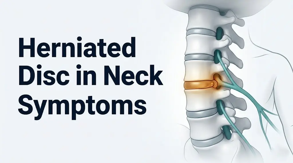
Herniated disc in neck symptoms include arm pain, hand numbness, tingling fingers, and muscle weakness, all produced by a damaged spinal disc pressing on or chemically irritating a nerve root in the cervical spine. The pattern of symptoms depends almost entirely on which cervical disc has herniated and which nerve root is affected, which means the location of your numbness or weakness is a clinically meaningful clue, not a random occurrence.
According to the National Institute of Neurological Disorders and Stroke (NINDS), disc herniations in the cervical spine are among the most common causes of neck pain radiating into the arm. Approximately 83 out of every 100,000 adults develop cervical radiculopathy each year, with men affected at roughly twice the rate of women, based on population data published in the Spine journal. The condition peaks in incidence between ages 45 and 54, though it occurs across a wide age range.
This guide covers every clinically recognized symptom of a herniated disc in the neck and lower back, explains the exact physiological mechanism behind each one, maps symptoms to specific nerve root levels, identifies which symptoms are self-limiting and which represent neurological emergencies, and tells you exactly which type of provider to see and when.
Herniated Disc in Neck Symptoms
Herniated disc in neck symptoms range from localized neck stiffness to severe radiating arm pain, and the specific pattern tells a clinician which cervical level is most likely involved before imaging is even performed.
The most frequently reported symptom is pain that travels from the neck into the shoulder, arm, or hand, following a predictable path dictated by the compressed nerve root’s territory. This radiating pain, called radicular pain, is not simply back pain that has spread. It is a distinct neurological signal generated at the point where the herniated disc material contacts the nerve root, then transmitted along the full length of that nerve’s distribution.
A second major symptom category is sensory change: numbness, tingling, or burning in a specific part of the arm, forearm, or hand. Unlike the vague “pins and needles” of a sleeping limb, the sensory changes from a cervical herniated disc follow a dermatomal pattern tied to the embryological territory of the affected nerve root.
Muscle weakness is the third core symptom group. When a nerve root is compressed enough to impair motor fiber function, the muscles that root controls begin to weaken. This can appear as difficulty gripping objects, trouble raising the arm overhead, or weakness when bending the elbow.
Key symptoms recognized clinically:
- Neck pain that worsens with head rotation or lateral bending toward the affected side
- Arm pain following a nerve-root-specific path from shoulder to specific fingers
- Numbness or tingling in a dermatomal distribution in the hand or forearm
- Muscle weakness in a specific muscle group controlled by the compressed root
- Diminished or absent deep tendon reflexes (bicep, tricep, or brachioradialis) at the affected level
- Pain aggravated by coughing, sneezing, or the Valsalva maneuver (which increases intradiscal pressure)
What Is a Herniated Disc and How Does It Produce Symptoms
A herniated disc is a structural failure of the intervertebral disc, the cartilaginous cushion that sits between each pair of adjacent vertebrae and absorbs spinal compression load.
Each disc has two components. The nucleus pulposus is the soft, hydrated gel core, composed primarily of water, Type II collagen, and proteoglycans. The annulus fibrosus is the outer shell of concentric fibrocartilage rings that contains the nucleus under pressure. Think of the disc like a sealed water balloon inside a multi-layered rubber ring: the outer ring holds the inner gel in place under load. When the outer rings crack or tear from repetitive stress, aging, injury, or a combination of all three, the inner gel can bulge outward or push through entirely.
When nucleus pulposus material escapes the annulus and contacts a spinal nerve root, two separate physiological processes produce symptoms simultaneously.
The first is mechanical compression. The herniated material physically narrows the space through which the nerve root exits the spinal canal through the intervertebral foramen, reducing blood flow to the nerve and disrupting axoplasmic transport, the process by which nutrients and signaling proteins travel along the nerve fiber. This produces pain, numbness, and weakness in the distribution of that nerve.
The second is chemical irritation, which is less well-known but equally important. Research published in the Spine journal has shown that nucleus pulposus tissue releases phospholipase A2, an enzyme that catalyzes the production of prostaglandin E2 via the arachidonic acid pathway. The immune cells that respond to the extruded disc material also release tumor necrosis factor alpha (TNF-alpha) and interleukin-1 beta, which directly sensitize the nerve root to mechanical stimuli. This explains why even a small herniation can produce severe radicular pain: the chemical environment alone can make the nerve hypersensitive, generating pain without requiring significant mechanical compression.
Quick Tip:
- A herniated disc does not always produce symptoms. The North American Spine Society (NASS) notes that MRI studies in asymptomatic adults frequently reveal disc herniations, particularly in people over 40. The presence of a herniation on imaging alone does not confirm it is the source of a person’s symptoms.
- People with pre-existing spinal stenosis (narrowing of the spinal canal) have less anatomical reserve, meaning a smaller herniation produces proportionally more severe nerve compression than in a person with a wide, uncompromised canal.
Symptoms of Herniated Disc in Neck
Symptoms of herniated disc in neck consistently involve some combination of neck pain, radiating arm pain, sensory changes in a hand or forearm, and muscle weakness in the arm, with the specific combination depending on which cervical disc level has herniated.
The cervical spine runs from the base of the skull to the top of the thoracic spine and contains seven vertebrae, labeled C1 through C7. The intervertebral discs sit between adjacent vertebrae: C2-C3, C3-C4, C4-C5, C5-C6, C6-C7, and C7-T1. Disc herniations in the neck most frequently occur at C5-C6 and C6-C7, based on published data from the Journal of Bone and Joint Surgery, because these levels bear the greatest combined load and have the highest range of motion in the lower cervical spine.
When a disc herniates posterolaterally (backward and to one side, which is the most common direction), it compresses the nerve root exiting at that level on the affected side. The nerve root being compressed determines every downstream symptom: where the pain goes, where the numbness appears, and which muscle weakens.
According to the American Academy of Orthopaedic Surgeons (AAOS), neck pain accompanied by arm symptoms that change with head position, particularly pain that worsens when tilting the head toward the painful side, is one of the most consistent clinical indicators of cervical nerve root involvement.
| Symptom | Physiological Mechanism | Clinical Significance |
|---|---|---|
| Radiating arm pain | Nociceptive signal transmission along compressed nerve root | Indicates active nerve root irritation or compression |
| Hand or finger numbness | Impaired sensory fiber conduction in affected dermatomal territory | Localizes the affected cervical level |
| Muscle weakness in arm | Motor fiber dysfunction from nerve root ischemia or compression | May indicate need for surgical evaluation if progressive |
| Diminished deep tendon reflex | Interrupted motor reflex arc at the compressed nerve root level | Objective neurological finding on clinical examination |
| Pain with coughing or sneezing | Transient intradiscal pressure spike compresses nerve root further | Confirms disc-mediated (not purely muscular) pain origin |
| Neck stiffness with limited rotation | Paraspinal muscle guarding protecting the inflamed disc level | Nonspecific; present in many cervical conditions |
Herniated Disc Neck Symptoms: Pain, Numbness, and Arm Weakness
Herniated disc neck symptoms of pain, numbness, and arm weakness each arise through distinct but related mechanisms, and understanding the difference helps explain why some people respond to anti-inflammatory treatment while others need more targeted intervention.
Neck pain from a herniated disc typically originates from two sources. The disc itself has a blood supply and nerve supply only in its outermost annular fibers, so annular tears produce localized neck pain through nociceptors in the outer annulus. When the herniated material additionally irritates the posterior longitudinal ligament, which is densely innervated, this adds a deeper, aching quality to the neck pain that many patients describe as pressure or heaviness in the cervical region.
Numbness in a specific part of the hand is often the most diagnostically informative symptom. Patients frequently report that one or two fingers feel persistently numb or tingly, even when the neck itself feels relatively comfortable. This happens because sensory fibers, which are thinner and metabolically more sensitive than motor fibers, lose function earlier under nerve root compression or chemical irritation. The specific fingers involved point directly to the compressed cervical level.
Arm weakness from a cervical herniation is the symptom that most reliably signals the need for timely professional evaluation. Motor fibers require sustained, adequate blood flow and axoplasmic transport. When compression is significant or prolonged, these fibers begin losing function. A patient notices they cannot grip a coffee mug firmly, cannot button a shirt with one hand, or cannot hold their arm at shoulder height for more than a few seconds. The AAOS notes that weakness affecting grip strength or shoulder abduction that persists beyond a few weeks warrants a clinical examination including motor strength grading.
People with rheumatoid arthritis affecting the cervical spine face a distinct risk: inflammatory joint destruction in this population can mimic or mask disc herniation symptoms, making clinical differentiation more difficult and increasing the importance of imaging to sort out the true cause of radiculopathy.
Cervical Radiculopathy Symptoms by Nerve Root Level
Cervical radiculopathy symptoms follow a predictable anatomical map based on which nerve root is affected, and this map is specific enough that an experienced clinician can often identify the likely herniated disc level from symptoms alone before ordering an MRI.
Each nerve root exits the spinal canal between adjacent cervical vertebrae and travels to a specific region of the arm, forearm, or hand. This territory, called a dermatome for sensory distribution and a myotome for motor distribution, is consistent enough across individuals that its pattern is taught as a diagnostic framework.
The Spurling maneuver, a clinical test in which the examiner extends the patient’s neck, rotates it toward the painful side, and applies gentle downward pressure on the head, temporarily narrows the affected intervertebral foramen and reproduces the arm pain or numbness if a nerve root is truly compressed. Research published in the Journal of Bone and Joint Surgery has shown the Spurling maneuver has approximately 93% specificity for cervical radiculopathy, meaning a positive result is highly reliable for confirming nerve root involvement.
| Nerve Root | Disc Level Most Commonly Responsible | Pain and Sensory Pattern | Motor Weakness | Reflex Affected |
|---|---|---|---|---|
| C4 | C3-C4 | Neck and shoulder, top of the shoulder blade | Shoulder shrug weakness (trapezius) | None reliably testable |
| C5 | C4-C5 | Outer shoulder and upper arm | Deltoid: arm elevation; Bicep: elbow flexion | Bicep reflex |
| C6 | C5-C6 | Outer forearm, thumb, index finger | Wrist extension weakness | Brachioradialis reflex |
| C7 | C6-C7 | Middle finger, back of forearm | Tricep: elbow extension; Wrist flexion | Tricep reflex |
| C8 | C7-T1 | Ring and little finger, inner forearm | Hand intrinsic muscles: grip and finger spread | None reliably testable |
Older adults over 65 often present with C3-C4 and C4-C5 level involvement secondary to cervical spondylosis (bony osteophyte formation from age-related disc degeneration) rather than from a soft disc herniation. Their symptom pattern tends to involve shoulder and upper arm more than hand and fingers, and the mechanism is more mechanical foraminal narrowing than chemical inflammation.
Key Takeaway: The location of your numbness or finger tingling is not random. It directly reflects which cervical nerve root is being compressed, and a clinician uses this pattern, along with reflex testing and motor strength assessment, to identify the disc level before imaging.
C5-C6 and C6-C7 Herniated Disc Symptoms Explained
C5-C6 and C6-C7 are the two most commonly herniated disc levels in the cervical spine, and they produce the most frequently encountered arm and hand symptoms seen in clinical practice.
A C5-C6 disc herniation compresses the C6 nerve root. The C6 root travels from the spine down the outer forearm to the thumb and index finger. Patients with C5-C6 herniation typically describe a burning or aching pain that starts at the base of the neck, travels through the shoulder, down the outer forearm, and settles into the thumb and index finger. The thumb-side of the hand goes numb or tingly first. Weakness shows up most noticeably in wrist extension (difficulty pulling the wrist back) and in the brachioradialis muscle. The brachioradialis reflex, tested by tapping the forearm just above the wrist, is commonly diminished or absent at this level.
A C6-C7 disc herniation compresses the C7 nerve root, which is the most frequently involved nerve root in cervical radiculopathy based on data from the Spine journal. The C7 root travels to the middle finger. Patients report pain extending from the neck through the back of the upper arm, through the back of the forearm, and concentrating in the middle finger. Tricep weakness is the characteristic motor finding: patients notice difficulty pushing objects overhead or difficulty straightening the elbow against resistance. The tricep reflex, tested at the back of the elbow, is the most reliable reflex sign at this level.
To describe C6-C7 symptoms accurately to a provider:
- Note which specific fingers feel numb, tingly, or burning
- Note whether the pain goes down the front or back of the arm
- Test grip strength by squeezing your provider’s fingers with both hands simultaneously to compare sides
- Note any specific movement that makes arm symptoms worse or better
- Report whether coughing, sneezing, or bearing down reproduces or worsens the arm symptoms
Women experiencing C6-C7 symptoms during pregnancy may find them more pronounced because the combined effects of postural change from an expanding abdomen, fluid retention compressing peripheral nerve channels, and the inflammatory environment of pregnancy can amplify nerve root sensitivity at an already compromised level.
Bulging Disc Neck Symptoms Versus Fully Herniated Disc
Bulging disc neck symptoms and herniated disc symptoms exist on a spectrum of the same pathological process, but they differ in severity, mechanism, and the likelihood of producing nerve root involvement.
A bulging disc occurs when the nucleus pulposus pushes the intact annulus fibrosus outward symmetrically or asymmetrically, without rupturing through the annular fibers. The disc wall remains intact. A herniated disc (also called herniated nucleus pulposus, or HNP) involves the nucleus pulposus pushing through a tear in the annular fibers, either partially (protrusion) or fully through (extrusion), and in some cases breaking off as a free disc fragment (sequestration) in the spinal canal.
The clinical difference matters because a bulging disc typically does not expose the nucleus pulposus material to the immune system, so the chemical inflammatory mechanism, the phospholipase A2 and TNF-alpha cascade, tends to be less prominent. A true herniation that breaches the annulus exposes nucleus pulposus to the epidural space and initiates an immune response that amplifies nerve root sensitization.
| Feature | Bulging Disc | Herniated Disc (Protrusion or Extrusion) |
|---|---|---|
| Annulus fibrosus integrity | Intact but deformed | Torn or breached |
| Chemical inflammatory mechanism | Minimal to none | Present and often significant |
| Likelihood of nerve root compression | Lower; depends on size | Higher, especially with extrusion |
| Typical symptom severity | Neck pain and stiffness, often no radiculopathy | Neck pain plus radicular arm/hand symptoms |
| Natural history | Usually resolves with posture and activity modification | 85 to 90% resolve without surgery over 6 to 12 weeks |
| MRI appearance | Disc margin extends beyond vertebral body edge symmetrically | Focal extension of disc material beyond annular margin |
The natural history difference is clinically important. A bulging disc that produces only axial neck pain (pain at the neck itself, not radiating into the arm) has a good prognosis with conservative management. A herniated disc with frank radiculopathy in younger and middle-aged adults still resolves without surgery in the majority of cases, based on data from the SPORT Trial published in JAMA.
Key Takeaway: A bulging disc and a herniated disc are structurally different lesions. A bulging disc keeps the annulus intact and rarely triggers the full inflammatory cascade. A herniated disc breaches the annulus, exposing nucleus pulposus to the immune system and producing the chemical radiculopathy component that makes even small herniations intensely painful.
Herniated Disc Lower Back Symptoms
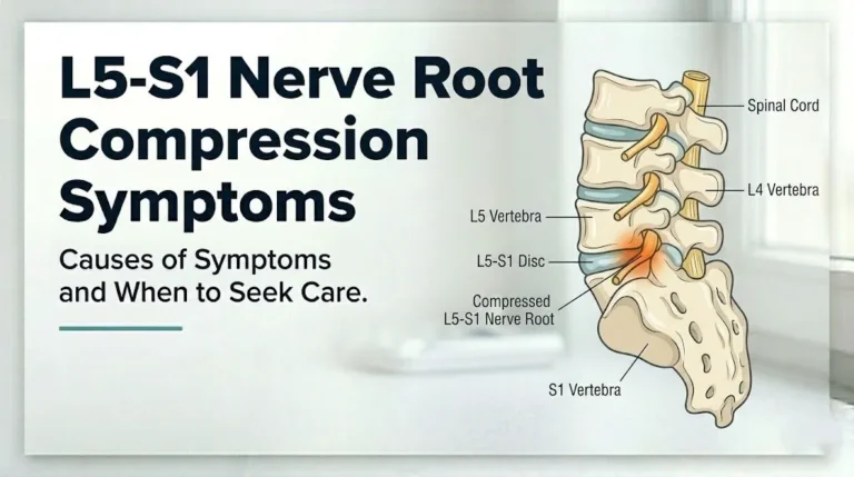
Herniated disc lower back symptoms include low back pain, leg pain following a specific nerve root path, foot or leg numbness, and in the most severe cases, weakness in the foot or loss of bowel or bladder control.
The lumbar spine contains five vertebrae (L1 through L5) and the intervertebral discs between them bear the greatest compressive load of any spinal region during lifting, bending, and prolonged sitting. The two most frequently herniated lumbar levels are L4-L5 and L5-S1, based on epidemiological data from the Spine journal, accounting for more than 90% of clinically significant lumbar disc herniations.
When a lumbar disc herniates posterolaterally, it compresses the nerve root that is traveling downward through that portion of the spinal canal before exiting at the next level down. At the L4-L5 level, the L5 nerve root is most commonly compressed. At the L5-S1 level, the S1 nerve root is most commonly compressed. This level-root offset is important: an L4-L5 herniation compresses L5, not L4.
The NINDS states that lumbar disc herniation is one of the most common causes of low back pain and leg pain combined, particularly in working-age adults between 30 and 50.
Key symptoms of lumbar herniated disc:
- Low back pain that may radiate across the buttock, hip, and down the leg
- Leg pain following a specific nerve root path (distinct from diffuse leg ache)
- Numbness or tingling in a specific part of the foot, outer leg, or inner thigh
- Muscle weakness in foot dorsiflexion (lifting the foot upward), calf strength, or quadricep strength
- Pain that worsens with sitting and prolonged flexion, and often improves temporarily with walking or lying flat
- Pain that spikes sharply with coughing, sneezing, or bearing down (Valsalva maneuver)
Pregnant women in the third trimester may experience exacerbated lumbar disc symptoms due to the combined effect of increased lumbar lordosis, elevated relaxin levels that reduce ligamentous spinal support, and the added compressive load on already vulnerable lumbar discs. A board-certified obstetrician-gynecologist should be involved in managing disc symptoms during pregnancy because some standard treatment approaches, including certain imaging modalities and medications, require modification.
L4-L5 and L5-S1 Herniated Disc Symptoms
L4-L5 and L5-S1 herniated disc symptoms are the specific presentations seen in the two most commonly herniated lumbar levels, and they produce identifiably different patterns of leg pain, foot numbness, and muscle weakness that help clinicians localize the affected disc before MRI.
An L4-L5 herniation typically compresses the L5 nerve root. The L5 root travels from the lumbar spine through the buttock and down the outer aspect of the thigh and lower leg to the top of the foot and the big toe. Patients report a burning or aching pain that travels from the low back through the outer thigh and lower leg to the top of the foot. Numbness or tingling concentrates on the top of the foot and in the great toe or second toe. The characteristic motor finding is weakness in foot dorsiflexion: the patient has difficulty lifting the front of the foot off the ground, a condition called foot drop in severe cases. Walking on the heels is painful or difficult.
An L5-S1 herniation typically compresses the S1 nerve root. The S1 root travels through the buttock, down the back of the thigh and calf, and to the outer edge of the foot and small toe. Patients describe pain from the low back through the center of the buttock and down the back of the leg to the outer heel and small toe. Numbness concentrates on the outer edge of the foot and the small toe side. Weakness affects calf strength and the ability to push off the toes during walking. The Achilles reflex, tested by tapping the heel cord, is commonly reduced or absent with S1 involvement.
| Disc Level | Nerve Root Compressed | Pain Pattern | Sensory Change | Motor Weakness | Reflex |
|---|---|---|---|---|---|
| L4-L5 | L5 | Outer leg to top of foot and great toe | Top of foot and great toe | Foot dorsiflexion (foot drop in severe cases) | No reliable reflex |
| L5-S1 | S1 | Back of thigh and calf to outer heel | Outer foot and small toe | Plantarflexion; calf; heel raise | Achilles reflex |
People with type 2 diabetes mellitus and pre-existing diabetic peripheral neuropathy present a clinical challenge at both of these levels. Their underlying sensory nerve damage means they may not feel the classic sharp radiating pain. Instead, they present with progressive foot weakness or new balance problems without significant pain. This atypical presentation often leads to delayed diagnosis. A neurologist should evaluate any diabetic patient with new foot weakness to differentiate worsening peripheral neuropathy from superimposed lumbar nerve root compression.
Sciatica and Leg Symptoms From a Lumbar Herniated Disc
Sciatica is the most widely recognized symptom of a lumbar herniated disc, and it describes the specific pattern of pain, numbness, or weakness traveling from the low back through the buttock and down one leg.
The sciatic nerve is the largest peripheral nerve in the body, formed by the union of the L4, L5, S1, S2, and S3 nerve roots from the lumbar and sacral spine. When a lumbar disc herniation compresses one or more of the nerve roots that form the sciatic nerve, the pain signal travels the full length of the nerve, producing the distinctive shooting or burning sensation that runs from the low back or buttock all the way down to the foot.
True sciatica from a disc herniation is unilateral (one leg) in the overwhelming majority of cases. Pain in both legs simultaneously from a disc herniation should be taken seriously and evaluated promptly, as bilateral leg symptoms from disc herniation can indicate a central disc herniation that compresses the cauda equina (the bundle of nerve roots at the base of the spinal cord), a condition discussed in detail in the emergency section below.
According to a 2021 systematic review published in the BMJ, approximately 40% of people with acute sciatica from lumbar disc herniation experience significant spontaneous improvement within 4 to 6 weeks, and up to 85% improve substantially within 12 weeks without surgical intervention. However, the same review noted that the subgroup with measurable motor weakness (foot drop, calf weakness) at presentation showed slower recovery rates and was more likely to benefit from earlier surgical evaluation.
Sciatica from a herniated disc typically worsens with:
- Sitting for extended periods (which increases intradiscal pressure and flexion load on the posterior disc)
- Bending forward at the waist
- Coughing, sneezing, or bearing down
- Getting up from sitting after a prolonged period
Key Takeaway: Sciatica is not a diagnosis but a symptom pattern produced when a lumbar disc herniation or other structural problem compresses a sciatic nerve root. Unilateral leg pain that travels from the buttock to the foot, changes with body position, and worsens with the Valsalva maneuver is a clinically distinctive pattern pointing to disc-related nerve root compression rather than muscle pain or hip joint pathology.
Symptoms of a Herniated Disc That Most People Miss
Several herniated disc symptoms are consistently underrecognized by patients and sometimes by clinicians, particularly when the presentation does not follow the textbook pattern of dramatic radiating pain.
Grip weakness without significant pain is frequently missed. A person may notice they are dropping things, struggling to open jars, or finding handwriting difficult, without making the connection to neck or spine pathology. When cervical nerve root compression impairs the C8 root, the intrinsic muscles of the hand (the muscles between the fingers responsible for grip and fine motor control) begin to weaken. Because the pain component may be mild, the hand weakness seems disconnected from the neck and is often attributed to overuse or “aging.”
Clumsiness walking or poor coordination in the legs can be an early sign of cervical myelopathy: compression of the spinal cord itself, rather than just a nerve root. Patients describe a subtle change in gait, difficulty with balance on uneven surfaces, or a tendency to trip. Because this symptom appears gradually, it is commonly attributed to inner ear problems, fatigue, or general aging before the spinal origin is identified.
Occipital headaches, the dull or aching pain at the base of the skull and the back of the head, can originate from a C2-C3 or C3-C4 disc problem, as the upper cervical nerve roots refer pain to the head. These headaches are easily mistaken for tension headaches or migraines and treated without identifying the underlying cervical disc pathology.
Pain that disappears but weakness persists is a missed warning sign. When radicular pain suddenly resolves but the arm or hand weakness simultaneously worsens, this does not indicate recovery. It can reflect nerve fiber deterioration progressing beyond the pain-processing threshold, and it warrants urgent neurological evaluation by a board-certified neurologist or neurosurgeon.
People who work at computers or desks for 8 or more hours daily often miss early cervical disc symptoms because the gradual onset of forearm numbness and wrist weakness is attributed to repetitive strain or carpal tunnel syndrome rather than cervical radiculopathy. The clinical distinction matters because carpal tunnel syndrome and cervical radiculopathy are treated differently, and treating one when the other is the true cause will not produce improvement.
How Herniated Disc Symptoms Progress Over Time
Herniated disc symptoms do not follow a single predictable course, but understanding the general natural history helps patients and clinicians distinguish appropriate watchful waiting from situations where delay worsens outcomes.
In the first 24 to 72 hours after an acute disc herniation, symptoms are typically at their most intense. The annular tear triggers local inflammation, and the fresh chemical contact between nucleus pulposus and the nerve root provokes maximum nociceptor sensitization. Pain can be severe, and sleep disruption is common during this phase.
Over the following 4 to 12 weeks, the majority of patients begin to improve. The body’s immune system recognizes the extruded nucleus pulposus as foreign material and begins to resorb it through a process involving macrophage infiltration and enzymatic degradation of the disc fragment. As the disc material reduces in volume, both the mechanical compression and the chemical irritation of the nerve root decrease. The SPORT Trial data published in JAMA showed that the majority of patients with lumbar disc herniation who chose conservative management over surgery demonstrated comparable functional outcomes at one and two years, though surgical patients reported faster early pain relief.
A minority of patients follows a chronic course in which symptoms persist beyond 12 weeks without substantial improvement. Chronic radiculopathy beyond this timeframe increases the risk of persistent motor fiber damage and warrants evaluation by a board-certified orthopedic surgeon or board-certified neurosurgeon to reassess surgical candidacy.
In some patients, particularly those over 60 with pre-existing degenerative disc disease and cervical spondylosis, the natural history does not involve a discrete acute event at all. Symptoms develop gradually over months to years as progressive foraminal narrowing from osteophytes slowly compresses nerve roots without a single precipitating herniation event.
| Phase | Typical Symptom Pattern | Appropriate Response |
|---|---|---|
| Acute (0 to 4 weeks) | Severe pain, radiating arm or leg symptoms, possible sensory change | Activity modification, NSAIDs if tolerated, evaluation by primary care physician |
| Subacute (4 to 12 weeks) | Gradual improvement in most; persistent radiculopathy in some | Physical therapy, repeat neurological assessment, imaging if not yet obtained |
| Chronic (beyond 12 weeks without improvement) | Persistent or worsening radiculopathy, possible progressive weakness | Referral to board-certified orthopedic surgeon or neurosurgeon; surgical discussion |
| Progressive neurological deficit (any phase) | Worsening weakness, reflex loss, new myelopathy signs | Urgent referral to neurosurgeon within days, not weeks |
How Symptoms Differ by Age, Sex, and Pre-Existing Condition
Herniated disc symptoms are not identical across all populations, and several biological and clinical factors predictably alter the presentation in ways that matter for diagnosis and management.
Age is the most powerful modifier. Adults under 40 with disc herniation almost always present with acute radiculopathy: sharp, well-localized arm or leg pain following a nerve root distribution, triggered by a specific activity or injury. Disc herniation in this age group involves a relatively healthy annulus that fails acutely under load, producing a soft disc herniation with a significant chemical inflammatory component.
Adults over 60 more commonly present with cervical myelopathy caused by combined disc degeneration, osteophyte formation, and ligamentum flavum hypertrophy gradually compressing the spinal cord rather than a single nerve root. Their symptoms include bilateral hand clumsiness, difficulty with fine motor tasks like buttoning, gait instability, and in more advanced cases, leg spasticity and sensory changes in the hands and feet. The absence of dramatic arm pain in older adults with myelopathy frequently delays diagnosis, as pain in this group is often milder than in younger radiculopathy patients.
Biological sex also influences presentation. Research published in the Spine journal indicates that men develop herniated discs at approximately twice the rate of women, likely related to occupational physical loading patterns, greater absolute disc volume placing higher hydrostatic pressure on the posterior annulus, and differences in body mechanics during lifting. Women who do develop symptomatic disc herniation, however, tend to report more diffuse pain patterns with greater central sensitization components, partially explained by sex hormone effects on the inflammatory mediator system. Estrogen has been shown in laboratory research to modulate prostaglandin synthesis and nociceptor sensitivity, which may contribute to the more widespread pain experience reported by some women with disc pathology.
Pre-existing conditions that alter the clinical picture:
- Diabetes mellitus: pre-existing peripheral neuropathy may mask radicular pain but allow motor weakness and balance problems to emerge early
- Rheumatoid arthritis: cervical joint inflammation and possible C1-C2 instability can confound or worsen disc herniation symptoms in the neck
- Obesity: adds axial compressive load on lumbar discs and accelerates degenerative disc disease, generally producing earlier and more severe lumbar herniation in this population
- Prior spinal surgery: scar tissue and altered spinal mechanics change the clinical symptom pattern and the interpretation of imaging findings
Key Takeaway: A 58-year-old with gradually worsening hand clumsiness and balance problems may have cervical myelopathy from degenerative disc disease, not a radiculopathy. An older adult presenting without the classic sharp arm pain should not have myelopathy ruled out on symptom grounds alone. The clinical picture changes substantially with age.
Herniated Disc Symptoms That Require Urgent Medical Evaluation
Certain herniated disc symptoms require medical evaluation within hours to days, not weeks, because delayed treatment can result in permanent neurological damage.
Progressive motor weakness is the most important signal that conservative management has reached its limit. When arm or leg weakness worsens over days to a week despite rest and anti-inflammatory measures, the nerve root is sustaining ongoing injury. Motor fiber damage that progresses to severe weakness (MRC grade 3 or below, meaning the patient cannot move the limb against gravity) may not fully recover even after surgical decompression. A board-certified orthopedic surgeon or board-certified neurosurgeon should evaluate progressive weakness within 1 to 3 days.
New gait disturbance or falls in a patient with known cervical disc problems should raise concern for developing cervical myelopathy. The spinal cord has limited capacity to recover from prolonged compression. Symptoms of myelopathy, including leg stiffness, hyperreflexia (overly brisk deep tendon reflexes), or a positive Babinski sign (the big toe extends upward rather than curling when the sole is stroked), warrant urgent evaluation by a neurosurgeon within days.
Bilateral arm or leg symptoms from a disc herniation are not the normal presentation of unilateral nerve root compression. Bilateral symptoms suggest either a large central disc herniation or, in the neck, developing myelopathy. Both presentations require prompt imaging and specialist assessment.
Specific symptoms requiring evaluation within 24 to 72 hours:
- Progressive arm or leg weakness worsening over days
- New onset of clumsiness, dropping objects, or coordination difficulties
- Bilateral limb symptoms beginning simultaneously
- Complete loss of sensation (dense numbness) in a limb rather than intermittent tingling
- Severe pain unresponsive to any position change or pain relief measure after 48 hours
Older adults with baseline cognitive changes from other conditions may not clearly report the onset of gait problems or hand clumsiness. Family members or caregivers who notice a sudden or gradual change in coordination, hand function, or walking pattern should bring this to the attention of the person’s primary care physician promptly.
Emergency Symptoms: When to Call 911 or Go to the ER
Certain symptoms associated with a herniated disc require immediate emergency evaluation. Do not wait to see if these resolve on their own.
Call 911 or go to the nearest emergency room immediately if you experience:
- Sudden loss of bowel or bladder control (inability to urinate or sudden inability to hold urine or bowel movements) in the context of back pain: this is the hallmark presentation of cauda equina syndrome, a surgical emergency in which the nerve bundle at the base of the spinal cord is being compressed and can be permanently damaged within hours
- Saddle anesthesia: numbness or loss of sensation in the inner thighs, groin, perineum, or the area that would contact a saddle when sitting; this is another defining sign of cauda equina syndrome
- Sudden onset of weakness in both legs simultaneously with or without back pain
- Rapid onset of inability to walk or stand without support
- Severe neck injury with sudden onset of arm or leg weakness, numbness, or paralysis: possible acute cervical disc herniation with cord compression requiring emergency decompression
- Progressive loss of function in arms or legs over hours in a patient with known cervical stenosis or cervical myelopathy
These presentations can indicate cauda equina syndrome, acute cervical cord compression, or rapidly progressing cervical myelopathy, all of which require emergency neurosurgical assessment, not an urgent care appointment or a scheduled clinic visit.
Cauda equina syndrome that progresses to complete bowel and bladder dysfunction and then receives surgical decompression within 24 to 48 hours of onset has substantially better neurological recovery rates than cases where surgery is delayed beyond 48 hours, based on clinical series data published in the Journal of Bone and Joint Surgery. Time is the critical variable.
How a Herniated Disc Is Diagnosed Based on Symptoms
Herniated disc diagnosis begins with a detailed clinical history and neurological examination, with symptoms directing imaging rather than replacing it.
A clinician evaluating suspected disc herniation will conduct a structured neurological examination that directly tests the nerve roots most likely to be involved based on symptom pattern. This includes testing dermatomal sensation (light touch and pinprick in specific hand or foot territories), myotomal motor strength (bicep, tricep, grip, wrist extension, ankle dorsiflexion, calf strength), and deep tendon reflexes (bicep, tricep, brachioradialis, patellar, Achilles). The pattern of findings across these three domains, sensory, motor, and reflex, tells the clinician which cervical or lumbar level is most likely affected.
The Spurling test for cervical radiculopathy and the straight leg raise (SLR) test for lumbar radiculopathy are the two most commonly used provocative clinical tests. The SLR test, in which the examiner raises the patient’s straight leg while the patient lies flat, reproduces sciatic nerve tension and provokes radicular leg pain if a lumbar nerve root is compressed. A 2023 clinical review in JAMA noted that the straight leg raise has approximately 80% sensitivity for lumbar disc herniation when it reproduces leg pain below the knee at 30 to 60 degrees of elevation.
Magnetic resonance imaging (MRI) is the gold-standard imaging tool for confirming herniated disc location, size, direction, and nerve root involvement. A contrast-enhanced MRI is not typically required for standard disc herniation evaluation. Electromyography (EMG) and nerve conduction velocity (NCV) testing are added when the clinical picture does not clearly match imaging findings, when peripheral neuropathy is suspected, or when surgical planning requires precise localization of the affected nerve root.
How the diagnostic process typically unfolds:
- Clinical history: onset, location, radiation pattern, aggravating and relieving factors, weakness, sensory change
- Neurological examination: sensory, motor, and reflex testing mapped to specific nerve root levels
- Provocative testing: Spurling maneuver (cervical) or straight leg raise (lumbar)
- Imaging: MRI of the relevant spinal region if symptoms persist beyond 4 to 6 weeks or if neurological deficits are present at first evaluation
- EMG/NCV testing: if imaging and clinical findings do not clearly correlate, or to differentiate radiculopathy from peripheral neuropathy
- Specialist referral: to a physiatrist, board-certified orthopedic surgeon, or board-certified neurosurgeon based on severity, duration, and neurological deficit status
People who have had prior spinal surgery should inform their evaluating clinician before MRI, as spinal implants from prior procedures may require modified imaging protocols or alternative imaging modalities such as CT myelography.
Frequently Asked Questions About Herniated Disc in Neck Symptoms
What does a herniated disc in the neck feel like?
A herniated disc in the neck typically feels like a sharp or burning pain that travels from the neck into the shoulder, arm, forearm, or fingers, following a path determined by which cervical nerve root is affected.
Many people also notice numbness or tingling in specific fingers, along with a sense of weakness when gripping or lifting with the affected arm.
The pain often worsens when turning the head to the affected side, coughing, sneezing, or sitting with poor posture, and may improve when lying down or holding the arm above the head to relieve tension on the nerve root.
Can a herniated disc in the neck cause headaches or dizziness?
A herniated disc at the upper cervical levels, particularly C2-C3 or C3-C4, can cause headaches at the base of the skull and the back of the head because the upper cervical nerve roots refer pain into the occipital region.
Dizziness from cervical disc pathology is less well-established but has been reported in association with cervical spondylosis and upper cervical dysfunction, likely related to altered proprioceptive input from the cervical joints and paraspinal muscles rather than direct nerve root compression.
If you are experiencing new or severe headaches with neck pain, a primary care physician should evaluate you to exclude other causes including vascular or intracranial pathology before attributing headaches to a disc problem.
How do I know if my neck pain is a herniated disc or just muscle strain?
Neck pain from a herniated disc is most reliably distinguished from muscle strain by the presence of radiating pain, numbness, or weakness in the arm or hand that follows a nerve root pattern.
Muscle strain typically produces pain localized to the neck and shoulder region that worsens with specific neck movements and does not cause numbness or tingling in the fingers or weakness in arm muscles.
If your neck pain is accompanied by any arm or hand symptoms, particularly constant numbness or grip weakness, a primary care physician or physiatrist should evaluate you to determine whether nerve root involvement is present.
How long do herniated disc in neck symptoms last?
Most cervical herniated disc symptoms improve substantially within 6 to 12 weeks with conservative management including activity modification, physical therapy, and anti-inflammatory treatment.
The SPORT Trial data published in JAMA demonstrated that the majority of patients with disc herniation who chose non-surgical management achieved comparable functional outcomes to surgically treated patients at one and two years, though improvement was slower in the early weeks.
Symptoms persisting beyond 12 weeks without meaningful improvement, or symptoms accompanied by progressive motor weakness at any point, warrant evaluation by a board-certified orthopedic surgeon or board-certified neurosurgeon.
What are the warning signs that a herniated disc is an emergency?
The most urgent warning signs are loss of bowel or bladder control, saddle anesthesia (numbness in the inner thighs and groin), or sudden weakness in both legs simultaneously, all of which suggest cauda equina syndrome, a neurosurgical emergency.
In the neck, rapidly worsening arm or leg weakness, sudden onset of gait instability, or bilateral limb symptoms from a cervical disc problem suggest cord compression that requires emergency evaluation.
Call 911 or go to the nearest emergency room immediately if any of these symptoms appear; these presentations require neurosurgical assessment within hours, not days.
Can you have a herniated disc in your neck with no symptoms?
Yes, a herniated disc in the neck can be entirely asymptomatic, and this is more common than most people realize.
MRI studies of asymptomatic adults published in the Spine journal have consistently found disc herniations in a substantial proportion of people over 40 who have no neck pain or arm symptoms, indicating that the presence of a herniation on imaging does not automatically mean it is the source of any given person’s symptoms.
The North American Spine Society (NASS) cautions that imaging findings must always be interpreted in the context of the clinical examination, because treating an incidentally found herniation rather than the actual source of symptoms leads to poor outcomes.
What Your Symptoms Are Telling You, and What to Do Next
The specific combination of symptoms you are experiencing with a herniated disc is not arbitrary. The finger that goes numb, the reflex that diminishes, the muscle that weakens: these follow anatomical logic that clinicians use to pinpoint the disc level before an MRI is ever ordered. Understanding this pattern gives you more useful information to bring to your first appointment.
If your symptoms involve arm or hand numbness, tingling, or weakness lasting more than a few days, a primary care physician or physiatrist is the right first point of contact. Bring a description of exactly which fingers are affected, whether the pain travels in a specific path, and whether coughing or sneezing makes it worse. That information narrows the diagnosis faster than any symptom description of “neck pain.”
If weakness is worsening, if you are losing balance or coordination, or if you have any change in bowel or bladder function alongside back or neck pain, do not schedule an appointment for next week. Those symptoms require evaluation now.