Dislocated Knee Symptoms: Full Clinical Warning Guide 2026
Dislocated knee symptoms range from severe pain, visible joint deformity, and complete inability to bear weight in the most serious cases, to a brief episode of the kneecap sliding out and back in during a pivoting movement, but even a mild-seeming presentation can indicate damage to structures that require urgent evaluation. The key distinction that every person reading this guide needs to understand first is that “dislocated knee” describes two anatomically different injuries with very different emergency profiles.
The American Academy of Orthopaedic Surgeons recognizes that true tibiofemoral knee dislocation, where the thighbone and shinbone separate, carries a 20 to 40 percent risk of injury to the popliteal artery, the major blood vessel running directly behind the knee. That rate comes from studies published in the Journal of Bone and Joint Surgery. Missing vascular injury at this site can result in limb loss within hours.
This guide separates the two injury types clearly, explains the physiological mechanism behind every recognized symptom, maps the population groups at highest risk, tells you which presentations require a 911 call versus an orthopedic office visit, and gives you the clinical framework to describe your injury accurately to an emergency medicine physician or orthopedic surgeon.
Dislocated Knee Symptoms: What the Injury Is Actually Doing to Your Joint
Dislocated knee symptoms are produced when the bones of the knee joint are forced out of their normal anatomical alignment, tearing the ligaments, capsule, and sometimes neurovascular structures that hold the joint together.
The knee joint is not a simple hinge. It is a complex structure where the femur (thighbone), tibia (shinbone), and patella (kneecap) must maintain precise positional relationships through range of motion. That precision depends on four major ligaments: the anterior cruciate ligament (ACL), the posterior cruciate ligament (PCL), the medial collateral ligament (MCL), and the lateral collateral ligament (LCL), as well as the posterolateral corner (PLC) complex. The patella is additionally stabilized by the medial patellofemoral ligament (MPFL) and the groove of the femoral trochlea.
When sufficient force disrupts any of these stabilizing structures enough to displace bones from their normal position, the resulting tissue damage triggers an immediate inflammatory cascade. Mast cells at the tear site release histamine and bradykinin, causing rapid vasodilation and increased vascular permeability. Prostaglandins from COX-1 and COX-2 enzymes sensitize local nociceptors (pain receptors). Blood from torn capillaries and ligamentous vessels fills the joint space, creating hemarthrosis that generates pressure-mediated pain on top of the direct injury pain.
The primary dislocated knee symptoms include:
- Sudden severe pain at the moment of injury
- Rapid swelling of the entire knee joint, often within minutes
- Visible abnormality in knee shape or position
- Complete or near-complete inability to bear weight or walk
- A sensation of instability, giving way, or the knee “not feeling right”
- In kneecap dislocation specifically: a visible lump lateral to the knee where the patella has migrated
What Is a Knee Dislocation and What Structures Are Involved?
A knee dislocation is an injury in which one or more bones of the knee joint are displaced from their normal anatomical position relative to the other joint bones, with the term encompassing both tibiofemoral dislocation (the tibia separating from the femur) and patellofemoral dislocation (the patella dislocating from the trochlear groove).
True tibiofemoral dislocation requires high-energy trauma in most cases: motor vehicle collisions, falls from height, or high-impact sports collisions. The exception is a subset of cases in patients with a body mass index above 40, where relatively low-energy pivoting mechanisms have produced tibiofemoral dislocation due to the additional joint loading, a pattern reported in case series published in the American Journal of Sports Medicine.
The structures at risk in tibiofemoral dislocation depend on the direction of displacement:
- Anterior dislocation (tibia displaced forward relative to femur): most common type; PCL always torn, ACL frequently torn; popliteal artery stretched at the posterior joint
- Posterior dislocation (tibia displaced backward): highest rate of popliteal artery injury because the vessel is directly compressed; PCL and ACL typically both torn
- Lateral or medial dislocation: MCL or LCL rupture; peroneal nerve at risk in lateral dislocation
- Rotatory dislocation: complex multiplanar displacement; highest risk of soft tissue entrapment preventing closed reduction
Patellofemoral dislocation involves the patella displacing out of the femoral trochlear groove, almost always in the lateral direction. The MPFL tears as the patella moves past the medial trochlear wall. The vastus medialis oblique (VMO) portion of the quadriceps muscle is stretched or partially avulsed. Bone contusions occur on the medial patellar facet and the lateral femoral condyle as the patella strikes the condyle during lateral displacement.
Adolescents and women generally have a shallower trochlear groove (trochlear dysplasia) and a wider Q-angle, both of which reduce patellar stability and explain the higher patellar dislocation rate in those demographics compared to adult men.
Tibiofemoral Knee Dislocation vs Patellofemoral Dislocation
Tibiofemoral knee dislocation and patellofemoral dislocation are anatomically and clinically distinct injuries that require different emergency responses, different imaging protocols, and different surgical management pathways.
Think of these as the difference between a car frame separating at the chassis (tibiofemoral: catastrophic, structural, vascular emergency) versus a wheel sitting in its hub at the wrong angle (patellofemoral: painful and damaging, but not typically limb-threatening in the acute phase). Both deserve serious medical attention. But only one of them can put a limb at risk within hours if vascular injury is not identified.
| Feature | Tibiofemoral Dislocation | Patellofemoral Dislocation |
|---|---|---|
| Bones involved | Femur and tibia separate | Patella slides out of trochlear groove |
| Energy required | Usually high-energy trauma | Lower-energy; pivoting, twisting, direct blow |
| Ligaments torn | Minimum 2 (ACL and PCL); often 3 to 4 | MPFL primarily; medial retinaculum |
| Vascular risk | Popliteal artery injury in 20 to 40% | Minimal vascular risk |
| Nerve risk | Common peroneal nerve in 10 to 40% | Minimal nerve risk |
| Visible deformity | Severe; knee grossly abnormal in shape | Lateral lump where patella has migrated |
| Self-resolution | Rarely reduces spontaneously | Frequently reduces spontaneously with leg straightening |
| Emergency level | Immediate: call 911 | Urgent: emergency room or orthopedics same day |
| Primary imaging | X-ray first; CTA for vascular assessment | X-ray; MRI for MPFL assessment |
| Most common in | Adults in high-energy trauma | Adolescents, female athletes, hypermobile patients |
The overlap in terminology creates genuine confusion. A patient who says “my knee dislocated” at an emergency department may have experienced either injury. The first priority of the examining physician is to determine which type has occurred, because the immediate management pathway diverges completely at that point.
Key Takeaway: True tibiofemoral knee dislocation is a vascular emergency that can threaten limb survival within hours due to popliteal artery injury. Patellar dislocation is painful and damaging but rarely immediately life- or limb-threatening. The distinction determines whether you call 911 or drive to urgent care.
Symptoms of a Dislocated Knee: The Complete Clinical Picture
Symptoms of a dislocated knee include severe immediate pain, rapid joint swelling, visible or palpable joint deformity, complete loss of normal range of motion, and instability, with the specific symptom pattern varying by whether the tibiofemoral joint or the patellofemoral joint has dislocated.
The most complete clinical picture across both injury types includes both the immediate symptoms (present within seconds to minutes of injury) and the subsequent symptoms that develop over the first hours as inflammation and swelling progress.
| Symptom | Mechanism | Present In | Urgency |
|---|---|---|---|
| Sudden severe pain at injury moment | Ligament and capsule rupture; nociceptor activation | Both types | Immediate evaluation |
| Rapid diffuse knee swelling | Hemarthrosis from torn ligament vessels; synovial inflammatory response | Both types | Same-day evaluation |
| Visible joint deformity or abnormal knee shape | Bone displacement from normal alignment | Tibiofemoral especially; patella displacement may be visible | 911 for tibiofemoral |
| Complete inability to bear weight | Loss of joint structural integrity; pain inhibition of quadriceps | Both types | Immediate evaluation |
| Lateral lump at the knee | Displaced patella sitting lateral to the trochlear groove | Patellofemoral specifically | Emergency room same day |
| Knee instability or “giving way” sensation | Ligament disruption with residual joint laxity | Both types, especially after spontaneous reduction | Orthopedic evaluation |
| Absent or diminished pulses below the knee | Popliteal artery injury compromising distal circulation | Tibiofemoral (20 to 40% of cases) | Emergency: call 911 |
| Foot drop or loss of ankle dorsiflexion | Common peroneal nerve injury | Tibiofemoral, especially posterior and lateral types | Emergency: call 911 |
| Paresthesia or numbness below the knee | Peroneal or tibial nerve stretch or compression | Tibiofemoral | Emergency evaluation |
| Cold, pale, or mottled foot | Distal ischemia from popliteal artery injury | Tibiofemoral | Emergency: call 911 immediately |
The National Institute of Arthritis and Musculoskeletal and Skin Diseases states that multi-ligament knee injuries represent among the most complex musculoskeletal injuries encountered in orthopedic practice, requiring evaluation by a board-certified orthopedic surgeon with experience in multi-ligament reconstruction.
Patients on anticoagulant medications (warfarin, apixaban, rivaroxaban) will develop faster and more severe hemarthrosis after any knee dislocation. This can mask or exaggerate swelling patterns and should be disclosed immediately to the treating emergency medicine physician.
Dislocated Kneecap Symptoms
Dislocated kneecap symptoms are produced when the patella slides laterally out of the femoral trochlea (the groove on the front of the femur that normally houses the kneecap), tearing the medial patellofemoral ligament and generating immediate pain, swelling, and visible lateral displacement of the kneecap.
The patella sits in the trochlear groove like a train on a track. When it derails, it almost always goes to the outside (laterally). This happens because the iliotibial band and the lateral retinaculum pull the patella outward, and in many people, the medial restraint (the MPFL) is not strong enough to counterbalance that lateral pull, especially under sudden twisting or pivoting forces.
The specific symptoms of patellar dislocation that distinguish it from other knee injuries:
- A visible lump or bulge on the outer (lateral) side of the knee where the patella has migrated
- A flat or abnormal contour in the center-front of the knee where the patella is normally visible
- Immediate severe pain, often described as “electric” or “burning” at the inner side of the kneecap (the MPFL tear site)
- A sensation that the knee “popped” or “gave way” during a twisting movement
- Inability to straighten the leg fully at first (the dislocated patella blocks full extension mechanically)
- Rapid swelling, typically less dramatic than in tibiofemoral dislocation but still visible within 30 minutes
- Tenderness specifically along the medial border of the patella and the inner side of the knee
Cleveland Clinic notes that first-time patellar dislocators who receive proper imaging and rehabilitation have lower recurrence rates than those who receive only rest and ice. MRI evaluation of the MPFL is recommended after a first patellar dislocation to assess whether surgical reconstruction is indicated.
Patients with trochlear dysplasia, a condition in which the trochlear groove is shallow or flat rather than concave, are at substantially higher risk for recurrent patellar dislocation even with relatively minor twisting movements. This anatomical variant can be identified on plain lateral X-ray or MRI.
Knee Dislocation Pain and Swelling Symptoms
Knee dislocation pain is generated by simultaneous activation of multiple tissue nociceptors at the moment of ligament rupture, joint capsule tearing, and bone surface contusion, producing a pain intensity that most patients describe as among the worst they have experienced.
The pain is not uniform across the knee. The location of maximum tenderness provides clinical information about which structures have been injured. Medial joint line tenderness corresponds to MCL or medial capsule injury. Posterior joint line tenderness implicates PCL disruption or posterior capsule rupture. Tenderness at the medial patellar border, specifically at the patellar insertion of the MPFL, is the hallmark of patellar dislocation.
Swelling after a knee dislocation follows a predictable timeline driven by two overlapping processes. The first is hemarthrosis: blood enters the joint space within minutes from torn ligament vessels, filling the suprapatellar bursa and joint recess. This produces a tense, firm swelling that makes the knee look like a balloon compared to its normal contour. The second is inflammatory edema: over the subsequent 12 to 24 hours, the prostaglandin-mediated inflammatory response increases vascular permeability, drawing protein-rich fluid into the periarticular soft tissues. This produces the softer, more diffuse swelling that spreads to the surrounding structures.
Quick Tip:
- Apply ice wrapped in cloth for no more than 20 minutes at a time to limit inflammatory edema while awaiting transport to a medical facility
- Do not apply direct ice to bare skin, particularly in anyone with diabetes or peripheral vascular disease who may have reduced sensation
- Elevate the limb at or above heart level to reduce hemarthrosis expansion while awaiting evaluation
- Do not attempt to walk on a suspected dislocated knee, as weight bearing on an unstable joint risks further ligament and vascular injury
Patients with diabetes mellitus may experience altered pain perception due to peripheral neuropathy and could underestimate the severity of a knee dislocation. A diabetic patient with a significantly swollen knee and only mild pain still requires the same emergency evaluation as any other patient.
Key Takeaway: The combination of severe, immediate-onset knee pain with rapid swelling that makes the knee look like a balloon within minutes of injury is highly consistent with hemarthrosis from a dislocation-level injury and warrants same-day emergency evaluation at minimum.
Visible Deformity and Knee Instability Symptoms
Visible deformity of the knee joint following injury is the most diagnostically reliable physical sign that a dislocation rather than a sprain has occurred, and its presence requires emergency evaluation without any further self-assessment.
In a true tibiofemoral dislocation, the normal smooth contour of the knee is replaced by a grossly abnormal shape. The tibia may be visibly displaced forward (anterior dislocation) or backward relative to the femur, creating a step-off deformity at the posterior aspect of the joint. In some cases, skin tenting occurs: the displaced bone presses against the skin from the inside, creating a white or blanched area where skin perfusion is compromised. Skin tenting is a clinical emergency indicator, as skin necrosis can occur if reduction is delayed.
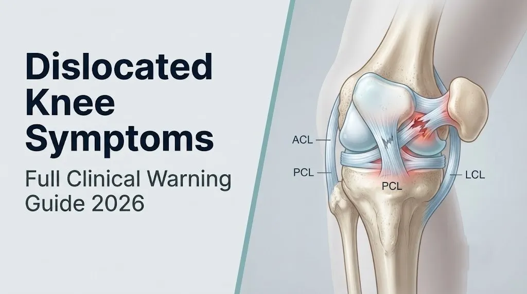
Knee instability is a different but equally important symptom. In a patient whose knee dislocation has spontaneously reduced (gone back into place) before clinical evaluation, there may be no visible deformity at all. What remains is ligamentous laxity: the sensation that the knee is moving in directions it should not, or that it will give way with any attempt at weight bearing.
Instability symptoms to report to your physician:
- The knee feels “loose” or “wobbly” when attempting to straighten or bend it
- A sensation that the tibia slides forward relative to the thigh with knee bending (anterior cruciate insufficiency pattern)
- A feeling that the outer side of the knee buckles outward under load (posterolateral corner insufficiency pattern)
- The knee gives way without warning during attempted walking
- A visible step-off at the back of the knee when viewed from the side
Mayo Clinic notes that clinical instability testing in a severely swollen, painful knee immediately after injury is limited in accuracy, which is why imaging (MRI and stress radiographs) is required for definitive ligamentous assessment.
Patients who have had prior ACL reconstruction surgery may experience combined instability patterns that are difficult to assess clinically. The prior reconstruction may partially mask ligamentous laxity findings on physical examination, making MRI even more important in that population.
Minor Dislocated Knee Symptoms and Partial Dislocation
Minor dislocated knee symptoms occur when a joint subluxation (partial dislocation, where the joint surfaces lose full contact but do not completely separate) or a spontaneously reduced patellar dislocation produces a clinical picture that is less dramatic than a complete dislocation but still indicates ligamentous and structural damage requiring evaluation.
The term “minor” is used here to describe the apparent severity at the time of presentation, not the severity of underlying structural damage. A knee that subluxates and immediately reduces may look surprisingly normal on visual inspection while harboring a complete MPFL tear, a substantial osteochondral fracture fragment, or early ligamentous compromise.
Partial subluxation symptoms:
- A brief sensation of the knee “slipping” or “going wrong” during a movement, followed by immediate resolution
- Dull aching pain in and around the knee joint after the episode
- Mild to moderate swelling that develops over one to four hours (less than the rapid balloon-swelling of complete dislocation with hemarthrosis)
- A sense of apprehension when the knee is put in the same position that caused the episode (apprehension sign)
- Occasional clicking or catching sensations during movement after the episode (from osteochondral fragments or loose bodies within the joint)
A 2020 study published in Knee Surgery, Sports Traumatology, Arthroscopy found that osteochondral fractures (bone and cartilage chips released into the joint) occur in approximately 40 to 70 percent of first-time patellar dislocations, even in cases where the patella reduced spontaneously and symptoms appeared minor. These fragments require MRI identification and, in some cases, surgical removal or fixation.
Individuals who have experienced what feels like a “minor” kneecap slip should be evaluated by a board-certified orthopedic surgeon within 48 hours of the episode and should specifically request MRI of the knee to assess for MPFL integrity and osteochondral injury.
Knee Dislocation Symptoms in Teenagers and Young Athletes
Patellar dislocation is disproportionately common in adolescents, particularly females aged 10 to 17, because skeletal growth, hormonal influences on ligament laxity, and anatomical Q-angle differences combine to make the patellofemoral joint less stable during this developmental period.
The Q-angle is the angle formed between the line of pull of the quadriceps muscle group (from the anterior superior iliac spine to the center of the patella) and the line of the patellar tendon (from the patella to the tibial tuberosity). In the female pelvis, the wider inter-acetabular distance (the distance between the hip sockets) increases this angle, typically placing it between 15 and 20 degrees in adolescent females versus 10 to 15 degrees in males of the same age. A wider Q-angle increases the lateral component of the quadriceps pull on the patella, making lateral patellar subluxation and dislocation more likely under the same loading conditions.
The American Journal of Sports Medicine reports that patellar dislocation occurs at an annual rate of approximately 29 per 100,000 persons overall, with the highest rates in adolescents aged 10 to 17. Recurrence rates after first-time patellar dislocation in adolescents range from 30 to 50 percent, higher than in adult populations, particularly in those with trochlear dysplasia, elevated tibial tuberosity-trochlear groove (TT-TG) distance, or ligamentous hypermobility.
Symptoms in teenagers that differ from adult presentation:
- Higher likelihood of spontaneous reduction without medical intervention (the patella pops back in as the leg straightens)
- More dramatic immediate apprehension and fear of movement after the episode than the pain level alone might suggest
- Rapid return of some mobility that can mislead parents into thinking the injury is minor
- Bone contusions visible on MRI at the lateral femoral condyle and medial patellar facet even when plain X-rays appear normal
Any teenager who reports a first episode of the kneecap visibly sliding sideways, with or without spontaneous reduction, should be evaluated by a board-certified orthopedic surgeon within 24 to 48 hours and receive MRI evaluation of the knee.
Key Takeaway: Adolescent females have the highest patellar dislocation rates of any demographic group due to wider Q-angle anatomy, and up to half of first-time teenage dislocators will experience a repeat episode without proper evaluation and treatment.
Popliteal Artery and Nerve Injury Symptoms in Knee Dislocation
Popliteal artery injury symptoms following true tibiofemoral knee dislocation represent the most dangerous aspect of this injury, as compromised blood flow to the lower leg produces ischemia that, if not surgically corrected within 6 to 8 hours, can lead to permanent muscle and nerve damage or limb loss.
The popliteal artery is particularly vulnerable in knee dislocation for a precise anatomical reason. It enters the popliteal fossa (the space behind the knee) through the adductor hiatus proximally and divides into the anterior and posterior tibial arteries distally, where it is tethered by the fibrous arch of the soleus muscle. These two tethering points mean the artery has very limited ability to shift away from a dislocating tibia. When the joint surfaces separate, the artery stretches. Even if it does not rupture immediately, stretch injury can cause an intimal tear, a microscopic disruption of the inner arterial wall that triggers platelet aggregation and progressive thrombotic occlusion over hours.
This is the clinical danger of what surgeons sometimes call a “warm ischemia window.” A patient may arrive with palpable pulses in the foot, because the popliteal artery is partially injured but still patent. Over the next two to four hours, thrombosis at the intimal tear site progresses until the artery occludes completely. By the time signs of ischemia become obvious, the window for successful arterial repair with full limb salvage has narrowed.
The five classic signs of arterial insufficiency after knee dislocation (the “Five Ps” of vascular injury):
- Pain disproportionate to what the knee injury itself should produce, particularly calf pain
- Pallor: the foot or lower leg appears pale or white compared to the uninjured side
- Paresthesia: numbness or tingling in the foot or lower leg indicating ischemic nerve dysfunction
- Paralysis: inability to move the toes or foot, indicating ischemic or direct nerve injury
- Poikilothermia: the foot is cold to touch compared to the uninjured extremity
The common peroneal nerve (common fibular nerve) is the most frequently injured neural structure in knee dislocation, affected in approximately 10 to 40 percent of cases according to data from the Journal of Bone and Joint Surgery. It wraps around the fibular head at the level of the lateral knee joint, making it vulnerable to stretch in lateral and posterolateral dislocations. Peroneal nerve injury produces foot drop (inability to lift the foot at the ankle, causing the toes to drag when walking) and numbness over the dorsum (top) of the foot and the outer lower leg.
Spontaneous Reduction: When a Dislocated Knee Goes Back In by Itself
Spontaneous reduction occurs when a dislocated joint returns to its normal anatomical position without manual assistance, and it is far more common with patellar dislocations than with tibiofemoral dislocations, but it does not mean the injury was minor or that medical evaluation can be skipped.
In patellar dislocation, spontaneous reduction is the rule rather than the exception. The displaced patella typically snaps back into the trochlear groove when the patient straightens the leg, because quadriceps tension pulls the patella proximally back toward its anatomical seat. This often happens before the patient has time to look at their knee or before bystanders understand what they are seeing. The patient knows something “went wrong” but may present to a medical facility with a knee that appears anatomically normal.
The clinical trap here is underestimation. The structures that tore during the dislocation (the MPFL, the medial retinaculum, the joint capsule, and articular cartilage) are still torn after spontaneous reduction. The hemarthrosis still develops. The osteochondral fracture fragments still float within the joint.
To describe a spontaneous reduction event accurately to a clinician:
- Describe the position the knee was in when the event occurred (twisting, pivoting, direct blow to the outer knee).
- Describe exactly what you saw or felt: did you see the kneecap slide to the outside? Did you feel a pop?
- State whether the knee straightened and the sensation resolved, or whether you required someone to help straighten the leg.
- Note the exact time of the episode, since this determines the timeline for imaging and swelling assessment.
- Describe the current symptoms: residual pain location, swelling volume and speed of onset, ability or inability to bear weight.
Spontaneous tibiofemoral reduction is less common and, when it occurs, is even more deceptive. The patient may have experienced a complete tibiofemoral dislocation with popliteal artery intimal injury, and the joint may now appear reasonably normal. All patients with suspected spontaneous tibiofemoral reduction require vascular assessment including ankle-brachial index (ABI) measurement and, if ABI is below 0.9, computed tomography angiography (CTA) of the popliteal vessel.
Dislocated Knee Symptoms vs Sprained Knee Symptoms
The most reliable clinical differentiators between a dislocated knee and a severe knee sprain are the presence of gross joint deformity, complete ligamentous instability in multiple planes, and neurovascular compromise, all of which point to dislocation rather than sprain.
A severe knee sprain (Grade III ligament tear) and a reduced knee dislocation can produce overlapping symptoms that make clinical differentiation genuinely difficult without imaging. Both produce severe pain, swelling, and instability. The critical differences are in degree and combination.
| Feature | Dislocated Knee (reduced) | Severe Knee Sprain (Grade III) |
|---|---|---|
| Number of ligaments involved | Minimum 2; often 3 to 4 | Typically 1 to 2 |
| Instability direction | Multiple planes simultaneously | Usually one primary plane |
| Vascular compromise signs | Possible (popliteal artery) | Essentially never |
| Neurological symptoms | Possible (peroneal or tibial nerve) | Essentially never |
| Visible deformity at time of injury | Common in tibiofemoral type | Rarely present |
| Speed of swelling | Rapid hemarthrosis: minutes | More gradual: 1 to 4 hours |
| Weight bearing immediately | Almost never possible | Sometimes possible (partial) |
| MRI findings | Multiple complete ligament tears; often bone contusions | Typically 1 to 2 ligament tears |
A useful clinical framework: if a patient reports that the knee “looked wrong” or “was pointing the wrong direction” at any point after the injury, even if it now looks normal, treat this as a potential reduced dislocation until proven otherwise by vascular assessment and MRI. The appearance of normality after spontaneous reduction does not exclude the catastrophic structural damage of a true dislocation.
OrthoInfo (American Academy of Orthopaedic Surgeons) recommends that any knee injury presenting with multiplanar instability on examination be evaluated with MRI to characterize the full ligamentous injury pattern, as clinical examination alone is insufficient for surgical planning.
Older adults with preexisting knee osteoarthritis may present with apparent gross instability from arthritic bone loss rather than ligamentous injury. A plain X-ray can differentiate these presentations by identifying the bony architecture of the joint.
Key Takeaway: If you are not sure whether a knee injury is a dislocation or a severe sprain, assume dislocation until proven otherwise by vascular assessment, because the consequence of missing a popliteal artery injury in a reduced dislocation is limb loss.
How a Dislocated Knee Is Diagnosed
A dislocated knee is diagnosed through a combination of clinical history, physical examination, vascular assessment, and imaging, with the specific sequence determined by whether vascular compromise is suspected, because arterial injury assessment takes priority over all other diagnostic steps.
The diagnostic sequence for suspected true tibiofemoral dislocation unfolds in this order:
- Immediate neurovascular check: Palpate posterior tibial and dorsalis pedis pulses. Check capillary refill time in the toes. Assess foot sensation and motor function (toe and ankle dorsiflexion). Any abnormality in this step triggers immediate vascular surgery consultation before imaging.
- Ankle-brachial index (ABI) measurement: Compare systolic blood pressure at the ankle to the arm. An ABI below 0.9 on the injured side indicates significant arterial compromise and requires CTA or surgical exploration.
- Plain X-ray (anteroposterior and lateral views): Confirms joint position, identifies fractures (including the Segond fracture, an avulsion of the lateral tibial plateau associated with ACL disruption and dislocation), and assesses bone architecture.
- Computed tomography angiography (CTA) of the popliteal vessels: Ordered when ABI is below 0.9 or when clinical signs of vascular injury are present. Identifies intimal tears, thrombosis, or complete arterial transection.
- MRI of the knee: Performed after the acute vascular situation is managed. Provides definitive assessment of all ligament injuries, meniscal tears, osteochondral fractures, and bone contusions. Essential for surgical planning in multi-ligament reconstruction.
For patellar dislocation, the diagnostic sequence is:
- Plain X-ray (including a skyline/sunrise view to assess patella position and trochlear groove morphology)
- MRI to assess MPFL integrity, identify osteochondral fractures, and measure the tibial tuberosity-trochlear groove (TT-TG) distance
Bring the following information to the emergency department or orthopedic appointment:
- Exact mechanism of injury (direction of force, body position at time of injury)
- Whether visible deformity was observed before any reduction occurred
- Any neurological symptoms below the knee (numbness, tingling, foot weakness)
- Any vascular symptoms below the knee (pallor, coldness, pain in the calf)
- Prior knee injuries or surgeries on the same knee
- Current medications, especially anticoagulants and anti-platelet agents
Knee Dislocation Symptoms by Severity: Urgency Framework
The urgency of medical response to knee dislocation symptoms is determined by the specific combination of signs present, ranging from the immediate 911 emergency level of a frank tibiofemoral dislocation with vascular compromise to the 24 to 48-hour orthopedic evaluation level of a spontaneously reduced patellar dislocation with intact neurovascular status.
| Symptom Pattern | Injury Type Suggested | Urgency Level | Appropriate Action |
|---|---|---|---|
| Gross knee deformity, pallor or coldness of foot, absent pulses | Tibiofemoral dislocation with vascular injury | Immediate emergency | Call 911. Do not move. |
| Gross knee deformity, intact pulses, unable to walk | Tibiofemoral dislocation without vascular signs | Emergency | Call 911 or emergency room immediately |
| Foot drop or numbness in outer foot after knee injury | Peroneal nerve injury in dislocation | Emergency | Emergency room immediately |
| Visible patella displacement to outside of knee | Patellar dislocation, unreduced | Urgent | Emergency room same day |
| Kneecap “popped back in” during injury, swollen, tender | Spontaneous patellar reduction | Urgent | Emergency room or orthopedic surgeon within 24 hours |
| Knee “slipped” briefly, mild swelling, able to walk with difficulty | Patellar subluxation | Semi-urgent | Orthopedic evaluation within 48 hours; MRI indicated |
| Severe knee pain, swelling, unable to bear weight, no gross deformity | Possible reduced dislocation or severe sprain | Urgent | Emergency room same day; needs vascular check and imaging |
Patients who are pregnant should note that swelling assessment may be confounded by physiological fluid retention, and standard dosing decisions for imaging contrast agents require obstetric team coordination. All suspected knee dislocations in pregnant patients should be evaluated immediately by an emergency medicine physician.
Emergency Symptoms: When a Dislocated Knee Requires Immediate Care
Certain presentations associated with knee dislocation indicate a vascular or neurological emergency. Do not wait to see if these improve on their own.
Call 911 or go to the nearest emergency room immediately if a knee injury is accompanied by any of the following:
- Gross visible deformity of the knee joint where the tibia or femur appears displaced from normal position (true tibiofemoral dislocation until proven otherwise)
- Pallor, whiteness, or blue-grey discoloration of the foot or lower leg on the injured side (signs of popliteal artery compromise)
- Coldness of the foot compared to the uninjured side, detectable by touching both feet simultaneously (sign of ischemia)
- Absent or markedly diminished pulse at the ankle compared to the uninjured side (confirming vascular disruption)
- Foot drop: inability to lift the front of the foot or extend the toes after a knee injury (common peroneal nerve injury)
- Numbness, tingling, or complete loss of sensation in the foot or outer lower leg (nerve compression or ischemic neuropathy)
- Severe calf pain disproportionate to the apparent knee injury, developing within minutes to hours (compartment syndrome risk or posterior tibial artery compromise)
- Skin tenting: an area of skin over the knee that appears white, blanched, and stretched tightly over a bony prominence (indicates risk of skin necrosis and requires urgent reduction)
These presentations indicate popliteal artery injury, common peroneal nerve injury, or impending compartment syndrome, all of which require emergency surgical assessment within 6 to 8 hours to prevent permanent disability or limb loss. These are not injuries to monitor overnight or evaluate at an urgent care clinic in the morning.
Dislocated Knee Recovery and What to Expect After Treatment
Recovery from a dislocated knee varies dramatically based on injury type, ranging from 4 to 8 weeks for a first-time patellar dislocation managed non-surgically to 9 to 18 months or longer for a tibiofemoral multi-ligament reconstruction with associated vascular repair.
Patellar dislocation recovery after non-surgical management involves three phases. The first phase (weeks 1 to 3) focuses on reducing hemarthrosis and effusion, restoring normal quadriceps activation, and protecting the healing MPFL with a brace that limits flexion past 30 degrees. The second phase (weeks 4 to 8) progresses to full range of motion exercises, quadriceps strengthening with emphasis on the VMO, and proprioception training. The third phase (weeks 8 to 16) involves sport-specific rehabilitation and return-to-activity clearance by a board-certified orthopedic surgeon or sports medicine physician.
Patients who undergo MPFL surgical reconstruction typically require 4 to 6 months before return to competitive sport, with full outcomes often assessed at 9 to 12 months.
Recovery expectations for multi-ligament knee reconstruction after tibiofemoral dislocation:
- Weeks 1 to 6: Protected weight bearing; management of swelling, pain, and range of motion
- Months 2 to 4: Progressive weight bearing, quadriceps and hamstring strengthening
- Months 4 to 9: Advanced strengthening, neuromuscular training, single-leg exercises
- Months 9 to 18: Return-to-sport testing and clearance; some patients never return to pre-injury sport level
OrthoInfo notes that outcomes after tibiofemoral knee dislocation are heavily influenced by the presence of vascular or nerve injury at the time of the acute event, with peroneal nerve injury in particular carrying a variable recovery prognosis. Approximately 50 to 70 percent of peroneal nerve injuries from knee dislocation show partial recovery, but complete recovery is not guaranteed.
Older adults who sustain tibiofemoral dislocation may be candidates for total knee arthroplasty rather than multi-ligament reconstruction, particularly if preexisting severe osteoarthritis is present. This decision is made by a board-certified orthopedic surgeon based on imaging findings, functional demands, and overall health status.
Key Takeaway: A first-time patellar dislocation that is treated seriously from day one, with MRI evaluation and structured rehabilitation targeting the MPFL and VMO, has a much lower recurrence rate than one managed with rest and ice alone. The initial response determines the long-term outcome.
Frequently Asked Questions About Dislocated Knee Symptoms
What does a dislocated knee feel like?
A dislocated knee feels like sudden, severe pain at the moment of injury combined with a sensation that the joint has “gone wrong” or is positioned abnormally.
In a patellar dislocation, patients often describe a feeling of the kneecap sliding to the outside of the knee, followed by an electric or burning pain at the inner side of the joint where the MPFL has torn.
In a true tibiofemoral dislocation, the pain is typically more severe and is accompanied by a sense of gross joint instability that makes any attempted movement feel unsafe or mechanically unsupported.
Can a dislocated knee go back into place on its own?
A dislocated kneecap (patellar dislocation) very frequently reduces spontaneously when the leg is straightened, because quadriceps tension pulls the patella back into the trochlear groove.
A true tibiofemoral knee dislocation rarely reduces spontaneously and almost always requires emergency closed or open reduction by an emergency medicine physician or orthopedic surgeon.
Spontaneous reduction of either type does not indicate a minor injury; all spontaneous reductions require same-day or next-day evaluation because the structural damage (MPFL tear, ligament rupture, osteochondral fracture) remains even after the bones return to position.
How do I know if my knee is dislocated or just sprained?
A dislocated knee typically produces more severe swelling within a shorter time frame (minutes rather than hours), visible or felt joint deformity, and instability in multiple directions simultaneously.
A severe knee sprain produces similar pain and swelling but typically without gross deformity, without multi-directional instability, and without any neurovascular symptoms below the knee.
Both severe sprains and reduced dislocations require same-day emergency or urgent care evaluation, because differentiating them clinically requires physical examination and MRI.
Is a dislocated knee an emergency?
True tibiofemoral knee dislocation is always a medical emergency because of the 20 to 40 percent risk of popliteal artery injury that can result in limb loss if not surgically addressed within 6 to 8 hours.
Patellar dislocation that has not spontaneously reduced is an urgent condition requiring same-day emergency room evaluation and closed reduction under sedation.
Even a spontaneously reduced patellar dislocation warrants evaluation within 24 to 48 hours, as MRI is needed to assess for MPFL tears and osteochondral fractures that require surgical intervention in a proportion of cases.
What are the symptoms of a dislocated kneecap in a teenager?
Dislocated kneecap symptoms in a teenager include a visible lump on the outer side of the knee, sudden severe pain with a twisting movement, and immediate inability to bear weight, often followed by spontaneous resolution when the leg is straightened.
Adolescents are the highest-risk demographic for patellar dislocation due to wider Q-angle anatomy, and recurrence rates are 30 to 50 percent after first-time dislocation in this age group without proper treatment.
Any teenager with a first episode of kneecap dislocation should be evaluated by a board-certified orthopedic surgeon within 24 to 48 hours and receive MRI evaluation of the medial patellofemoral ligament and articular surfaces.
How long does it take to recover from a dislocated knee?
Recovery from a first-time patellar dislocation managed without surgery typically takes 4 to 8 weeks for basic mobility, with full return to competitive sport usually requiring 3 to 4 months of structured rehabilitation.
Recovery from a surgically managed MPFL reconstruction after patellar dislocation typically requires 4 to 6 months before return to sport.
Recovery from a tibiofemoral multi-ligament knee reconstruction after true knee dislocation takes 9 to 18 months, and outcomes are substantially influenced by whether vascular or nerve injury occurred at the time of the original dislocation.
Closing
The most important thing to take from this guide is the separation between two injuries that share a name but have entirely different urgency profiles. A patellar dislocation deserves prompt orthopedic evaluation, proper imaging, and structured rehabilitation. A true tibiofemoral dislocation requires 911 activation and emergency vascular assessment, because the six-to-eight-hour window for popliteal artery repair closes faster than most people expect.
If you are reading this after a knee injury, check for the five vascular signs right now: pulse at your ankle, temperature of your foot, color of your lower leg, sensation in your foot, and ability to lift your toes. Any of those signs being abnormal is a 911 call, not a phone consult.
If your kneecap slipped sideways and came back on its own, do not assume you are fine. Book an orthopedic evaluation within 24 to 48 hours, ask specifically for an MRI, and give your surgeon the full description of the mechanism: exactly what position you were in and exactly what you saw and felt when it happened.