C5-C6 Disc Herniation Symptoms: Complete 2026 Guide
C5-C6 disc herniation symptoms include neck pain, arm pain radiating from the shoulder to the thumb and index finger, numbness or tingling in the thumb and first two fingers, biceps and wrist extensor weakness, and a depressed biceps tendon reflex, all produced by compression of the C6 nerve root as it exits the spinal cord between the fifth and sixth cervical vertebrae. The specific pattern of symptoms maps precisely to the C6 nerve root’s anatomical territory and is distinct from every other cervical disc herniation level.
The C5-C6 disc level is the most frequently herniated segment in the entire cervical spine. According to epidemiological data published in Spine, C5-C6 accounts for a substantial proportion of all cervical disc herniations, exceeded only marginally by C6-C7 in some populations. What most generic content fails to explain is that symptoms arise not just from mechanical pressure on the nerve, but from a chemical inflammatory cascade driven by enzymes released from the herniated disc material itself.
This article covers the complete C5-C6 herniation symptom picture with the anatomical and physiological mechanism behind each finding, explains why the direction of herniation matters for which symptoms develop, distinguishes radiculopathy from the more serious myelopathy, names the specific red flags requiring emergency evaluation, and outlines what the diagnostic process and realistic recovery look like.
C5-C6 Disc Herniation Symptoms: What You Need to Know
The hallmark C5-C6 disc herniation symptoms are unilateral neck pain, pain radiating down one arm from the shoulder to the thumb side of the hand, numbness or tingling in the thumb and index finger, weakness of the biceps and wrist extensors, and a reduced or absent biceps tendon reflex on the affected side.
These symptoms form a recognizable clinical pattern because they all reflect dysfunction of a single nerve root: C6. The C6 nerve root carries sensory fibers to the thumb and index finger (its dermatomal territory), motor fibers to the biceps brachii and wrist extensor muscles (its myotomal territory), and contributes to the biceps tendon reflex arc. Damage or irritation at C6 affects all three simultaneously in varying degrees.
Not everyone with a C5-C6 herniation experiences all of these symptoms at once. Some present with predominantly pain. Others report numbness without pain. The severity and combination depend on whether the herniated disc material is compressing the nerve mechanically, irritating it chemically through inflammatory mediators, or doing both.
Key symptoms by category:
- Neck pain: localized to the lower cervical spine, often worse with extension or ipsilateral lateral rotation
- Shoulder and periscapular pain: aching between the shoulder blades or deep in the deltoid region
- Arm pain: shooting or burning pain traveling from the shoulder down the lateral forearm to the thumb and index finger
- Sensory changes: numbness, tingling, or burning in the thumb, index finger, and the lateral forearm
- Motor changes: weakness opening jars, doing bicep curls, or extending the wrist
- Reflex change: depressed or absent biceps tendon reflex on the affected side
Individual variation note: People with pre-existing cervical spondylosis (age-related bony degenerative changes at the uncovertebral joints and facet joints) may have a narrower baseline neuroforamen at C5-C6 before any disc herniation occurs. In these individuals, even a small disc protrusion can produce severe C6 nerve root symptoms because the foramen has less reserve space.
Anatomy of the C5-C6 Spinal Segment
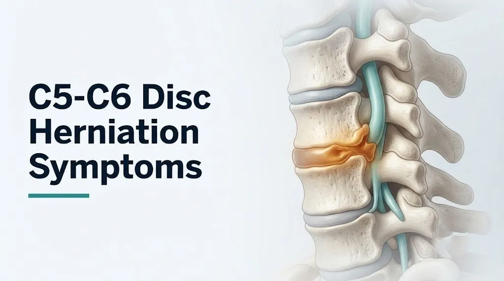
The C5-C6 intervertebral disc sits between the fifth and sixth cervical vertebral bodies and serves as the pivot point for a large proportion of cervical flexion and extension movement, making it the most mechanically stressed disc in the lower cervical spine.
Each cervical intervertebral disc has two structural components. The nucleus pulposus is the gel-like inner core, composed predominantly of water and proteoglycans (aggrecan) held together by a loose collagen II matrix. The annulus fibrosus is the tough outer ring, built from concentric layers of collagen I fibers oriented at alternating angles to resist rotational and compressive forces. The posterior aspect of the annulus fibrosus, closest to the spinal canal, is the thinnest and weakest zone, which is why most disc herniations project posteriorly or posterolaterally.
The C6 nerve root exits the spinal canal through the C5-C6 neuroforamen, the bony channel formed between the inferior articular process of C5 and the superior articular process of C6, bordered anteriorly by the uncinate process of C6 and the posterolateral disc. This foramen is narrow by design, leaving the C6 nerve root with minimal space to accommodate any additional volume from a herniated disc fragment.
Think of the neuroforamen like a narrowly fitted pipe through which the C6 nerve root runs. The disc sits directly against the front wall of that pipe. When disc material herniates posterolaterally, it acts like a stone dropped into a tight pipe, pressing the nerve root against the bony posterior wall. That pressure on the dorsal root ganglion (the cluster of sensory neuron cell bodies housed just inside the foramen) is particularly painful because the ganglion lacks the protective perineurium that shields distal nerve segments.
Individual variation note: Women have statistically smaller neuroforaminal dimensions at C5-C6 than men on average, according to morphometric studies of cervical spine anatomy. This means equivalent disc herniation volume can produce more severe nerve compression in women than in men of the same age and disc degeneration stage.
How a C5-C6 Disc Herniation Compresses the C6 Nerve Root
A C5-C6 disc herniation compresses the C6 nerve root through two simultaneous mechanisms: direct mechanical displacement of the herniated nucleus pulposus against the nerve root in the neuroforamen, and a neurochemical inflammatory cascade driven by phospholipase A2 (PLA2) and tumor necrosis factor-alpha (TNF-alpha) released from the herniated disc material.
Most people understand the mechanical part. What few realize is that the chemical mechanism is equally important in producing radicular pain. Research by Saal et al., published in Spine in 1990 and subsequently confirmed by multiple studies, identified high concentrations of PLA2 in herniated nucleus pulposus material. PLA2 cleaves membrane phospholipids in the nerve root sheath to release arachidonic acid, which is then metabolized into prostaglandins (particularly prostaglandin E2) and leukotrienes. These mediators directly sensitize C6 nociceptors, producing pain and allodynia even in regions where mechanical compression is minimal.
Simultaneously, macrophages recruited to the disc-nerve interface release interleukin-1 beta (IL-1 beta) and IL-6, which amplify nociceptor sensitivity and promote the production of substance P in C6 dorsal root ganglion neurons. Substance P is released at both the central end of the ganglion (within the spinal cord, amplifying pain signal transmission) and the peripheral end (in the nerve root sheath, producing local neurogenic vasodilation and edema that further compresses the nerve in the confined foramen).
This neurochemical cascade explains a clinically important observation: patients with small disc herniations on MRI can have severe radicular pain, while patients with large herniations sometimes have minimal symptoms. The size of the disc fragment matters less than the degree of inflammatory response it triggers. This also explains why oral corticosteroids (which suppress the phospholipase A2 and prostaglandin cascade) and epidural steroid injections can provide meaningful symptom relief without decompressing the disc mechanically.
Individual variation note: People who smoke have accelerated avascular degeneration of the nucleus pulposus through reduced nutrient delivery via the vertebral endplate capillary network. The degenerated nucleus pulposus of a chronic smoker releases higher concentrations of proinflammatory cytokines on herniation than a healthier disc, potentially explaining the association between smoking and worse radiculopathy severity documented in observational data published in Spine.
Key Takeaway: C5-C6 radiculopathy is driven by both mechanical compression of the C6 nerve root and a phospholipase A2-mediated neurochemical inflammatory cascade, which is why anti-inflammatory treatments can reduce pain even without surgical decompression.
Neck and Shoulder Pain from C5-C6 Herniation
Neck pain from a C5-C6 disc herniation originates from nociceptive fibers innervating the posterior annulus fibrosus, the posterior longitudinal ligament, and the facet joint capsules at C5-C6, producing a deep, aching pain localized to the lower cervical spine that worsens with neck extension and ipsilateral lateral bending.
The posterior annulus fibrosus and posterior longitudinal ligament are densely innervated by the sinuvertebral nerve (recurrent meningeal nerve), which carries pain signals back into the spinal cord. When a disc herniates posteriorly, tearing through the annular fibers, these nociceptive nerves are directly activated, producing the local neck pain component. This pain is typically worse with neck extension because that position narrows the posterior disc space and neuroforamen further, increasing pressure on both the annular tear and the nerve root.
Shoulder and periscapular pain in C5-C6 herniation reflects referred pain traveling along the C6 nerve root’s sensory territory. The deep, aching quality of referred pain from the disc itself, without radiation to the arm, can be easily confused with shoulder pathology such as rotator cuff tendinopathy or acromioclavicular joint degeneration. The clinical distinction matters because these conditions require different management. Shoulder joint pathology worsens with shoulder movement independent of neck position. C5-C6 referred shoulder pain typically worsens with neck movements rather than isolated shoulder abduction or internal rotation.
The Mayo Clinic notes that cervical radiculopathy pain commonly presents as a sharp, burning, or shooting sensation that begins in the neck and radiates into the shoulder, arm, and hand along a dermatomal path, distinguishing it from the more diffuse ache of rotator cuff or acromioclavicular disorders.
Individual variation note: People with concurrent ipsilateral shoulder pathology (rotator cuff tears, biceps tendon pathology) can have both sources of pain active simultaneously. In these cases, a Spurling’s test (cervical compression test eliciting radicular symptoms) and shoulder-specific provocative tests should both be performed to avoid attributing all pain to a single source.
Arm Pain and Radiculopathy from C5-C6 Nerve Root Compression
Cervical radiculopathy from C5-C6 nerve root compression produces a shooting, burning, or electric-shock-quality pain that travels from the lower neck and shoulder down the lateral aspect of the upper arm, continues along the lateral forearm, and terminates in the thumb and index finger, following the C6 dermatomal distribution precisely.
The word “radiculopathy” combines the Latin radix (root) with the Greek pathos (disease). It describes pathology of the nerve root itself rather than the nerve at its distal distribution. This distinction matters clinically: the pain is perceived along the entire length of the C6 nerve root’s territory because the brain maps all pain signals from C6 sensory fibers as originating in the arm and hand, even though the actual injury is at the root level in the neck.
Radicular arm pain is characteristically worsened by specific cervical positions. Extension of the neck, lateral bending toward the affected side, or direct ipsilateral neck rotation reduces the C5-C6 foraminal diameter by approximately 20% to 30%, according to anatomical studies of cervical foraminal dimension with positional change. These movements reliably reproduce arm pain in true radiculopathy. Conversely, the shoulder abduction relief sign (raising the affected arm overhead, which reduces longitudinal traction on the nerve root) often partially relieves radicular arm pain and is a clinically useful diagnostic observation.
Radicular pain from C5-C6 compression has a characteristic quality that distinguishes it from musculoskeletal arm pain. It is frequently described as burning, electric, or shooting, rather than the deep, constant ache of muscle or joint pain. It may be present at rest and worsen with Valsalva maneuver (coughing, sneezing, or bearing down), because increased intrathecal pressure from Valsalva augments the forces acting on an already compressed nerve root.
Individual variation note: Athletes involved in contact sports (football, wrestling, rugby) who experience acute arm pain after a cervical impact should be evaluated by an emergency medicine physician before returning to play, because the symptom pattern of a “stinger” (transient brachial plexopathy from cervical nerve root stretch) can overlap with acute C5-C6 disc herniation and requires imaging to distinguish.
Numbness and Tingling in the Hand from C5-C6 Compression
Numbness, tingling, or a pins-and-needles sensation in the thumb, index finger, and the lateral aspect of the forearm is the most specific sensory symptom of C6 nerve root compression and reflects disrupted sensory axon conduction in the cutaneous branches of the C6 dermatomal territory.
The dermatomal distribution of C6 is clinically predictable. The C6 sensory root innervates the lateral forearm, the thumb, and the index finger, with some overlap into the middle finger. When the C6 root is compressed, the affected sensory axons cannot conduct electrical impulses normally. In the early phase, this produces paresthesia (the tingling, pins-and-needles sensation) as mildly damaged axons fire spontaneously and irregularly. With more sustained compression, sensory axons begin to fail entirely, producing hypesthesia (reduced sensation) or full anesthesia (absent sensation) in the C6 dermatomal territory.
The specific fingers affected are diagnostically useful. Numbness predominantly in the thumb and index finger, with relative sparing of the ring and little fingers, strongly suggests C6 involvement rather than C7 (which would affect the middle finger more prominently) or carpal tunnel syndrome (which affects the thumb, index, middle, and half the ring finger due to median nerve compression at the wrist).
Distinguishing C6 radiculopathy from carpal tunnel syndrome (median nerve compression at the wrist) based on finger numbness alone can be clinically misleading, because both conditions affect the thumb and index finger. The distinguishing features are: carpal tunnel syndrome numbness is worse at night with wrist flexion and is not associated with neck pain or worsened by cervical movements; C6 radiculopathy numbness is reproduced by neck extension and ipsilateral cervical lateral bending (positive Spurling’s test) and accompanied by neck or shoulder pain.
The North American Spine Society (NASS) clinical guidelines for cervical radiculopathy recommend electrodiagnostic testing (EMG and nerve conduction velocity study) when the clinical presentation doesn’t clearly differentiate C6 radiculopathy from peripheral entrapment neuropathy such as carpal tunnel syndrome, because both can coexist (a condition called double crush syndrome).
Individual variation note: Older adults over 65 may have baseline peripheral neuropathy from diabetes, vitamin B12 deficiency, or prior chemotherapy that produces hand numbness independent of any cervical pathology. In this population, the dermatomal distribution of numbness is a less reliable diagnostic indicator, and electrodiagnostic testing plays a more important role in determining the level and source of nerve involvement.
Key Takeaway: Numbness in the thumb and index finger with neck pain worsening on extension is the most specific sensory indicator of C6 nerve root compression, but it must be distinguished from carpal tunnel syndrome and peripheral neuropathy through clinical examination and electrodiagnostic testing.
Muscle Weakness from C5-C6 Herniated Disc
Muscle weakness from a C5-C6 herniated disc affecting the C6 nerve root involves the biceps brachii, brachioradialis, extensor carpi radialis longus, and extensor carpi radialis brevis, producing detectable weakness in elbow flexion, forearm supination, and wrist extension on the affected side.
These muscles form the C6 myotome, the specific group of muscles primarily innervated by motor axons traveling in the C6 nerve root. Motor axons exit the dorsal root ganglion in the C6 root and travel distally in the brachial plexus before reaching their target muscles. When these axons are compressed or chemically irritated at the C5-C6 disc level, the muscles they supply receive fewer functional motor impulses, producing weakness that is typically graded from mild (detectable only on formal strength testing) to moderate (affecting daily activities such as opening jars, lifting, or writing) to severe (inability to flex the elbow or extend the wrist against gravity).
In clinical practice, C6 weakness is tested by asking the patient to flex the elbow against resistance (testing the biceps brachii, the primary C5-C6 elbow flexor) and to extend the wrist against resistance (testing the extensor carpi radialis muscles, primary C6 wrist extensors). Weakness in both of these movements, combined with sensory changes in the C6 dermatomal territory and a depressed biceps reflex, forms the classic C6 radiculopathy triad that strongly points to C5-C6 disc pathology.
The American Academy of Orthopaedic Surgeons (AAOS) evidence-based clinical practice guidelines note that progressive motor weakness, meaning weakness that is worsening over days to weeks rather than remaining stable, is one of the primary indications for expedited specialist evaluation and potential surgical intervention, even when pain levels are manageable.
Individual variation note: People with dominant arm involvement (right-arm weakness in right-handed individuals, for example) experience more functional impairment from equivalent C6 weakness than those with non-dominant arm involvement, because the dominant arm performs the fine motor tasks most sensitive to early weakness. This population should be prioritized for specialist evaluation without waiting the full 6-week conservative management period if weakness is affecting their ability to work or perform essential daily activities.
Biceps Reflex Changes from C5-C6 Disc Herniation
A reduced or absent biceps tendon reflex on the side of the herniation is one of the most objectively reliable signs of C6 nerve root dysfunction from C5-C6 disc herniation, because the reflex arc runs specifically through the C5-C6 nerve roots and the musculocutaneous nerve to the biceps brachii.
The biceps tendon reflex arc works this way: when a reflex hammer strikes the biceps tendon at the antecubital fossa, a stretch signal travels up the musculocutaneous nerve to the C5-C6 spinal cord segments. There, an interneuron synapses on anterior horn motor neurons that send a signal back down to the biceps brachii, producing the reflex contraction. If the C6 nerve root is compressed and cannot conduct this signal normally, the arc is interrupted, and the reflex is diminished or absent on that side.
A depressed or absent biceps reflex on one side, with a normal or hyperactive reflex on the other, is a clinically meaningful finding that strongly suggests ipsilateral C6 nerve root dysfunction. It’s a fact of spinal anatomy that no amount of muscle soreness, guarding, or pain independently suppresses a deep tendon reflex. A reduced reflex indicates actual nerve root or motor neuron involvement, not just pain.
The brachioradialis reflex (C6) is often also reduced or absent in C5-C6 herniation. A particularly important finding is the inverted radial reflex: when tapping the brachioradialis tendon elicits finger flexion rather than the normal brachioradialis contraction, it indicates both a depressed C6 reflex arc and hyperreflexia of the finger flexors from cord compression just below the level. The inverted radial reflex is a sign of concurrent myelopathy and should prompt urgent specialist evaluation.
Quick Tip:
- A reflex that is present but asymmetrically reduced (compared to the opposite side) is as clinically meaningful as an absent reflex.
- Bilateral reflex depression in both biceps tendons is not typical of a single-level disc herniation and suggests a systemic neuromuscular condition, polyneuropathy, or multi-level cervical pathology.
- People taking muscle relaxants or central nervous system depressants (opioids, benzodiazepines) may have globally reduced deep tendon reflexes that obscure the asymmetric finding a clinician is looking for.
Individual variation note: Older adults may have globally reduced deep tendon reflexes due to age-related peripheral nerve changes, making the asymmetric depression less obvious than in younger patients. In this population, the Spurling’s test and EMG/nerve conduction velocity study carry more diagnostic weight than the clinical reflex examination alone.
Central vs. Posterolateral vs. Foraminal C5-C6 Herniation Symptoms
The direction of disc herniation at C5-C6 determines which structures are compressed and which symptoms dominate, with posterolateral herniation producing classic unilateral C6 radiculopathy, central herniation risking both-sides arm symptoms and cervical myelopathy, and foraminal herniation producing pure unilateral radiculopathy with the most predictable surgical anatomy.
Think of the C5-C6 disc as a squeezed tube of toothpaste. The cap is open at the back, but the contents can exit slightly to the left, squarely in the center, or slightly to the right. Where the material exits determines what it touches.
Posterolateral herniation is the most frequent pattern. The nucleus pulposus protrudes posterolaterally (off-center to one side), pressing against the C6 nerve root as it enters the ipsilateral neuroforamen. Symptoms are unilateral: the arm pain, numbness, and weakness are all on one side only. The spinal cord itself is typically spared because the herniation is eccentric, away from the midline.
Central herniation is the pattern with the highest clinical urgency. The disc material protrudes directly posterior into the center of the spinal canal. At the C5-C6 level, the spinal cord occupies this space. Central herniation at C5-C6 compresses the anterior cervical spinal cord, particularly the corticospinal tracts and the anterior horn cells. This produces cervical myelopathy (upper motor neuron syndrome) rather than simple radiculopathy. Symptoms can be bilateral (both arms), involve the legs (spastic gait, leg stiffness), and include bowel or bladder dysfunction.
Foraminal herniation occurs entirely within the lateral recess or foramen itself, impacting the exiting nerve root with high specificity. Symptoms are identical to posterolateral herniation but often more severe and more precisely confined to the C6 dermatomal territory, because the compression is isolated to the nerve root without any canal compromise.
| Herniation Direction | Structures Compressed | Primary Symptoms | Myelopathy Risk |
|---|---|---|---|
| Posterolateral | C6 nerve root, partial canal | Unilateral arm pain, thumb/index numbness, biceps weakness | Low |
| Central | Anterior spinal cord, bilateral C6 roots | Bilateral arm symptoms, leg weakness, gait disturbance, bladder dysfunction | High |
| Foraminal | C6 nerve root in foramen | Pure unilateral radiculopathy, precise C6 dermatomal distribution | Very low |
| Far lateral (extraforaminal) | C5 nerve root (not C6) | Shoulder and lateral arm symptoms, deltoid weakness | Very low |
Individual variation note: People with congenitally narrow spinal canals (cervical canal stenosis) have less reserve space in the spinal canal. In these individuals, even a posterolateral herniation that would produce only unilateral radiculopathy in a person with a normal-sized canal can compress the lateral spinal cord, producing myelopathic features in addition to radiculopathy.
Key Takeaway: Central C5-C6 disc herniation is the most urgent presentation because it compresses the cervical spinal cord rather than just the C6 nerve root, producing myelopathy with bilateral symptoms, leg involvement, and bowel or bladder dysfunction that requires urgent surgical evaluation.
Cervical Myelopathy Symptoms from C5-C6 Central Disc Herniation
Cervical myelopathy from a C5-C6 central disc herniation occurs when herniated disc material compresses the anterior cervical spinal cord, injuring the corticospinal tracts and producing upper motor neuron dysfunction in the arms and legs, with symptoms that are fundamentally different from and more serious than simple radiculopathy.
Unlike radiculopathy (nerve root injury), myelopathy involves the spinal cord itself. The spinal cord at the C5-C6 level carries motor commands from the brain to every muscle below the neck via the corticospinal tracts, and sensory information from the legs, trunk, and arms up to the brain via the spinothalamic tracts and dorsal columns. Compression at this level produces a spectrum of upper motor neuron findings that affect not just the arms but the entire body below the lesion.
Symptoms of cervical myelopathy from C5-C6 central herniation include:
- Bilateral hand clumsiness or loss of fine motor coordination (difficulty buttoning shirts, typing, or holding small objects)
- Lhermitte’s sign: an electric-shock sensation running from the neck down the spine into the legs, triggered by neck flexion (reflects compression of the posterior columns of the spinal cord)
- Hoffmann’s sign: when the clinician flicks the patient’s middle fingernail downward, the thumb and index finger involuntarily flex; this is a pathological upper motor neuron reflex indicating corticospinal tract dysfunction
- Lower extremity stiffness, heaviness, or a “wooden legs” sensation
- Spastic gait: stiff-legged, wide-based walking from corticospinal tract injury to the legs
- Hyperreflexia: exaggerated deep tendon reflexes in the knees and ankles (paradoxically increased reflexes in the legs despite the neck being the site of pathology, because upper motor neuron injury removes inhibitory control over spinal reflexes)
- Urinary urgency, frequency, or incontinence from disruption of corticospinal tract control over the sacral bladder centers
- In severe cases: weakness in both arms and both legs (quadriparesis)
According to a clinical guideline published in Neurosurgery, cervical spondylotic myelopathy is the most common cause of spinal cord dysfunction in adults over 55, and C5-C6 is one of the most common levels involved. The condition can progress slowly and insidiously, with patients attributing their gait changes to aging before recognizing the neurological basis.
Individual variation note: Older adults with cervical spondylotic myelopathy (myelopathy from chronic bony and disc degeneration rather than acute herniation) may present with slowly progressive gait disturbance over months to years rather than the more acute presentation seen in younger patients with sudden disc herniation. The gradual onset makes early diagnosis harder and increases the risk of delayed surgical intervention after permanent spinal cord injury has occurred.
Red Flags and Emergency Symptoms of C5-C6 Disc Herniation
Certain symptoms associated with C5-C6 disc herniation require immediate emergency evaluation. Do not wait to see if these resolve on their own.
Call 911 or go to the nearest emergency room immediately if you experience:
- Sudden loss of bladder or bowel control in the setting of any neck or arm symptoms, because this indicates acute spinal cord compression affecting the sacral autonomic centers and constitutes a neurological emergency requiring emergency surgical decompression
- Sudden weakness or paralysis of both arms, both legs, or all four limbs (acute quadriparesis or quadriplegia), indicating acute central cervical spinal cord compression at or above the C5-C6 level
- Rapidly progressive leg weakness making walking impossible or unsafe within hours, reflecting acute corticospinal tract injury from central disc herniation at C5-C6
- A positive Lhermitte’s sign (electric shock sensation down the spine with neck flexion) accompanied by bilateral arm or leg symptoms, because this combination indicates active cervical myelopathy requiring urgent surgical evaluation within days, not weeks
- Sudden severe increase in cervical pain following trauma (car accident, fall, sports impact), even with pre-existing disc disease, because this may indicate acute disc herniation into a previously stable cord compression
- Fever with neck pain and arm symptoms together, because this combination raises concern for cervical epidural abscess, which mimics disc herniation but is an infectious emergency
- Bilateral arm weakness that has progressed over days to weeks, combined with any lower extremity changes, because this pattern indicates myelopathy progression that carries a risk of permanent spinal cord injury without surgical intervention
Red flags requiring urgent (same-day to 48-hour) evaluation by an orthopedic spine surgeon or neurosurgeon (not emergency room):
- Worsening motor weakness in the biceps, wrist extensors, or hand grip over days to weeks
- Any symptom pattern suggesting myelopathy (clumsiness, coordination changes, leg stiffness) without emergency features listed above
These presentations can indicate acute cervical myelopathy, acute cord compression, or cervical epidural abscess and require emergency medical assessment, not an urgent care appointment or a phone call to a primary care office.
How Age, Sex, and Occupation Affect C5-C6 Herniation Symptoms
The clinical presentation of C5-C6 disc herniation varies by age, biological sex, and occupational history in ways that determine both symptom severity and the dominant compressive mechanism.
Age profoundly changes the pathological mechanism. In adults under 45, acute C5-C6 symptoms most commonly reflect herniation of the nucleus pulposus through a tear in the annulus fibrosus: a relatively sudden event producing acute radiculopathy with a significant inflammatory component. This population tends to have a higher proportion of inflammatory (neurochemical) radiculopathy and responds better to anti-inflammatory interventions. In adults over 55, the dominant mechanism shifts to cervical spondylosis: osteophyte formation at the uncovertebral joints and posterior disc space, hypertrophy of the posterior longitudinal ligament, and facet joint arthrosis, all of which progressively narrow the neuroforamen. Symptoms develop more gradually and tend to be more chronic, with less acute-on-chronic inflammatory component.
Biological sex affects both the anatomy and the pain experience. Morphometric studies of cervical spine anatomy have documented smaller neuroforaminal dimensions in women than men at equivalent disc degeneration grades, meaning that women with equivalent disc herniation size may experience more severe nerve root compression. Separately, observational data published in Spine found that women with cervical radiculopathy report higher pain intensity scores on the Visual Analog Scale than men with equivalent MRI findings, a pattern attributed to multiple factors including sex-based differences in central pain processing and peripheral nociceptor density.
Occupation is one of the most modifiable risk factors for C5-C6 disc herniation and symptom severity. Sustained cervical flexion during desk or screen work, heavy manual labor involving overhead lifting, repetitive vibration exposure (construction equipment operators, forklift drivers), and high-impact contact sports all accelerate cervical disc degeneration at C5-C6 and increase herniation risk, according to occupational epidemiology data published in Spine.
Individual variation note: People with rheumatoid arthritis affecting the cervical spine have a higher baseline risk of atlantoaxial instability (C1-C2) that can co-exist with C5-C6 disc disease. In this population, any cervical intervention (physical therapy, manipulation) requires prior imaging of the entire cervical spine, not just the symptomatic C5-C6 level, to ensure there is no occult instability at higher cervical segments.
How Doctors Diagnose a C5-C6 Disc Herniation
The diagnosis of a C5-C6 disc herniation is established through a combination of clinical history, physical examination findings specific to C6 nerve root dysfunction, and magnetic resonance imaging (MRI) of the cervical spine, with electrodiagnostic testing added when the clinical and imaging findings don’t fully align.
The diagnostic process unfolds in a structured sequence:
- Clinical history: The clinician documents the onset, quality, radiation pattern, and aggravating/relieving factors of pain and sensory changes, asking specifically about positions that worsen arm symptoms (neck extension, ipsilateral lateral bending) and the shoulder abduction relief sign.
- Physical examination: Evaluation of the biceps tendon reflex (C5-C6 arc), strength testing of the biceps and wrist extensors (C6 myotome), sensory testing of the C6 dermatomal territory (thumb, index finger, lateral forearm), and provocative tests including Spurling’s test (downward axial compression with ipsilateral lateral bending reproducing radicular symptoms) and the distraction test (upward distraction of the head temporarily relieving radicular symptoms by enlarging the foraminal diameter).
- MRI cervical spine: The gold standard imaging study for soft tissue disc pathology. MRI identifies the location, direction, and size of disc herniation, the degree of foraminal compromise, and the presence of spinal cord signal changes (indicating myelopathy). The American Academy of Orthopaedic Surgeons (AAOS) recommends MRI as the imaging of choice for suspected cervical radiculopathy when neurological deficits are present or conservative management has failed after 4 to 6 weeks.
- Cervical spine X-rays with flexion-extension views: Used to assess for instability, degenerative changes, disc space narrowing, and foraminal osteophytes. Less sensitive than MRI for disc herniation but accessible and useful as a first-line study.
- EMG and nerve conduction velocity (NCV) study: Ordered when MRI and clinical findings don’t clearly distinguish C6 radiculopathy from carpal tunnel syndrome, other peripheral entrapment neuropathies, or brachial plexopathy. EMG identifies the specific muscle groups showing denervation, confirming the nerve root level involved.
- CT myelogram: Reserved for patients who cannot undergo MRI (pacemaker, certain implants) or when MRI is inconclusive. CT myelogram provides excellent bony detail of the foraminal anatomy.
Individual variation note: Pregnant women who develop C5-C6 symptoms during pregnancy cannot undergo MRI with gadolinium contrast. Non-contrast MRI is generally preferred and should be discussed with an obstetrician and spine specialist jointly before ordering.
Key Takeaway: MRI of the cervical spine is the diagnostic gold standard for C5-C6 disc herniation when neurological deficits are present, but EMG and nerve conduction velocity studies are essential when symptoms overlap with carpal tunnel syndrome or when MRI findings don’t match the clinical picture.
Natural History and Recovery from C5-C6 Disc Herniation
The natural history of C5-C6 disc herniation with C6 radiculopathy is generally favorable: approximately 75% to 90% of patients with acute cervical radiculopathy improve with conservative management within 4 to 6 weeks, according to prospective observational data cited in the North American Spine Society (NASS) clinical guidelines for cervical radiculopathy.
This favorable natural history reflects a well-documented biological process. Over weeks to months, herniated nucleus pulposus material undergoes progressive desiccation (water loss) and resorption by macrophages. The disc fragment literally shrinks. As the volume of herniated material decreases, the mechanical pressure on the C6 nerve root reduces, and the neurochemical inflammatory cascade subsides as the source of PLA2 and cytokines is removed. MRI studies tracking disc herniation size over time have shown that large herniations, paradoxically, tend to show greater resorption than small protrusions, because larger fragments expose more nucleus pulposus to macrophage access.
The natural history is not uniformly favorable for all presentations. The specific factors that predict a worse prognosis for spontaneous recovery include:
- Progressive motor weakness (rather than stable weakness or predominant pain)
- Central herniation with myelopathic features (which does not resolve with conservative management and tends to progress without surgical intervention)
- Large disc extrusions or sequestrations (free disc fragments in the canal), which have higher rates of nerve root adherence and slower resorption
- Patients over 65 with cervical spondylotic myelopathy rather than soft disc herniation, where the bony and ligamentous compressive structures do not spontaneously reduce
A landmark randomized trial comparing surgical vs. conservative management of cervical radiculopathy, summarized in the New England Journal of Medicine, found that while surgical patients improved faster in the first few months, conservative patients reached equivalent outcomes at 12 to 24 months in the absence of progressive neurological deficits.
Individual variation note: Patients with diabetes mellitus have slower peripheral nerve recovery from compressive injury because hyperglycemia impairs Schwann cell function and axonal remyelination. A diabetic patient with C6 radiculopathy may take substantially longer to recover motor and sensory function than a non-diabetic patient with equivalent disc herniation.
Treatment Approaches for C5-C6 Disc Herniation Symptoms
Treatment of C5-C6 disc herniation symptoms ranges from structured conservative management as the first-line approach for most patients to surgical decompression for those with progressive neurological deficits or myelopathy, with the specific treatment pathway guided by symptom severity, neurological findings, and imaging characteristics.
Conservative management options for C6 radiculopathy without progressive deficits:
- Physical therapy: Cervical traction (manual or mechanical, which increases foraminal diameter and reduces nerve root compression), cervical stabilization exercises, and postural retraining; the NASS guidelines support physical therapy as a first-line treatment
- Activity modification: Avoiding sustained cervical flexion, prolonged screen work without neck support, and overhead activities that worsen foraminal narrowing
- Cervical soft collar: Short-term use (days, not weeks) to rest the cervical spine during acute severe pain; prolonged collar use causes cervical muscle deconditioning
- Oral nonsteroidal anti-inflammatory drugs (NSAIDs): Suppress the prostaglandin component of the neurochemical inflammatory cascade at the nerve root; named for clinical context only, not as a specific recommendation
- Oral corticosteroids (short-course): More potent suppression of the PLA2/prostaglandin cascade; named for clinical context, not recommended without physician guidance
- Gabapentinoids (gabapentin, pregabalin): Target central sensitization at the dorsal horn, reducing the neuropathic pain component; named for clinical context only
- Epidural steroid injection (ESI): Delivers corticosteroids directly to the epidural space adjacent to the compressed C6 nerve root, suppressing local inflammation; the NASS evidence-based guidelines support ESI as an option for patients with inadequate relief from oral medications
Surgical options when conservative management fails or neurological deficits are progressive:
- Anterior cervical discectomy and fusion (ACDF): Removes the herniated disc from a front-of-neck incision, decompresses the C6 nerve root and spinal cord, and stabilizes the C5-C6 segment with a bone graft and plate; the most commonly performed surgery for C5-C6 herniation
- Posterior cervical foraminotomy: A keyhole approach from the back of the neck that enlarges the C5-C6 foramen to decompress the C6 nerve root; preserves motion but doesn’t address central herniation
- Cervical disc arthroplasty: Replaces the degenerated disc with an artificial disc, preserving C5-C6 motion; appropriate for specific patients without significant adjacent-level spondylosis
The AAOS recommends surgical consultation for any patient with progressive motor weakness, signs of myelopathy, or radiculopathy that hasn’t responded to 6 weeks of structured conservative management.
Individual variation note: Older adults with multilevel cervical spondylotic myelopathy may require surgical decompression at multiple levels (C4-C5 and C5-C6, for example) rather than a single-level procedure. A board-certified orthopedic spine surgeon or neurosurgeon should evaluate the complete imaging picture before any surgical planning is undertaken.
Frequently Asked Questions About C5-C6 Disc Herniation Symptoms
What are the main symptoms of a C5-C6 disc herniation?
The main symptoms are neck pain, arm pain radiating from the shoulder to the thumb and index finger, numbness or tingling in the thumb and lateral forearm, weakness in the biceps and wrist extensors, and a reduced biceps tendon reflex on the affected side.
These symptoms all reflect compression or irritation of the C6 nerve root, which exits the spinal canal through the C5-C6 neuroforamen.
The specific combination and severity of symptoms depend on the size and direction of the herniation and the degree of inflammatory response it triggers.
What does C5-C6 nerve pain feel like in the arm?
C5-C6 nerve pain in the arm is typically described as a burning, shooting, or electric-shock sensation traveling from the lower neck and shoulder down the lateral forearm to the thumb and index finger.
It often worsens with neck extension, ipsilateral lateral bending, or Valsalva maneuver (coughing or sneezing), and may partially improve when the affected arm is raised overhead (the shoulder abduction relief sign).
The burning, radiating quality distinguishes C6 radiculopathy from the deep, constant ache of shoulder or muscle pain.
Can a C5-C6 herniated disc cause shoulder blade pain?
A C5-C6 herniated disc can cause deep aching pain in the shoulder blade (periscapular region) as a referred pain pattern from the C6 nerve root and from nociceptive fibers in the posterior annulus fibrosus.
This referred shoulder blade pain is typically accompanied by neck stiffness and worsens with cervical movements rather than with isolated shoulder movement.
Pain that increases with shoulder abduction or internal rotation independent of neck position is more likely from rotator cuff pathology and should be evaluated by an orthopedic surgeon with attention to both the shoulder and the cervical spine.
How do I know if my C5-C6 herniation is causing myelopathy?
Myelopathy from C5-C6 central disc herniation produces symptoms beyond the neck and one arm: bilateral hand clumsiness, leg stiffness or weakness, difficulty walking steadily, electric-shock sensation down the spine with neck flexion (Lhermitte’s sign), and bladder urgency or incontinence.
A neurologist or orthopedic spine surgeon diagnoses myelopathy through physical examination findings including hyperreflexia in the legs, a positive Hoffmann’s sign, and spastic gait, confirmed with MRI showing spinal cord signal changes.
Any symptom pattern suggesting myelopathy warrants urgent evaluation by a neurosurgeon or orthopedic spine surgeon, not watchful waiting.
Can a C5-C6 disc herniation heal without surgery?
Approximately 75% to 90% of patients with C5-C6 radiculopathy improve with conservative management within 4 to 6 weeks, according to prospective data cited in the North American Spine Society clinical guidelines.
The herniated nucleus pulposus undergoes progressive desiccation and macrophage-mediated resorption over weeks to months, reducing mechanical and chemical nerve root irritation without surgical intervention.
Conservative healing is appropriate only for radiculopathy without progressive neurological deficits; myelopathy from central herniation does not reliably improve without surgical decompression and carries a risk of progressive permanent spinal cord injury.
When is a C5-C6 herniation a medical emergency?
A C5-C6 herniation becomes a medical emergency when it causes sudden loss of bladder or bowel control, bilateral arm weakness, sudden quadriparesis, or rapidly progressive leg weakness, all of which indicate acute spinal cord compression.
Call 911 or go to the nearest emergency room immediately for any of these symptoms because emergency surgical decompression may be required to prevent permanent neurological disability.
Worsening but not acute motor weakness, progressive numbness, or increasing pain without the above features warrants urgent (same-day to 48-hour) evaluation by a neurosurgeon or orthopedic spine surgeon rather than an emergency room visit.
The single most important thing to take from this article is the distinction between radiculopathy and myelopathy. Radiculopathy from a C5-C6 herniation, with arm pain, thumb numbness, and biceps weakness on one side, is typically manageable with conservative care and carries a favorable natural history. Myelopathy, with bilateral symptoms, leg involvement, coordination changes, or bowel and bladder dysfunction, is a different condition entirely and requires urgent surgical evaluation.
If you have been told you have a C5-C6 disc herniation and you are experiencing primarily one-sided arm pain and numbness, schedule an evaluation with a primary care physician who can coordinate imaging and refer you to a physiatrist, orthopedic spine surgeon, or neurosurgeon based on your trajectory. If your symptoms include both arms, any leg symptoms, coordination problems, or any bowel or bladder changes, do not wait for a scheduled appointment.
You now have the clinical anatomy and the physiological mechanisms to bring a specific, informed conversation to whatever provider you see next. Ask specifically about the direction of your herniation on MRI, whether there are any spinal cord signal changes, and what the 6-week conservative management plan involves and what the criteria are for escalating to specialist care.