Failed Root Canal: Symptoms, Warning Signs, and Next Steps
Failed root canal symptoms include returning or persistent pain in a previously treated tooth, swelling around the root, a pimple-like bump on the gum, tooth darkening, and pain when biting, all produced by bacterial reinfection or structural failure within the root canal system. Recognizing these signs early determines whether the tooth can be saved or whether the infection has progressed to an emergency.
Root canal treatments have a well-documented success rate, but failure does occur. The American Association of Endodontists (AAE) reports that root canal treatment has an overall success rate of approximately 85 to 97% when performed by an endodontist, but failure rates rise when initial treatment misses canal anatomy, when coronal restorations leak over time, or when specific bacteria such as Enterococcus faecalis establish a persistent biofilm in the root system. Understanding why failure happens, and what it produces as observable symptoms, is the first step toward getting the right treatment before a localized infection becomes a systemic one.
This guide covers every recognized symptom of root canal failure with the specific anatomical and microbiological mechanism behind each one, distinguishes between early and late failure presentations, identifies which signs are dental office emergencies and which require a hospital emergency room, and explains your treatment options clearly.
Failed Root Canal Symptoms
Failed root canal symptoms are the clinical signs produced when bacteria persist within or re-enter the root canal system after treatment, causing ongoing or new periapical tissue inflammation, bone resorption, and infection in a tooth that was expected to remain non-painful and non-infectious after the procedure.
The core symptoms span a wide spectrum. Some patients experience severe, unmistakable pain. Others have an active infection with no pain at all, discovered only on a routine dental radiograph. Both presentations represent failure, and both require professional evaluation.
Recognizing the full range of failed root canal symptoms matters because the specific combination of signs determines which type of failure is occurring, how urgently it needs to be addressed, and which treatment is appropriate.
Key symptoms of a failed root canal, ordered from most to least commonly reported:
- Returning or persistent pain in a tooth that was previously pain-free after root canal treatment
- Swelling of the gum, jaw, or face near the treated tooth
- A raised, pimple-like bump on the gum near the tooth root (sinus tract or fistula)
- Increased sensitivity to hot or cold temperatures in the treated tooth
- Pain when biting down or pressing on the tooth
- Darkening or graying of the tooth crown
- A bad taste in the mouth from drainage of infection through a sinus tract
- Recurrence of abscess after previous drainage
- Loose feeling in the treated tooth in advanced cases with significant bone loss
People with Type 2 diabetes mellitus often show faster symptom progression and a higher likelihood of abscess formation with less localized swelling than immunocompetent patients, because impaired neutrophil chemotaxis reduces the containment of periapical infection.
What Is a Failed Root Canal and How Common Is It
A failed root canal is a root canal treatment that has not achieved or maintained the elimination of periapical infection and inflammation, resulting in persistent or new pathology in the periapical tissues surrounding the root tip of the treated tooth.
Root canal treatment works by mechanically removing the infected or necrotic pulp tissue from the pulp chamber and root canals, chemically disinfecting the canal space with sodium hypochlorite irrigant, and sealing the cleaned space with a biocompatible material (typically gutta-percha) to prevent bacterial recolonization. When any of these steps fails to adequately eliminate bacteria or prevent reinfection, the periapical tissues remain in a state of chronic or acute inflammation.
According to a systematic review published in the International Endodontic Journal, the reported failure rate of primary root canal treatment ranges from 3% to 15% over a 4 to 10 year follow-up period, with higher failure rates observed in teeth with pre-existing periapical lesions before treatment and in teeth restored with inadequate coronal coverage. Endodontist-performed treatments consistently show lower failure rates than those performed by general dentists, reflecting the difference in training, instrumentation, and magnification use.
| Treatment Scenario | Reported Success Rate Range | Primary Failure Risk Factor |
|---|---|---|
| Root canal by endodontist, no pre-existing lesion | 92 to 97% | Coronal microleakage over time |
| Root canal by endodontist, pre-existing periapical lesion | 85 to 92% | Persistent biofilm in apical ramifications |
| Root canal by general dentist | 74 to 88% | Missed canals, incomplete debridement |
| Root canal retreatment | 75 to 85% | Residual infection in dentinal tubules |
Older adults above age 65 face a higher rate of missed canals during initial treatment because secondary dentin deposition progressively narrows the canal space with age, making canal identification and instrumentation technically more challenging.
How Do I Know If My Root Canal Failed
You may be experiencing root canal failure if you have new or returning pain, swelling, or a gum bump in a tooth that previously had root canal treatment, particularly if these symptoms develop weeks to months after the procedure or years later after a period of being completely asymptomatic.
The challenge is that failed root canals do not always produce obvious symptoms. A periapical granuloma or a low-grade chronic abscess with a draining sinus tract may cause little to no pain. The tooth may feel entirely normal to the patient while an active infection slowly destroys the surrounding alveolar bone. This is why the National Institute of Dental and Craniofacial Research (NIDCR) emphasizes the importance of regular dental radiographic monitoring of root canal-treated teeth, even when the patient has no symptoms.
To assess whether your symptoms match a failed root canal presentation, consider the following systematic approach:
- Note the timing: symptoms appearing within the first 1 to 2 weeks post-treatment often represent normal post-procedure inflammation; symptoms appearing or returning after 3 or more months suggest treatment failure rather than healing variation.
- Assess pain character: post-procedure sensitivity from a successful root canal typically decreases week by week; pain that stays the same or increases over time is a failure signal.
- Check for swelling: any swelling of the gum tissue directly over or near the root of the treated tooth is not normal at any time point after root canal treatment and warrants prompt evaluation.
- Look for a gum bump: a raised, firm, or draining bump on the gum tissue near the root tip suggests a sinus tract connected to a periapical infection.
- Evaluate bite comfort: the treated tooth should not produce pain when you bite on it firmly after the first 2 to 4 weeks of post-procedure tenderness have resolved.
- Schedule a dental evaluation with your treating dentist or a board-certified endodontist if any of the above criteria are met, and bring any prior dental radiographs for comparison.
Pregnant women who suspect root canal failure should not delay dental evaluation: untreated dental infection in pregnancy carries risks for both maternal and fetal health, and most diagnostic radiographs with appropriate shielding are considered safe during the second trimester per American College of Obstetricians and Gynecologists (ACOG) guidance.
Key Takeaway: A failed root canal does not always hurt; a pimple-like gum bump, jaw swelling, or pain when biting on a previously root-canal-treated tooth are the three most clinically actionable warning signs that warrant prompt evaluation by a board-certified endodontist.
Failed Root Canal Pain
Pain from a failed root canal is produced when persistent bacteria in the root canal system release lipopolysaccharide (LPS) and other virulence factors into the periapical periodontal ligament, triggering innate immune activation that generates prostaglandin E2 and interleukin-1 beta, which sensitize TRPV1 and TRPA1 nociceptors in the surviving nerve fibers near the apical foramen.
Think of the periapical tissues surrounding a failed root canal like a sealed room with a slow gas leak. The root canal-treated tooth has no living pulp, so the tooth itself does not feel pain. But the periodontal ligament surrounding the root, and the alveolar bone beyond it, retain fully functional sensory nerve fibers. When bacteria at the root tip release inflammatory toxins, those fibers become sensitized and produce pain even though the tooth’s interior is non-vital.
This explains two patterns that confuse patients. First, the pain from root canal failure is typically felt not as a toothache inside the tooth but as a deep aching pressure or throbbing sensation at the root of the tooth or in the jaw below it. Second, tapping the tooth gently on the biting surface (a percussion test) produces pain disproportionate to the pressure applied, because the inflamed PDL is mechanically sensitive.
| Pain Pattern | Clinical Significance | Urgency |
|---|---|---|
| Spontaneous throbbing pain, worse at night | Active acute periapical abscess with increasing pressure | Same-day dental evaluation |
| Pain only when biting or pressing the tooth | Periapical inflammation without frank abscess | Dental appointment within 24 to 48 hours |
| Dull ache at root that comes and goes over weeks | Chronic low-grade periapical infection | Dental appointment within days to 1 week |
| No pain at all, asymptomatic | Chronic periapical granuloma or radicular cyst | Detected on radiograph; scheduled endodontic evaluation |
Patients taking NSAIDs (nonsteroidal anti-inflammatory drugs) long-term for arthritis or chronic pain conditions may have their pain perception masked by the prostaglandin inhibition these drugs provide, making them more likely to have advanced periapical lesions discovered only on radiograph without preceding pain symptoms.
Failed Root Canal Swelling
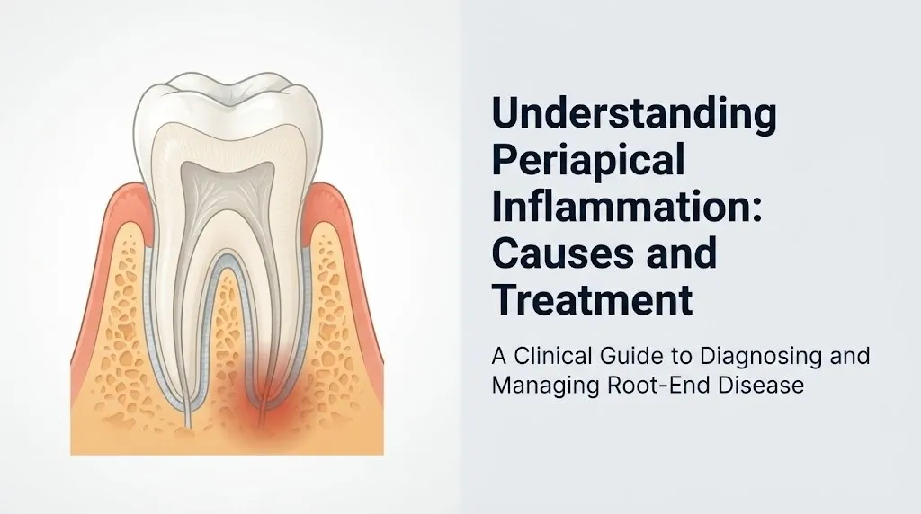
Swelling from a failed root canal occurs when the immune system’s response to periapical bacterial infection produces a local accumulation of inflammatory exudate, white blood cells, and tissue fluid in the soft tissues surrounding the root, with the location and severity of swelling directly reflecting the drainage pathway available to the abscess.
The periapical abscess begins at the root tip where bacteria are stimulating the immune response. From there, purulent material (pus) follows the path of least resistance through the cancellous bone toward the cortical plate. Where the cortical bone is thinnest, typically the buccal (cheek-side) plate in most teeth, the infection perforates and spreads into the adjacent soft tissue, producing visible and palpable swelling.
Swelling patterns from failed root canal infection tell the clinician where the infection is tracking:
- Localized gingival swelling (soft bump directly on the gum over the root): indicates abscess contained within the alveolar bone or just beneath the gingiva; typically not yet spreading
- Vestibular swelling (puffiness of the cheek mucosa or lip area): indicates the infection has penetrated the cortical bone and entered the vestibular soft tissues; more advanced
- Facial swelling (visible puffiness of the cheek, lower jaw, or submandibular region): indicates spreading cellulitis; requires urgent same-day evaluation
- Submandibular or neck swelling: indicates spread below the jaw; this is a potential emergency (see the emergency section below)
Swelling that appears and disappears cyclically, over days to weeks, without frank pain is often associated with a chronic abscess that has established a sinus tract for drainage. When the tract drains, pressure and swelling decrease; when it temporarily closes, swelling recurs.
Immunocompromised individuals including those on biologic medications (TNF-alpha inhibitors, IL-6 inhibitors) for autoimmune conditions may develop rapidly spreading dental infections with less localized swelling and fewer systemic warning signs, because their impaired immune response cannot mount a typical containment reaction.
Failed Root Canal Abscess Symptoms
A failed root canal abscess is a localized collection of pus in the periapical tissues or surrounding bone, produced by neutrophils attempting to destroy bacterial colonies in the infected periapical space, and it produces distinct symptoms that differ depending on whether the abscess is acute or chronic.
An acute periapical abscess develops rapidly, often within 24 to 48 hours, and produces intense throbbing pain that is continuous rather than intermittent, worsens when lying down (because elevation of the head reduces venous drainage and increases tissue pressure at the site), and is exquisitely tender to any percussion or touch. Patients often report that the tooth feels like it has “grown” or become slightly raised in the socket, because inflammatory exudate in the PDL physically lifts the tooth out of its normal occlusal position.
A chronic periapical abscess develops over weeks to months and may produce minimal pain because the slower accumulation of purulent material allows a drainage pathway (sinus tract) to form, decompressing the abscess before pressure reaches the pain threshold.
According to clinical guidance published by the American Association of Endodontists (AAE), the systemic signs of an acute periapical abscess requiring urgent attention include:
- Fever above 100.4°F (38°C) with localized dental pain and swelling
- Malaise and general feeling of illness accompanying localized dental signs
- Regional lymphadenopathy (swollen, tender lymph nodes in the submandibular or cervical chain)
- Trismus (reduced mouth opening, typically below 20mm interincisal distance) indicating spread to the masticator space
Patients with a history of rheumatic heart disease or prosthetic heart valves should communicate this to their dentist before any drainage procedure, as bacteremia from dental abscess drainage warrants antibiotic prophylaxis consideration per the American Heart Association (AHA) guidelines on infective endocarditis prevention.
Key Takeaway: An acute periapical abscess from root canal failure producing continuous throbbing pain, a raised tooth, facial swelling, and fever is a dental emergency requiring same-day evaluation by a dentist or endodontist, not a condition to manage at home with over-the-counter pain medications while waiting for a scheduled appointment.
Sinus Tract and Draining Fistula After Root Canal
A sinus tract after a root canal is a narrow channel connecting the periapical abscess at the root tip to the surface of the gingiva or, rarely, the facial skin, appearing as a small raised bump (often called a “gum boil” or “parulis”) that may intermittently release a salty or bitter-tasting fluid when pressed.
The formation of a sinus tract is the body’s way of decompressing a chronic abscess. When periapical infection cannot spontaneously resolve and cannot spread through bone rapidly enough, an epithelium-lined channel gradually forms through the path of least resistance in the cancellous bone and gingival tissue, allowing pus to drain continuously or intermittently. This drainage paradoxically reduces pain, which is why patients with a sinus tract often feel less pain than patients with an undraining acute abscess, despite having an equally serious infection.
The presence of a sinus tract is a reliable clinical indicator of a failed root canal. A tract can be traced back to its origin by gently inserting a size 20 or 25 gutta-percha point into the opening and taking a radiograph; the opaque point shows exactly which root is the source. This procedure, performed by a dentist or endodontist, is called gutta-percha tracing and is one of the most diagnostically useful tools for identifying the specific failed tooth when multiple teeth have been root-canal-treated in the same quadrant.
Quick Tip:
- Do not attempt to squeeze or puncture a gum boil at home; disrupting the external surface of a sinus tract does not treat the underlying infection and can introduce oral bacteria into the tract channel
- A sinus tract that closes (stops draining) does not mean the infection has resolved; it often means the drainage pathway has temporarily obstructed, causing internal pressure to build again
- Sinus tracts on facial skin rather than oral gingiva are rare but can be confused with a skin cyst or infection; any new skin opening on the cheek or jaw below a root canal-treated tooth warrants dental evaluation before dermatological management
Children with baby teeth that had root canal treatment (pulpotomy or pulpectomy) may develop sinus tracts that open near the gingival margin of the affected tooth; this pattern indicates failure of the primary tooth treatment and may require extraction to protect the developing permanent tooth below.
Tooth Discoloration After Failed Root Canal
Tooth discoloration in a root canal-treated tooth is produced by the degradation of residual pulp tissue hemoglobin within the dentinal tubules, by the leaching of iron sulfide compounds from decomposing blood cells into the surrounding dentin, and in some cases by the penetration of dye-like bacterial metabolites from the infected root canal space through the dentinal tubules into the crown dentin.
A successfully root canal-treated tooth may naturally darken slightly over time due to the aging of the residual dentin in the absence of vital pulp hydration. This normal physiological graying is gradual and uniform. Discoloration associated with root canal failure tends to progress more rapidly and appears alongside other clinical signs of infection.
The most common discoloration pattern from root canal failure is a progressive graying or brownish-gray darkening of the crown, starting from the coronal access restoration and spreading into the visible crown surfaces. This occurs because bacteria in a failing root canal produce metabolic byproducts that diffuse through the gutta-percha seal or through coronal leakage pathways into the dentinal tubules of the crown, where they interact with residual iron in hemosiderin deposits from pulp degradation to produce iron sulfide, a dark pigment.
Discoloration alone does not confirm root canal failure: many root canal-treated teeth darken without any active infection. However, discoloration that appears or accelerates alongside other symptoms, particularly pain, swelling, or a gum bump, should prompt radiographic evaluation by the treating dentist or a board-certified endodontist.
Patients with thin or translucent enamel (more common in individuals with amelogenesis imperfecta or with aging-related enamel thinning) may notice discoloration at lower severity levels of dentin pigmentation, making the sign appear earlier clinically than in patients with thicker enamel coverage.
Bite Pain and Sensitivity After Root Canal Failure
Pain when biting down on a root canal-treated tooth occurs because the periodontal ligament (PDL), the fibrous connective tissue suspension connecting the root to the alveolar bone socket, becomes inflamed by the prostaglandin E2 and interleukin-1 beta released from the periapical infection, making the mechanoreceptors and nociceptors within the PDL hyperresponsive to the compressive forces of occlusion.
Think of the periodontal ligament like shock absorbers in a car suspension system. In a healthy tooth, the PDL fibers cushion and distribute biting forces across the alveolar bone in a controlled, pain-free way. When the PDL becomes inflamed by a periapical infection tracking from a failed root canal, those shock absorbers become hypersensitive. Even light biting force that would normally be imperceptible becomes painful because the mechanoreceptors in the inflamed PDL are firing at much lower thresholds than normal.
Bite pain from root canal failure has a specific character that differentiates it from other causes of post-procedure sensitivity. It is usually felt upon initial contact with the opposing tooth, peaks immediately at the moment of occlusal contact, and does not linger for minutes afterward the way cold sensitivity from a cracked tooth does. Patients often describe it as a sharp, localized jolt felt specifically in the jaw below the tooth rather than inside the tooth itself.
Cold sensitivity in a root canal-treated tooth requires a careful clinical interpretation. A successfully root canal-treated tooth has no living pulp and should not be sensitive to cold. Cold sensitivity in a previously treated tooth may indicate:
- An accessory canal or missed canal that retains vital pulp tissue: still responding to cold
- Cracked tooth syndrome extending below the original root canal treated area: crack allows thermal stimulus to reach the PDL
- Root canal failure with periapical inflammation that creates heightened PDL sensitivity to thermal changes transmitted through the root
Patients with severe bruxism (nocturnal or diurnal tooth grinding) face a higher rate of post-root-canal bite pain because the excessive occlusal forces applied to the treated tooth amplify PDL inflammation at a periapical infection site that would otherwise be tolerable.
Key Takeaway: Cold sensitivity in a tooth that has already been root canal treated is never a normal finding and always warrants dental evaluation, because a successfully treated tooth should have no vital pulp tissue left to respond to thermal stimulus.
Can a Root Canal Fail Years Later
Yes, a root canal can fail years or even decades after apparently successful treatment, most often through the mechanism of coronal microleakage, in which the crown restoration overlying the root canal access gradually develops microscopic gaps that allow oral bacteria to penetrate the sealed root canal space and recolonize it.
Root canal treatment seals the cleaned canal from the inside with gutta-percha and sealer, but the tooth depends on an intact coronal restoration (filling, crown, or both) for protection from the oral environment above. When that restoration cracks, wears, or loses its marginal seal over time, oral microorganisms gain access to the root canal through the access cavity. Once inside, they can traverse the length of the canal and re-establish a periapical biofilm within weeks to months of coronal seal failure.
According to research published in the Journal of Endodontics, the coronal seal provided by a tooth’s crown or restoration deteriorates significantly over 3 to 10 years in many patients, with the rate of degradation accelerating in patients who grind their teeth, in patients with high-acid oral environments from frequent carbonated beverage consumption or gastroesophageal reflux disease, and in patients who delay crown placement after root canal completion.
Late failure symptoms (appearing years after treatment) tend to follow the same pattern as early failure symptoms, but the clinical history is different: the patient recalls a period of complete comfort following treatment that can span years before symptoms returned. This period of comfort, followed by new onset of symptoms, is a hallmark of coronal microleakage-driven late failure rather than incomplete initial treatment.
Patients who had root canal treatment on a molar without subsequent crown placement (contrary to standard recommendations) face a substantially higher rate of late failure, both from coronal leakage and from vertical root fracture due to unprotected cyclic biting forces on the brittle, non-vital root dentin.
Vertical Root Fracture Symptoms After Root Canal
Vertical root fracture (VRF) after root canal treatment produces a symptom pattern that is distinctly different from microbial failure: it typically causes pain that is specific to biting on one particular cusp of the tooth, a narrow periodontal pocket on one root surface that doesn’t match adjacent teeth, and a J-shaped or halo-shaped radiolucency extending along the lateral aspect of the root rather than solely at the apex.
Root canal treatment removes the vital pulp that normally hydrates and slightly flexes root dentin during function. The obturated root dentin becomes more brittle over time without this hydration. Cyclic loading from biting, particularly in back teeth (premolars and molars) without crown protection, creates compressive and tensile stresses within the root walls that can propagate a fracture longitudinally along the length of the root.
Vertical root fracture is a failure mode that no amount of retreatment can resolve. Once the root has fractured longitudinally, the fracture line provides a permanent colonization space for bacteria along its entire length, and the bacteria produce a destructive wedge-shaped bone defect along the fracture that is visible on a cone-beam computed tomography (CBCT) scan as a characteristic halo or J-shaped defect not seen with endodontic biofilm failure. Extraction of the affected root (or the whole tooth in single-rooted teeth) is the definitive treatment.
Characteristics that distinguish vertical root fracture from microbial root canal failure:
| Feature | Microbial Failure | Vertical Root Fracture |
|---|---|---|
| Pain location | Diffuse; felt at root tip and jaw | Sharp; specific to one cusp or biting direction |
| Periodontal probing | Broad, wide defect if bone loss present | Narrow, isolated deep pocket on one surface |
| Radiographic appearance | Periapical radiolucency at root tip | J-shaped or halo radiolucency along root lateral surface |
| Swelling pattern | Can swell anywhere around the root | Often narrow sinus tract directly over the fracture |
| Retreatment response | May respond to nonsurgical or surgical retreatment | Does not respond; extraction required |
Mandibular premolars and maxillary first premolars with two roots are at the highest anatomical risk for vertical root fracture because of their root morphology and the high biting forces concentrated at these positions in the dental arch.
Periapical Granuloma and Radicular Cyst Symptoms
A periapical granuloma is a chronic inflammatory lesion consisting of macrophages, lymphocytes, plasma cells, and fibroblasts enclosed in a fibrous connective tissue capsule at the root apex, and it is the most commonly encountered periapical lesion in failed root canals, often producing no symptoms while silently resorbing the surrounding alveolar bone.
The periapical granuloma develops when the acute immune response to periapical bacterial infection transitions to a chronic state. The acute neutrophil-dominant response is replaced by a macrophage and lymphocyte-dominated chronic granulomatous response aimed at walling off the persistent infection. This walled-off state is relatively stable: bone resorption occurs slowly, pain is often absent, and the lesion may remain static for months to years before either resolving (unlikely without treatment) or expanding.
When chronic periapical inflammation stimulates the epithelial rests of Malassez (remnant epithelial cells from the Hertwig epithelial root sheath that remain in the PDL throughout life), these cells can proliferate and form a cystic epithelial lining around the inflammatory exudate, converting the granuloma into a radicular cyst. This conversion is typically asymptomatic. Radicular cysts can grow slowly and expand into adjacent bone, occasionally becoming large enough to displace adjacent teeth or cause pathological fracture of the jaw if untreated over many years.
The distinction between a periapical granuloma and a radicular cyst cannot be made clinically or radiographically with certainty: both appear as periapical radiolucencies on a dental radiograph, and histopathological examination of the removed lesion tissue is the only definitive method of differentiation. A cone-beam computed tomography (CBCT) scan can characterize the three-dimensional extent of the lesion and assess cortical bone integrity, which informs the surgical approach when apicoectomy is planned.
Immunosuppressed patients (organ transplant recipients on calcineurin inhibitors, or patients on long-term corticosteroids) may develop unusually large periapical lesions from a relatively small periapical infection stimulus, because their reduced T-lymphocyte activity impairs the normal granulomatous containment response.
What Causes a Root Canal to Fail
Root canal treatment fails through five primary mechanisms: missed or untreated canal anatomy, incomplete debridement leaving viable bacterial biofilm, coronal microleakage allowing bacterial reentry, vertical root fracture of the brittle obturated root, and inadequate obturation (incomplete canal sealing) leaving spaces for residual bacteria.
Missed canals are the most common cause of root canal failure identified at retreatment. The root canal system is not a simple tube: it contains lateral canals, accessory canals, isthmuses connecting two canal spaces in a single root, and apical deltas where the main canal branches into multiple small foramina. Standard radiographs visualize only two dimensions. Canals oriented in the bucco-lingual plane may be superimposed on the main canal and missed entirely on a 2D radiograph. Maxillary first molars, for example, have a second canal in the mesiobuccal root (designated MB2) that historically was missed in approximately 40 to 60% of cases without magnification and CBCT-guided treatment, according to research published in the Journal of Endodontics.
Coronal microleakage has already been addressed in the years-later failure section, but as a primary cause: any failure to place an adequate coronal restoration promptly after root canal completion dramatically increases long-term failure risk.
Common causes of root canal failure:
- Missed lateral canals, accessory canals, or canal isthmuses not instrumented or disinfected during treatment
- Insufficient chemical disinfection (inadequate sodium hypochlorite concentration, volume, or contact time)
- Ledging, perforation, or instrument fracture blocking complete canal instrumentation
- Inadequate obturation: voids, short fills, or poor gutta-percha adaptation to canal walls
- Coronal microleakage from failed, cracked, or unplaced crown restoration
- Vertical root fracture from cyclic loading of unprotected non-vital root dentin
- Specific resistant bacteria: Enterococcus faecalis can survive in dentinal tubules and resist standard sodium hypochlorite disinfection due to its tolerance of alkaline environments and ability to survive in nutrient-poor conditions
Patients who had their root canal treated without a dental operating microscope are at higher risk of missed canals and incomplete debridement, since microscopic magnification directly improves the endodontist’s ability to identify canal orifices and apical anatomy.
Key Takeaway: The single most common identifiable cause of root canal failure found at retreatment is a missed canal, often the MB2 canal in maxillary first molars, that was never instrumented or disinfected during the original procedure.
How Is a Failed Root Canal Diagnosed
A failed root canal is diagnosed through a combination of clinical tests (percussion, palpation, periodontal probing, and temperature testing), periapical radiography, and in complex or anatomically challenging cases, cone-beam computed tomography (CBCT) imaging, which provides a three-dimensional view of the root canal system and periapical tissues.
The clinical examination begins with percussion: the endodontist taps the treated tooth with a mirror handle. Pain on percussion indicates periapical inflammation of the PDL, which is consistent with active periapical pathology. Palpation of the gingiva over the root tip produces pain in a periapical abscess and may reveal fluctuance (a fluid-filled feel) if an abscess is pointing to the surface. Periodontal probing assesses the sulcular depth around each root surface: a narrow, isolated deep pocket suggests vertical root fracture, while broad and shallow probing around all surfaces suggests diffuse bone loss from systemic rather than localized failure.
Periapical radiographs remain the primary imaging tool for diagnosis. A failing root canal produces a periapical radiolucency: a dark area at the root tip representing bone that has been resorbed by the chronic inflammatory response. The AAE recommends comparing current radiographs to the original post-treatment radiograph; an increasing or new radiolucency, or loss of the normal lamina dura (the white line representing the bony socket wall), confirms progressive failure.
Cone-beam computed tomography (CBCT) is indicated when conventional radiographs cannot fully characterize the lesion, when missed canals are suspected, when the anatomy of the root system is uncertain before retreatment or surgery, or when vertical root fracture is suspected. Research published in the Journal of Endodontics has shown that CBCT detects periapical pathology that is missed on conventional radiographs in approximately 30 to 35% of cases, particularly lesions in the early stages of bone resorption.
The endodontist will also review the patient’s dental history, the quality of the original root canal obturation visible on radiograph, and the integrity of the current coronal restoration, all of which contribute to the overall failure diagnosis.
Failed Root Canal Emergency Symptoms
Certain symptoms associated with a failed root canal require immediate emergency evaluation. Do not wait for a scheduled dental appointment or monitor these at home.
Call 911 immediately or go to the nearest emergency room if you experience:
- Difficulty breathing or swallowing: indicates that dental infection has spread to the deep neck spaces (parapharyngeal or retropharyngeal space) or has caused airway edema; this is a life-threatening emergency called Ludwig’s angina or a deep neck space abscess
- Swelling extending from the jaw into the neck or below the chin: indicates spread of infection below the mandible into the submandibular or submental spaces; spread here can compress the airway within hours
- Rapidly increasing facial swelling over less than 24 hours with fever above 101°F (38.3°C) and a general feeling of severe illness: indicates spreading cellulitis requiring intravenous antibiotics and possible surgical drainage, not oral antibiotics
- Trismus (inability to open the mouth) combined with swelling and fever: indicates masticator space involvement; airway access for emergency care becomes increasingly difficult as trismus worsens
- Confusion, stiff neck, or severe headache alongside dental infection: rare but indicates possible intracranial spread of infection; this requires immediate neurological and infectious disease evaluation
- High fever (above 103°F / 39.4°C) with shaking chills and dental infection: possible bacteremia or early sepsis from a dental source, requiring emergency evaluation and blood cultures
Go to an emergency dental clinic the same day (not the ER unless the above apply) if you experience:
- Severe continuous throbbing tooth pain uncontrolled by over-the-counter analgesics
- A visibly swollen cheek or gum that is enlarging
- Fever of 100.4°F (38°C) or higher with localized dental pain and swelling
These presentations can indicate Ludwig’s angina, a parapharyngeal space abscess, sepsis from a dental source, or a spreading dentoalveolar cellulitis, all of which require emergency medical or dental management, not a monitored wait.
Root Canal Retreatment Versus Extraction for Failed Root Canal
For a failed root canal, the two primary treatment options are nonsurgical retreatment (re-entering and re-cleaning the root canal system), surgical retreatment (apicoectomy with root-end resection and retrofill), or extraction of the tooth followed by replacement with a dental implant, bridge, or partial denture, with the choice depending on the specific failure mechanism, the restorability of the tooth, and the patient’s systemic health and treatment goals.
Nonsurgical retreatment is the first choice when the failure is caused by missed canals, incomplete debridement, or coronal microleakage, because these failures are theoretically addressable by re-accessing the canal system, identifying missed anatomy with a dental operating microscope, repeating disinfection with sodium hypochlorite and EDTA irrigation, and replacing the obturation. The Cochrane Database of Systematic Reviews has evaluated retreatment outcomes and found that nonsurgical retreatment has success rates of approximately 75 to 85% at 4-year follow-up, though success rates decline in teeth that have already undergone one previous retreatment.
Apicoectomy (periapical surgery, also called root-end resection) is indicated when nonsurgical retreatment is not feasible because of a post or dowel obstructing canal access, because of a persistent periapical lesion that fails to heal after retreatment, or because the apex requires direct surgical debridement and sealing with a biocompatible retrofill material.
Extraction is the definitive treatment for vertical root fracture, for teeth with inadequate remaining tooth structure to support a new restoration, or for patients in whom the financial or procedural complexity of retreatment is not appropriate. A dental implant placed in a healed extraction site offers a predictable long-term replacement option for most patients.
Patients taking bisphosphonate medications (alendronate, zoledronic acid) for osteoporosis or oncological bone disease should discuss this history with their endodontist before any periapical surgical procedure, because these medications impair osteoclast-mediated bone remodeling and increase the risk of bisphosphonate-related osteonecrosis of the jaw (BRONJ) following surgical manipulation of the jaw bone.
Frequently Asked Questions About Failed Root Canal Symptoms
How do I know if my root canal is failing?
The most reliable signs that a root canal is failing are new or returning pain in the treated tooth, swelling of the gum or jaw near the tooth, and a raised, pimple-like bump on the gum near the root tip.
These symptoms, particularly if they develop after a period of complete comfort following treatment, indicate that bacteria have persisted in or re-entered the root canal system and are producing periapical inflammation.
Any of these findings warrants evaluation by a board-certified endodontist, who will perform clinical tests and take a periapical radiograph or CBCT to assess the periapical tissues.
How long after a root canal can it fail?
A root canal can fail at any time from days after the procedure to decades later, with early failures (within the first year) most often caused by incomplete debridement or missed canals, and late failures (years after treatment) most often caused by coronal microleakage from a deteriorated restoration.
According to research published in the International Endodontic Journal, root canals in teeth with pre-existing periapical lesions have a higher failure rate and may show failure signs within 2 to 4 years if the infection was not fully eliminated.
Any new symptom in a previously asymptomatic root canal-treated tooth, regardless of how long ago the treatment was performed, warrants radiographic evaluation.
What does a failed root canal feel like?
A failed root canal typically produces a deep, aching or throbbing sensation at the root of the tooth or in the jaw, rather than a sharp pain inside the tooth itself, because the tooth has no living pulp but the surrounding periodontal ligament retains functioning pain fibers that become inflamed.
Some patients describe the sensation as pressure in the jaw, sensitivity when biting on that specific tooth, or a constant dull ache that worsens at night or when lying down.
A failed root canal with a draining sinus tract may actually cause minimal or no pain, despite an active infection, because the drainage pathway decompresses the abscess.
Can a failed root canal heal on its own?
A failed root canal cannot heal on its own, because the underlying cause is either a persistent bacterial biofilm within the root canal system or a physical structural failure (root fracture) that neither the immune system nor any healing process can eliminate without professional intervention.
The body’s immune response can partially contain a periapical infection in a chronic state (granuloma formation), but it cannot eliminate bacteria sheltered within the root canal’s physical space, which is inaccessible to circulating immune cells and antibiotics at bactericidal concentrations.
Delaying treatment allows the periapical infection to slowly expand, destroying alveolar bone and reducing the likelihood that retreatment will preserve the tooth.
Is a failed root canal an emergency?
A failed root canal is a dental emergency requiring same-day care if it is accompanied by facial swelling that is enlarging, fever above 100.4°F (38°C), severe continuous pain uncontrolled by over-the-counter analgesics, or any difficulty swallowing or breathing.
If swelling extends below the jaw into the neck, or if breathing becomes difficult, call 911 immediately and go to the nearest emergency room, because these signs indicate a potentially life-threatening spread of infection to the deep neck spaces.
A failing root canal without these signs should still be evaluated within 24 to 48 hours, not monitored at home while waiting for a routine appointment.
What happens if a failed root canal is left untreated?
A failed root canal left untreated will continue to resorb alveolar bone around the root apex, progressively reduce the structural support for the tooth, and in some cases allow the periapical infection to spread through fascial planes to adjacent spaces in the jaw, neck, or (rarely) intracranially.
The American Association of Endodontists notes that a periapical lesion that enlarges over time without treatment can eventually involve adjacent teeth, requiring more extensive treatment than would have been necessary if addressed promptly.
Systemic spread of a dental infection, while uncommon, has been documented to cause life-threatening conditions including Ludwig’s angina, cavernous sinus thrombosis, and sepsis, all of which are preventable with timely endodontic or surgical management.
Pain returning to a previously root-canal-treated tooth is not something to monitor for weeks while hoping it resolves. It means bacteria are active, the periapical tissues are inflamed, and the window for preserving the tooth with retreatment rather than extraction is closing. The sooner a board-certified endodontist evaluates the tooth with current radiographs and clinical tests, the more options remain on the table.
If your symptoms include facial swelling that is growing, fever, difficulty opening your mouth fully, or any change in your ability to swallow or breathe, do not drive to a dental office. Go directly to an emergency room or call 911 immediately.
For every other presentation of suspected root canal failure, contact a board-certified endodontist and ask specifically for a periapical radiograph compared against your original post-treatment film, and ask whether a CBCT scan is indicated to detect missed canals or early bone loss before it is visible on conventional imaging.