Pulled Hamstring Symptoms: Complete 2026 Clinical Guide
Pulled hamstring symptoms range from a mild aching tightness in the back of the thigh to a sudden, severe tearing sensation that stops you mid-stride and makes weight-bearing immediately impossible. The severity depends on how many muscle fibers tore, exactly which muscle and which region of that muscle was injured, and whether the injury involved the muscle itself, the tendon attachment at the bone, or both.
Hamstring injuries are among the most frequently reported soft tissue injuries in sport. Research published in the British Journal of Sports Medicine by Ekstrand and colleagues, drawing from the UEFA Elite Club Injury Study, found that hamstring strains account for approximately 12 percent of all muscle injuries in professional football players, with re-injury rates between 12 and 34 percent in athletes who return to play. The National Institute of Arthritis and Musculoskeletal and Skin Diseases (NIAMS) identifies hamstring injuries as one of the most common causes of posterior thigh pain in both athletic and physically active populations.
This article covers every layer of the pulled hamstring symptom picture: the anatomy and physiology behind each symptom, the grading system that determines severity, the specific presentations that differ by location in the thigh, the populations who face different injury patterns, the distinguishing features from sciatic nerve pain, and the red flags that require immediate professional evaluation rather than home monitoring.
What Is a Pulled Hamstring and How Does It Cause Symptoms
A pulled hamstring is a partial or complete tear of one or more of the three muscles forming the hamstring muscle group at the back of the thigh, occurring when the force applied to the muscle during eccentric loading exceeds the tensile strength of the myotendinous junction, the transition zone between muscle fiber and tendon where mechanical stress concentrates most intensely.
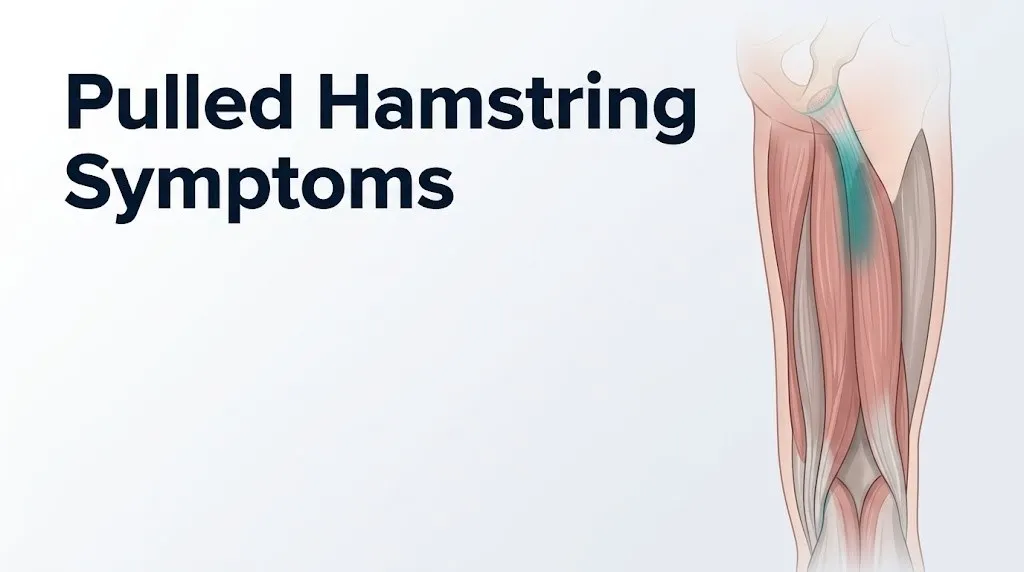
The hamstring group consists of three muscles. The biceps femoris, which has a long head originating from the ischial tuberosity (the bony prominence at the base of the pelvis) and a short head originating from the linea aspera of the femur, is the most commonly injured, accounting for approximately 80 percent of hamstring strains in sprinting sports according to prospective cohort data in the British Journal of Sports Medicine. The semimembranosus and semitendinosus originate from the ischial tuberosity and share a common origin via the conjoint tendon with the biceps femoris long head.
Think of the myotendinous junction like the splice point between two ropes of different elasticity. The muscle belly stretches readily; the tendon resists stretch. The junction between them takes the full impact when the pull is sudden. Most hamstring tears happen at this splice point, not in the middle of the muscle belly.
All three hamstring muscles cross both the hip and the knee. They extend the hip (pulling the thigh backward) and flex the knee (bending it). The injury mechanism that produces most hamstring tears is the late swing phase of sprinting: the hamstring is simultaneously stretched (lengthening as the leg swings forward) while actively contracting to decelerate the leg before foot strike. This eccentric contraction under high load is the specific biomechanical scenario that concentrates tensile stress at the myotendinous junction beyond its failure point.
When myofibrillar disruption occurs, the endomysium (connective tissue around individual muscle fibers) and perimysium (connective tissue around fiber bundles) tear along with the muscle fibers. Both structures contain dense microvascular networks. Their disruption allows blood to accumulate within the fascial compartment of the posterior thigh, forming an intramuscular hematoma that is responsible for several of the most recognizable pulled hamstring symptoms.
Pulled Hamstring Symptoms: The Full Clinical Picture
Pulled hamstring symptoms, taken as a complete inventory, include sudden posterior thigh pain, a possible audible or felt popping sensation at the moment of injury, swelling and warmth in the back of the thigh, bruising that develops over 24 to 72 hours, weakness in knee flexion and hip extension, reduced range of motion in the hip and knee, and in more severe injuries, an inability to bear weight.
Not every patient experiences every symptom. The pattern and severity depend on the grade of injury, the specific muscle affected, and the injury location along the muscle’s length. The American Academy of Orthopaedic Surgeons (AAOS) notes that the classic presentation of a hamstring strain involves a sudden onset of posterior thigh pain during high-speed or high-load activity, which distinguishes it from the gradual aching of delayed onset muscle soreness (DOMS) or overuse tendinopathy.
The full symptom inventory in clinical order of appearance:
- Sudden posterior thigh pain: the most universal symptom, present in virtually all grades; described as sharp, stabbing, or tearing in acute presentation
- Popping or snapping sensation: present in moderate to severe tears when myofibrillar bundles fail rapidly; not present in every Grade I strain
- Immediate muscle weakness: reduced ability to flex the knee against resistance or extend the hip; severity directly correlates with the grade
- Swelling and warmth: appears within minutes to hours at the injury site from local inflammatory vasodilation and histamine release
- Bruising (ecchymosis): develops 24 to 72 hours after injury as the hematoma tracks along fascial planes toward the skin surface
- Muscle spasm: protective reflex contraction of surrounding muscle fibers, generating secondary cramping pain distinct from the primary tear pain
- Tightness and reduced range of motion: hip flexion and knee extension are restricted because stretching the hamstring in either direction lengthens the torn site and triggers pain
Individual variation is immediate and clinically relevant. Patients taking anticoagulant medications (warfarin or direct oral anticoagulants such as rivaroxaban or apixaban) may develop dramatically larger hematomas from the same degree of fiber disruption that would produce only mild bruising in a person not on blood thinners. This population requires evaluation within 24 hours regardless of perceived injury severity.
What Does a Pulled Hamstring Feel Like
A pulled hamstring feels like a sudden, sharp pain or burning sensation at the back of the thigh, often accompanied by a feeling of “giving way” in the leg, and in moderate to severe tears, a distinct pop or snap felt inside the thigh at the moment of injury.
The sensory experience is driven by simultaneous activation of multiple pain pathways. Bradykinin, released within seconds from damaged tissue through the kinin cascade, directly stimulates nociceptors (pain-sensing nerve endings) in the muscle and surrounding connective tissue. Prostaglandin E2, produced by COX-2 enzymes in the injured tissue within minutes, sensitizes nociceptors so they respond more intensely to subsequent stimuli. The result is both immediate sharp pain from bradykinin and a heightened, spreading ache from prostaglandin sensitization.
The popping sensation that many patients describe at the moment of injury represents rapid mechanical failure of myofibrillar bundles. When a large group of muscle fibers fail simultaneously at the myotendinous junction, the stored elastic energy in the stretched muscle is suddenly released. That release is fast enough to be both felt and sometimes audibly heard. It’s a similar principle to snapping a rubber band that’s been stretched to its limit.
Within the first 30 to 60 minutes after injury, the affected area feels warm to the touch. This warmth is produced by the acute inflammatory response: mast cells release histamine, which dilates local blood vessels and increases blood flow to the injury site. The tissue feels firm because of swelling from increased capillary permeability and early hematoma formation.
Patients frequently describe the day-after experience as a deep, diffuse aching throughout the back of the thigh, worse when sitting (which stretches the proximal hamstring against the ischial tuberosity), when climbing stairs, and when getting up from a seated position. The distinction from delayed onset muscle soreness (DOMS) is important: DOMS produces bilateral, diffuse soreness peaking 24 to 48 hours after exercise, and it does not include the immediate sharp onset pain, the popping sensation, or the rapid swelling that characterizes an acute hamstring strain.
Quick Tip:
- If the pain came on suddenly during a specific movement rather than building over hours or the day after exercise, that pattern is more consistent with an acute strain than DOMS.
- If you felt or heard a pop and have immediate difficulty walking, seek evaluation by a sports medicine physician or primary care physician within 24 hours.
- Older adults may not experience the classic sharp pop because degenerative tendon changes alter the injury mechanism, producing a more gradual failure pattern with less acute onset.
Key Takeaway: The combination of sudden posterior thigh pain during activity, a felt or heard pop, and immediate difficulty bearing weight distinguishes an acute hamstring strain from muscle soreness and warrants professional assessment within 24 to 48 hours at minimum.
Grade 1 Hamstring Strain Symptoms
Grade 1 hamstring strain symptoms involve mild posterior thigh pain with minimal functional loss, representing a tear of less than approximately 10 percent of muscle fibers at the myotendinous junction with no palpable defect in the muscle.
The pain of a Grade 1 strain is often described as a “twinge” or “pull” rather than a sharp tear. It may not stop you from completing your activity immediately, though continued high-intensity performance typically worsens it. The injury site is tender to direct palpation but the tenderness is localized to a small area. Range of motion at the hip and knee is preserved or only mildly reduced.
According to the American Academy of Orthopaedic Surgeons, Grade 1 hamstring strains typically produce no visible bruising within the first 24 hours (though mild discoloration can develop later), no significant swelling, and no loss of normal walking gait. A person with a Grade 1 strain can usually walk without a visible limp, though jogging and sprinting will reproduce pain.
| Feature | Grade 1 Presentation | Clinical Implication |
|---|---|---|
| Pain quality | Aching, tightness, mild “pull” sensation | Does not require emergency evaluation |
| Ability to walk | Normal or nearly normal | Can bear weight without a limp |
| Swelling | Minimal or absent | No hematoma formation of clinical concern |
| Bruising | Usually absent or minor after 48 hours | Mild ecchymosis may appear; not alarming |
| Muscle tenderness | Mild, localized | Tenderness to firm palpation only |
| Range of motion | Preserved or mildly reduced | Hip flexion and knee extension near normal |
| Estimated fiber disruption | Less than 10 percent | Muscle continuity intact |
Adolescent athletes with a Grade 1 presentation in the proximal posterior thigh deserve special clinical consideration. In athletes under approximately 16 years of age, the apophysis (the growth plate at the ischial tuberosity) has not fully fused to the pelvis. What presents clinically like a Grade 1 strain can actually be an avulsion fracture of the ischial tuberosity apophysis, a bone injury rather than a muscle tear. An X-ray of the pelvis is recommended for any young adolescent with proximal posterior thigh pain after a high-speed activity, regardless of apparent symptom severity.
Grade 2 Hamstring Strain Symptoms
Grade 2 hamstring strain symptoms involve moderate posterior thigh pain, visible swelling, limited ability to walk without a noticeable limp, and a partial tear of between approximately 10 and 90 percent of muscle fibers at the myotendinous junction.
The pain of a Grade 2 strain is clearly more intense than Grade 1. Most patients describe it as a sharp, burning sensation that prevents immediate continuation of the activity that caused it. There is a distinct moment of injury followed by rapidly worsening pain over the next 15 to 30 minutes. Walking is possible but produces a visible antalgic gait: the patient shortens their stride on the injured side and avoids full hip extension to limit hamstring stretch.
The American College of Sports Medicine identifies Grade 2 as the most commonly mismanaged severity in recreational athletes, primarily because early pain reduction with anti-inflammatory measures leads patients to underestimate the injury and return to full activity too soon, significantly increasing re-injury risk at the same myotendinous junction site.
Muscle spasm is a prominent and often distressing feature of Grade 2 strains. The surrounding intact muscle fibers contract reflexively to protect the injured site, producing a hard, palpable, painful band in the posterior thigh that can be mistaken for a more severe tear. This protective spasm is mediated by the reflex arc through the spinal cord’s alpha motor neuron pool, triggered by nociceptive input from the injured area.
Bruising in Grade 2 injuries typically becomes visible within 24 to 48 hours and may spread well below the injury site, appearing behind the knee or even into the upper calf, because blood from the hematoma tracks distally along the fascial planes of the posterior thigh compartment. The size of the bruised area does not directly reflect the severity of the tear, because hematoma tracking depends on fascial anatomy and tissue pressure rather than simply the volume of blood.
Patients with Grade 2 strains should be evaluated by a primary care physician or sports medicine physician within 24 to 48 hours of injury. At that appointment, ask specifically about imaging (musculoskeletal ultrasound or MRI) to confirm grade, and a referral to a physical therapist for a structured rehabilitation protocol, because returning to activity without guided eccentric strengthening substantially increases re-injury probability.
Grade 3 Hamstring Tear Symptoms
Grade 3 hamstring tear symptoms involve severe, immediate posterior thigh pain with near-complete or complete loss of knee flexion strength, often a palpable defect or gap in the muscle belly, and an inability to walk normally or bear full weight.
The onset of a Grade 3 tear is typically unmistakable. The pain is severe enough to stop movement immediately and most patients describe it as the worst muscle pain they have ever experienced. The popping sensation, if present, is often described as more pronounced than in lower-grade injuries. Some patients report a sensation of the back of the thigh “giving way” or a sudden collapse of the leg.
A palpable muscle defect (a gap or concavity in the posterior thigh tissue where the torn muscle has retracted) is a critical physical examination finding that distinguishes Grade 3 from Grade 2. In Grade 3 tears involving the proximal musculotendinous junction or the proximal tendon itself, the retracted muscle belly may be palpable as a firm, distal mass in the posterior thigh. This “balling up” of the retracted muscle is analogous to a snapped elastic waistband rolling away from the torn end, pulling the intact portion of the muscle toward the knee.
According to research published in the American Journal of Sports Medicine, Grade 3 hamstring tears with complete disruption of the muscle-tendon unit are associated with a prolonged recovery of 6 to 12 months without surgery in some cases, and with persistent strength deficits and high re-injury risk on return to sport if rehabilitation is not comprehensive.
Neurovascular symptoms are possible in severe Grade 3 injuries. The sciatic nerve travels in close anatomical proximity to the proximal hamstring tendons in the posterior thigh. Large hematomas, significant tissue retraction, or direct injury to the nerve at the time of tear can produce numbness, tingling, or weakness in the lower leg and foot, referred along the sciatic nerve’s distribution. If any lower leg neurological symptoms are present after a severe posterior thigh injury, evaluation at a hospital emergency room the same day is the appropriate step, not monitoring at home.
Key Takeaway: A palpable gap or defect in the posterior thigh after a hamstring injury, combined with severe weakness and an inability to flex the knee against resistance, indicates a Grade 3 tear requiring same-day evaluation by a board-certified orthopedic surgeon.
Pulled Hamstring Pain Location and Why It Spreads
Pulled hamstring pain is located primarily in the posterior thigh, but its exact position, whether proximal (near the buttock crease), mid-thigh, or distal (near the back of the knee), directly reflects which part of which muscle was injured and determines important differences in recovery timeline and complication risk.
The most common pain location is the proximal posterior thigh, just below the gluteal fold and centered toward the lateral side, which corresponds to the proximal myotendinous junction of the biceps femoris long head. This is the anatomical site most frequently involved in sprint-related hamstring injuries. Pain here is typically reproduced by firm palpation 5 to 10 centimeters below the ischial tuberosity along the posterior lateral thigh.
Pain at the ischial tuberosity itself (the bony prominence felt deep in the buttock when sitting) indicates proximal tendon involvement rather than muscle belly injury. This is a different and more serious anatomical zone, associated with longer recovery and higher complication risk. The distinction matters because proximal tendon injuries at or near the ischial tuberosity are the zone where avulsion injuries occur.
Mid-thigh pain from a biceps femoris short head injury or a semimembranosus belly injury produces a more centrally located posterior thigh ache. These injuries are less common and carry a somewhat shorter recovery timeline than proximal myotendinous junction injuries.
Distal hamstring pain, located just above or behind the knee, reflects injury to the distal musculotendinous junctions where the hamstring tendons approach the knee. The semitendinosus and semimembranosus tendons insert at the medial tibial head; the biceps femoris tendon inserts at the fibular head. Distal injuries are less common than proximal ones and may be confused with posterior knee pathology.
Pain spreading beyond the posterior thigh to the lower leg, foot, or lateral calf signals possible sciatic nerve involvement and should not be assumed to be simple radiated muscle pain. Any lower limb neurological symptoms following a posterior thigh injury require evaluation by a sports medicine physician or, if severe, by an emergency medicine physician.
Pulled Hamstring Swelling, Bruising, and Hematoma
Swelling in a pulled hamstring appears within minutes to hours of injury as the acute inflammatory response increases capillary permeability and allows plasma proteins and fluid to accumulate in the injured tissue, while bruising develops over 24 to 72 hours as blood from the disrupted microvasculature tracks toward the skin surface.
The swelling of an acute hamstring strain is produced by the rapid release of histamine from mast cells at the injury site, which dilates local arterioles and venules and increases capillary wall permeability. Plasma, proteins, and white blood cells move from the capillary lumen into the interstitial space, producing visible and palpable tissue swelling within the posterior thigh. The thigh may feel firm, warm, and tender to touch within an hour of injury.
Bruising (ecchymosis) in a pulled hamstring almost always appears later than the swelling, often 24 to 72 hours after the injury. This delay confuses many patients who see no bruising immediately after a tear and conclude the injury is minor. The delay occurs because blood from the intramuscular hematoma must track through tissue planes and ascend to the superficial skin layers before becoming visible as discoloration. The bruising can appear well away from the actual tear site: a proximal hamstring hematoma commonly produces bruising visible behind the knee or even in the upper calf, because blood tracks distally along the fascial planes of the posterior compartment.
According to the National Institute of Arthritis and Musculoskeletal and Skin Diseases, the extent of bruising visible on the skin surface is not a reliable indicator of injury severity because hematoma size depends on the depth of the tear, the local fascial containment, and the patient’s clotting status, not simply the number of fibers torn.
Patients on warfarin or direct oral anticoagulants face a disproportionate hematoma risk. Even a Grade 1 or Grade 2 strain can produce a large intramuscular hematoma in this population, raising the possibility of compartment syndrome (discussed in the emergency section below). Any patient on blood-thinning medication who sustains a posterior thigh injury should contact their primary care physician or anticoagulation clinic the same day and monitor the thigh circumference (measured at mid-thigh) every few hours; a circumference increase of more than 2 to 3 centimeters within 6 hours warrants emergency evaluation.
Pulled Hamstring Weakness and Inability to Walk
The degree of weakness following a pulled hamstring injury is the single most clinically reliable indicator of injury severity, because strength loss reflects the proportion of muscle fibers that have failed and are no longer generating contractile force.
The primary functional loss in a pulled hamstring is weakness in knee flexion (bending the knee against resistance) and weakness in hip extension (pulling the thigh backward against resistance), because these are the two primary actions of the hamstring muscle group. The severity of weakness maps directly onto the grade classification: Grade 1 shows minimal weakness with near-normal function; Grade 2 shows moderate weakness with an antalgic limp; Grade 3 shows severe weakness with inability to flex the knee at full effort.
Walking ability is a practical clinical proxy for severity:
- Grade 1: Normal or near-normal walking, no visible limp, pain reproduced only by jogging or fast walking
- Grade 2: Visible antalgic limp, shortened stride on the injured side, pain reproduced by normal walking pace and especially by stair climbing or walking on an incline
- Grade 3: Severe limitation of walking, inability to bear full weight on the injured leg, the foot may drag or require toe-walking to avoid full knee extension
The inability to walk normally after any posterior thigh injury is the threshold for same-day evaluation by a primary care physician, sports medicine physician, or emergency medicine physician. Do not wait several days to see if walking improves.
Weakness that extends to the foot, including foot drop (inability to lift the forefoot off the ground during walking), indicates sciatic nerve involvement rather than pure muscle weakness. The sciatic nerve runs in close proximity to the proximal hamstring tendons and can be compressed by a large hematoma or damaged directly in a severe proximal tear. Foot drop following a hamstring injury is a neurological emergency.
Older adults, particularly those over 60 with pre-existing degenerative changes in the proximal hamstring tendons, may experience complete functional loss from what initially appears to be a moderate mechanism of injury. Degenerative tendinopathy weakens the tendon-bone interface before any acute event, so lower-energy activities (such as stepping off a curb or reaching into a car trunk) can produce the same functional loss as a sprint injury in a younger athlete. This population should not attempt to “walk it off” but rather seek evaluation by a sports medicine physician or orthopedic surgeon within 24 hours.
Key Takeaway: Inability to walk without a noticeable limp after a posterior thigh injury indicates at least a Grade 2 strain and requires professional evaluation within 24 to 48 hours; foot drop indicates sciatic nerve involvement and is a neurological emergency requiring immediate care.
Proximal Hamstring Avulsion Symptoms
Proximal hamstring avulsion is a complete or near-complete tear of the hamstring tendons at their origin on the ischial tuberosity, producing immediate severe posterior thigh and buttock pain, a palpable proximal gap or deformity, and in many cases sciatic nerve irritation symptoms that distinguish it from a standard Grade 3 muscular tear.
A proximal hamstring avulsion is not simply a severe muscle strain. It is a tendon-level injury in which the conjoint tendon of the biceps femoris long head and semitendinosus, along with the tendon of the semimembranosus, completely or near-completely separates from the ischial tuberosity. In some cases, a small fragment of ischial bone avulses with the tendon. This is a distinct injury from a Grade 3 muscle belly tear and carries different surgical implications.
The mechanism is typically a forceful simultaneous hip flexion and knee extension: the classic scenario is water skiing, a splits maneuver in gymnastics or dance, or a high-speed fall. The sciatic nerve is located in immediate anatomical proximity to the proximal hamstring origin, and it can be tethered by scar tissue from a large hematoma or directly stretched by tendon retraction, producing posterior thigh pain that radiates into the lower leg in a sciatic distribution.
Research published in the American Journal of Sports Medicine (Lempainen et al., 2009, and subsequent studies) found that complete proximal hamstring avulsions treated non-surgically were associated with persistent strength deficits, chronic posterior thigh pain, and sciatic nerve symptoms in a substantial proportion of patients, whereas surgical repair within the first several weeks of injury was associated with better functional outcomes and lower rates of persistent neurological symptoms.
Specific symptoms that distinguish proximal avulsion from standard Grade III tear:
- Deep, severe aching in the buttock crease and ischial region (rather than mid-posterior thigh) immediately after injury
- A palpable gap or emptiness in the proximal posterior thigh just below the gluteal fold
- Difficulty sitting due to severe pain directly over the ischial tuberosity (the sitting bone)
- Numbness, tingling, or burning pain radiating down the back of the thigh to the calf (sciatic irritation)
- Visible retraction of the hamstring muscle belly into the mid-thigh, appearing as a distal bulge
Any patient with these features after a posterior thigh injury requires evaluation by a board-certified orthopedic surgeon with experience in sports medicine or hamstring repair within 24 to 48 hours, and an MRI of the posterior thigh and pelvis to confirm the diagnosis and determine the extent of tendon retraction before the surgical window for primary repair closes.
Pulled Hamstring Symptoms in Athletes, Teenagers, and Older Adults
Pulled hamstring symptoms present differently across age groups and activity levels in ways that have direct physiological explanations and require different clinical responses.
In sprinters and team sport athletes, hamstring injury almost always involves the biceps femoris long head at its proximal myotendinous junction, produced by the eccentric loading of the late swing phase. The onset is acute and high-velocity. According to the British Journal of Sports Medicine, elite football players who sustain a hamstring injury face a re-injury rate of approximately 12 to 34 percent on return to the same sport within the same season, with the re-injury occurring most often at the same myotendinous junction site where fibrotic scar tissue has replaced normal elastic muscle fiber.
In adolescent athletes (approximately 12 to 16 years old), the clinical concern that top competitors miss entirely is ischial tuberosity apophyseal avulsion fracture. In adolescents, the growth plate (apophysis) at the ischial tuberosity has not yet fused to the pelvis. The tendon-bone interface at this unfused apophysis is mechanically weaker than the muscle itself, so under high tensile load (sprinting, kicking, gymnastics splits), the apophysis avulses from the pelvis before the muscle tears. This presents almost identically to a proximal hamstring strain: sudden posterior hip pain, difficulty walking, buttock tenderness. An X-ray of the pelvis is the differentiating test. Missing this diagnosis risks improper management and potential non-union of the apophyseal fragment if the displacement is large enough to require surgical fixation.
In older adults (above 50 to 60 years of age), the injury mechanism changes. Chronic degenerative proximal hamstring tendinopathy weakens the tendon-bone interface over years, so avulsion-level injuries can occur with lower-energy mechanisms than would be expected. An older adult who describes a sudden pop in the posterior thigh while simply bending forward rapidly or stepping off a curb may have a complete proximal avulsion, not a mild strain. This population underreports injury severity because the pain sometimes plateaus quickly due to reduced acute inflammatory response associated with age-related changes in mast cell density and prostaglandin production.
Pregnant women with posterior thigh or buttock pain face a diagnostic challenge: posterior pelvic girdle pain from pubic symphysis diastasis or sacroiliac joint dysfunction during pregnancy produces posterior thigh and buttock aching that can be mistaken for a hamstring injury. Evaluation by a primary care physician or obstetric provider is appropriate before any musculoskeletal treatment is initiated during pregnancy.
Pulled Hamstring vs Sciatica: How to Tell the Difference
Pulled hamstring symptoms and sciatica share posterior thigh pain as their most prominent feature, but the distinguishing characteristics, including onset pattern, quality of pain, neurological features, and specific provocative tests, allow a clinician to differentiate the two in most cases.
Sciatica is posterior leg pain produced by irritation or compression of the sciatic nerve (formed by the L4, L5, S1, S2, and S3 nerve roots) at the level of the lumbar spine or piriformis muscle, not at the posterior thigh itself. Classic sciatica produces a burning or electric shock-type pain that radiates from the lower back or buttock down the posterior thigh, through the calf, and often into the foot. It is characteristically worsened by sitting, forward bending of the spine, and the straight leg raise test (which reproduces sciatic pain when the examiner raises the straightened leg past 30 to 70 degrees with the patient lying flat, stretching the nerve root).
A pulled hamstring produces posterior thigh pain that is reproduced by stretching the hamstring muscle (hip flexion with knee extended) or contracting it against resistance (knee flexion or hip extension). It does not typically radiate into the foot, does not produce the burning electric quality of nerve pain (unless the sciatic nerve is also involved in a proximal avulsion), and is not worsened by spinal movements.
| Clinical Feature | Pulled Hamstring | Sciatica |
|---|---|---|
| Onset | Sudden, during activity | Often gradual, or following a specific back event |
| Pain quality | Aching, tearing, or cramping | Burning, electric, shooting |
| Pain radiation | Posterior thigh only (usually) | Back to buttock to calf to foot |
| Foot/ankle symptoms | Absent unless sciatic nerve involved | Numbness, tingling, weakness in foot common |
| Tenderness to palpation | Directly in posterior thigh muscle | Usually not tender in thigh; lumbar spine may be tender |
| Worsened by knee extension with hip flexed | Yes, specifically | Variable |
| Straight leg raise provocation | Produces hamstring stretch pain, not radiating | Reproduces radiating leg pain past 70 degrees |
| Lumbar spine involvement | None | Primary source of pathology |
The overlap becomes genuinely difficult to distinguish when a proximal hamstring tear produces sciatic nerve irritation from hematoma compression or tendon retraction. In this case, both a muscular and a neurological component are present simultaneously. This mixed presentation requires MRI of the posterior thigh and pelvis, combined with clinical examination, to differentiate and is best evaluated by a sports medicine physician or orthopedic surgeon.
When Pulled Hamstring Symptoms Become a Medical Emergency
Certain presentations following a posterior thigh injury require immediate emergency evaluation and cannot be managed at home or with a scheduled clinic appointment.
Go to the nearest emergency room immediately, or call 911 if you cannot transport yourself, if you experience:
- A tense, hard posterior thigh compartment with pain that is disproportionately severe and worsening despite rest and ice: this pattern suggests developing compartment syndrome, in which pressure within the fascial compartment of the posterior thigh rises to a level that compromises blood flow to the muscle and nerve. Compartment syndrome requires emergency surgical fasciotomy; delay beyond 6 hours significantly increases the risk of permanent nerve and muscle damage.
- Foot drop after a posterior thigh injury: inability to lift the forefoot during walking indicates sciatic or peroneal nerve dysfunction and requires emergency neurological and orthopedic assessment the same day.
- Pain with passive stretch of the ankle and foot combined with a hard, swollen posterior thigh compartment: this is the clinical sign of compartment syndrome in the posterior thigh compartment and requires emergency room evaluation, not a wait-and-see approach.
- Rapidly expanding bruising with progressively increasing thigh circumference in a patient on blood-thinning medication: this indicates a large expanding hematoma with potential for compartment pressure. Go to the emergency room immediately.
- Complete inability to bear any weight on the leg combined with a visibly deformed or hollow proximal posterior thigh: this presentation in a high-mechanism injury suggests proximal hamstring avulsion, which requires same-day orthopedic surgical evaluation to determine if primary repair is possible before the surgical window closes.
- Numbness, tingling, or weakness in the foot or lower leg after a severe posterior thigh injury: any neurological deficit in the lower extremity distal to the injury site requires emergency evaluation to rule out sciatic nerve injury, compartment syndrome, or vascular compromise.
These presentations can indicate compartment syndrome, complete proximal hamstring avulsion with sciatic nerve involvement, or vascular injury, and they require emergency medical assessment at a hospital emergency room, not a next-day clinic appointment.
How a Pulled Hamstring Is Diagnosed
A pulled hamstring is diagnosed through a combination of clinical history, physical examination, and imaging, with the specific tests ordered depending on the suspected severity and location of the injury.
The clinical history focuses on the mechanism of injury, the onset (sudden vs. gradual), the initial symptom character, whether a pop was felt or heard, and the current functional status including walking ability. A sports medicine physician or orthopedic surgeon then performs a targeted physical examination.
How a typical diagnostic process unfolds from injury to confirmed diagnosis:
- The examiner palpates the posterior thigh systematically from the ischial tuberosity proximally to the popliteal fossa distally, identifying the point of maximum tenderness and any palpable muscle defect or gap.
- Range of motion is assessed: passive hip flexion with the knee straight (stretching the hamstring), and active knee flexion against resistance (contracting the hamstring), with pain reproduction and strength deficit noted.
- The straight leg raise test is performed to assess sciatic nerve involvement: positive for nerve symptoms (burning radiation to the calf or foot) distinguishes neurological from pure muscular pain.
- Neurovascular assessment is performed: sensation in the lower leg and foot, ankle reflexes, and capillary refill time in the toes to rule out sciatic nerve or vascular involvement.
- If Grade II or III injury is suspected, imaging is ordered.
Imaging options and their clinical applications:
| Imaging Test | What It Shows | When It Is Ordered |
|---|---|---|
| Musculoskeletal ultrasound | Real-time muscle fiber architecture, hematoma size and location, partial vs. complete tear | First-line for Grade II+ when immediate confirmation is needed; less detailed than MRI |
| MRI (magnetic resonance imaging) | Gold standard for soft tissue: identifies injury grade, exact anatomical location, tendon involvement, nerve proximity | Ordered for any suspected Grade III, proximal avulsion, or when return-to-sport timeline is needed |
| X-ray (pelvis and femur) | Bone integrity only; cannot show muscle or tendon | Ordered for adolescents to rule out ischial tuberosity apophyseal avulsion fracture; ordered for adults if bony avulsion is suspected |
According to the American Academy of Orthopaedic Surgeons, MRI of the hamstring provides the most accurate information for surgical planning in proximal avulsion injuries and for prognosis in high-level athletes requiring return-to-sport clearance. Routine MRI is not required for Grade I injuries managed conservatively, but becomes the standard of care when Grade II or III injury is suspected or when symptoms are not resolving as expected.
Key Takeaway: MRI of the posterior thigh is the gold standard for confirming Grade III tears and proximal avulsion injuries, and should be requested specifically when a palpable muscle gap, inability to walk, or sciatic symptoms are present after a hamstring injury.
Pulled Hamstring Recovery and What Symptoms to Expect Over Time
Pulled hamstring recovery follows a predictable biological timeline of inflammation, repair, and remodeling, with each phase producing distinct changes in symptoms that an informed patient can use to gauge whether their recovery is progressing normally.
The acute inflammatory phase (days 0 to 3) is characterized by increasing pain and swelling, warmth, and the appearance of bruising. This phase represents the body’s initial response: neutrophils and macrophages clear damaged tissue, and capillary permeability remains high. Pain is typically worst during this phase, and attempting to stretch or exercise the hamstring during this period risks extending the injury.
The proliferative phase (days 3 to 21) involves fibroblast migration into the damaged area and early scar tissue (collagen) formation. Pain typically begins to decrease during this phase, swelling gradually resolves, and the bruising progresses through yellow-green as hemoglobin is metabolized. Range of motion improves progressively. This is the phase when physical therapy, guided by a licensed physical therapist, should begin: structured progressive loading of the healing tissue promotes organized collagen fiber orientation rather than the disorganized, weaker scar that forms with immobilization.
The remodeling phase (weeks 3 to 12 or longer for Grade III) involves ongoing collagen crosslinking and maturation of the scar tissue. Clinically, symptoms in this phase are mild aching rather than acute pain, and functional strength progressively returns. Research published in the Journal of Orthopaedic and Sports Physical Therapy demonstrates that premature return to full activity before collagen remodeling is sufficient is the primary driver of hamstring re-injury in athletes.
Expected recovery timelines by grade:
- Grade I: 1 to 3 weeks for return to full activity with appropriate load management
- Grade II: 4 to 8 weeks for return to low-impact activity; 8 to 12 weeks for full return to sport
- Grade III: 3 to 6 months for non-surgically managed tears; 6 to 12 months post-surgery for proximal avulsion repair with structured rehabilitation
Persistent pain beyond 6 weeks that has not improved with appropriate management, particularly deep aching at the ischial tuberosity made worse by sitting, running, and forward bending, may indicate proximal hamstring tendinopathy rather than an unresolved muscle strain. This is a distinct pathological entity requiring different management and should prompt re-evaluation by a sports medicine physician.
Frequently Asked Questions About Pulled Hamstring Symptoms
How do I know if I pulled my hamstring or something more serious?
A pulled hamstring feels like sudden posterior thigh pain during activity, often with a pop and immediate difficulty walking.
If you have pain directly over the ischial tuberosity (sitting bone), a visible hollow in the upper thigh, sciatic symptoms in the lower leg, or complete inability to bear weight, these features suggest a more serious proximal avulsion injury rather than a muscular strain.
Evaluation by a sports medicine physician or board-certified orthopedic surgeon within 24 hours is the appropriate step when any of these features are present.
Can you walk with a pulled hamstring?
Walking ability after a pulled hamstring depends on the grade: Grade 1 strains typically allow normal or near-normal walking, while Grade 2 strains produce a visible antalgic limp, and Grade 3 tears often make full weight-bearing impossible.
The ability to walk without a limp does not rule out a clinically significant injury, because Grade 2 tears can produce deceptively tolerable pain in the first few hours before inflammation peaks.
Any injury that produces a noticeable limp or pain with each step warrants evaluation by a primary care physician or sports medicine physician within 24 to 48 hours.
Why does bruising appear days after a pulled hamstring?
Bruising in a pulled hamstring appears 24 to 72 hours after injury because blood from the intramuscular hematoma must track through fascial tissue planes before reaching the superficial skin layers where it becomes visible as discoloration.
The bruising often appears well below the actual tear site, sometimes behind the knee or in the upper calf, because blood travels distally along the fascial planes of the posterior thigh compartment.
The size of the visible bruising does not reliably indicate how severe the tear is, because hematoma tracking depends on fascial anatomy and the patient’s clotting status.
What does a Grade 2 hamstring strain feel like compared to Grade 1?
A Grade 2 hamstring strain produces sharper, more intense posterior thigh pain than Grade 1, with a visible limp, moderate swelling, and pain that prevents normal walking pace rather than just jogging.
Grade 1 feels like a mild tug or aching tightness that does not stop activity immediately; Grade 2 feels like a burning, more acute pain that stops activity at the moment of injury and does not allow comfortable normal gait.
Grade 2 strains also typically develop more noticeable bruising and muscle spasm within 24 to 48 hours than Grade 1, reflecting greater fiber disruption and hematoma formation.
How long do pulled hamstring symptoms last?
Grade 1 hamstring strain symptoms typically resolve within 1 to 3 weeks with appropriate management including rest and progressive loading.
Grade 2 symptoms last 4 to 8 weeks before return to low-impact activity, and 8 to 12 weeks before full return to sport with guided rehabilitation.
Grade 3 tears and proximal avulsions can produce symptoms lasting 3 to 12 months depending on whether surgical repair was performed and how structured the rehabilitation protocol is.
When should I go to the emergency room for a hamstring injury?
Go to the emergency room immediately if your posterior thigh feels hard and tense with rapidly worsening pain, your thigh is visibly swelling and you are on blood-thinning medication, or you develop foot drop or numbness in the lower leg after the injury.
These signs suggest compartment syndrome, a large expanding hematoma, or sciatic nerve injury, all of which require emergency medical evaluation and cannot be managed at home.
Call 911 immediately if you are unable to transport yourself and have any of these neurological or vascular symptoms.
Closing
The most practically useful thing this article gives you is a framework for grading your own injury before you see a provider, not to replace that evaluation but to arrive at it with the right information and the right questions. Can you walk without a limp? Is there a palpable gap in the back of your thigh? Is pain centered in the muscle itself or deep in the ischial bone where you sit? Are there any tingling or weakness symptoms in the lower leg?
Those answers tell a sports medicine physician or orthopedic surgeon what imaging to order and how urgently. Bring them clearly described, along with the exact mechanism of injury, when and how it happened, and your current functional status.
The hamstring that has torn once is the hamstring most likely to tear again. Getting the diagnosis right the first time, confirming the grade with imaging when the injury is moderate or severe, and completing the full rehabilitation protocol before returning to full activity, is what separates a 6-week recovery from a recurring problem that sidelines you for months