C5-C6 Foraminal Stenosis Symptoms: A Complete Guide
C5-C6 foraminal stenosis symptoms are produced by compression of the C6 nerve root within the narrowed intervertebral foramen at the C5-C6 spinal level, and they follow a predictable anatomical pattern: pain and numbness radiating from the neck through the lateral forearm into the thumb and index finger, weakness in the biceps and wrist extensors, and a reduced biceps tendon reflex. These symptoms reflect exactly which nerve root is compressed and which structures that nerve root supplies.
The North American Spine Society identifies cervical radiculopathy, of which C5-C6 foraminal stenosis is one of the most frequent causes, as one of the most prevalent spinal conditions in adults, with peak incidence between ages 40 and 60. The C5-C6 level is among the most commonly affected levels in the cervical spine because it bears the largest range of motion and the highest cumulative mechanical load across a lifetime.
This article covers every symptom of C5-C6 and C4-C5 foraminal stenosis with the precise anatomical and physiological reason it occurs, the specific dermatomal and myotomal patterns that distinguish these levels from each other, the critical distinction between radiculopathy and myelopathy, who is most at risk for severe symptoms, how this condition is diagnosed, and exactly when your symptoms require emergency evaluation rather than a scheduled appointment.
C5-C6 Foraminal Stenosis Symptoms: What Is Actually Happening
C5-C6 foraminal stenosis symptoms result from the physical compression of the C6 nerve root within the narrowed C5-C6 intervertebral foramen, producing a combination of pain, altered sensation, muscle weakness, and reflex changes distributed precisely in the anatomical territory supplied by that nerve root.
The intervertebral foramen at C5-C6 is a bony tunnel through which the C6 nerve root travels as it exits the spinal canal and enters the peripheral nervous system. This foramen is bounded on four sides by specific structures, each of which can narrow the tunnel if it enlarges, degenerates, or displaces. The anterior-lateral boundary is formed by the posterolateral edge of the C5-C6 disc and the uncovertebral joints (also called the joints of Luschka). The posterior boundary is the facet joint (zygapophyseal joint). The superior and inferior boundaries are the pedicles of C5 and C6 respectively.
When any of these boundaries encroaches on the foramen, the C6 nerve root bears the mechanical consequence. Compression of the nerve root produces three distinct physiological effects simultaneously. First, direct mechanical pressure on the axons disrupts electrical conduction, producing numbness and tingling. Second, compression of the vasa nervorum (the small blood vessels supplying the nerve root) produces ischemia of the axons, amplifying both pain and conduction failure. Third, the inflammatory response at the compression site, driven by tumor necrosis factor alpha (TNF-alpha) released from macrophages within the degenerating disc, directly sensitizes the C6 nerve root’s nociceptive afferents, making the nerve root electrically hyperexcitable.
| Structure Bounding C5-C6 Foramen | What Causes It to Narrow | Resulting Pathology |
|---|---|---|
| Posterolateral disc (anterior-lateral) | Disc herniation (nucleus pulposus extrusion) | Soft disc compressing C6 nerve root |
| Uncovertebral joints (anterior-lateral) | Osteophyte formation from joint arthritis | Hard bone spur compressing C6 nerve root |
| Facet joint (posterior) | Facet joint hypertrophy from degenerative arthritis | Posterior C6 nerve root compression |
| Ligamentum flavum (posterior) | Ligament buckling in extension | Dynamic posterior compression |
Women over 50 experience accelerated disc degeneration at C5-C6 because the decline in estrogen at menopause reduces the production of proteoglycans in the disc, decreasing the disc’s ability to retain water and maintain its height. As disc height decreases, the foramen narrows from above and below, compressing the C6 nerve root even in the absence of frank disc herniation.
What Is Cervical Foraminal Stenosis and How It Compresses the Nerve Root
Cervical foraminal stenosis is the narrowing of one or more intervertebral foramina in the cervical spine to a degree that mechanically compresses the nerve root passing through that opening, producing the characteristic arm and hand symptoms of cervical radiculopathy (ICD-10: M54.12).
The key anatomical distinction that defines foraminal stenosis as different from central canal stenosis is location. The intervertebral foramen sits at the lateral edge of the spinal canal, where each segmental nerve root exits. Foraminal stenosis compresses only the nerve root exiting at that level, producing unilateral symptoms in the arm and hand on the side of compression. Central canal stenosis compresses the spinal cord itself within the central canal, potentially producing bilateral symptoms in the arms and legs.
Think of the cervical spine as a high-rise building with two types of corridors. The central elevator shaft is the spinal canal, carrying the spinal cord. The side doors at each floor are the intervertebral foramina, through which individual nerve roots exit to reach the arm. Foraminal stenosis jams one side door, trapping one nerve root. Central canal stenosis constricts the elevator shaft, threatening everything passing through it. The symptoms produced by these two scenarios are categorically different, and the urgency of intervention differs accordingly.
The Mayo Clinic describes cervical foraminal stenosis as most frequently caused by two structural mechanisms in clinical practice: soft disc herniation in younger patients (typically under 50), where the gelatinous nucleus pulposus extrudes through a tear in the annulus fibrosus and compresses the nerve root; and hard disc or osteophytic stenosis in older patients (typically over 50), where degenerative bone spurs from the uncovertebral or facet joints gradually encroach on the foramen over years to decades.
People who have undergone previous cervical spine surgery at an adjacent level may develop accelerated degeneration at the adjacent level (adjacent segment disease), making them at higher risk for symptomatic foraminal stenosis developing at the next level above or below the surgical level within 5 to 10 years.
C4-C5 Foraminal Stenosis Symptoms and the C5 Nerve Root
C4-C5 foraminal stenosis symptoms are produced by compression of the C5 nerve root, and they affect a distinctly different anatomical region than C5-C6 stenosis: the lateral upper arm, deltoid area, and proximal shoulder, with weakness in shoulder abduction and biceps flexion and a reduced deltoid response to testing.
The C5 nerve root exits at the C4-C5 level and supplies the deltoid muscle (via the axillary nerve), the supraspinatus and infraspinatus muscles (via the suprascapular nerve), and contributes substantially to the biceps brachii (alongside C6). The sensory distribution of C5 covers the lateral upper arm from the shoulder to approximately the elbow, and the region over the deltoid. Crucially, C5 does not supply the forearm, the hand, or the fingers.
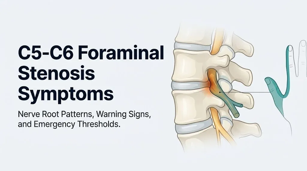
This is where C4-C5 and C5-C6 foraminal stenosis are most reliably distinguished clinically. C5 radiculopathy produces shoulder and upper arm symptoms. C6 radiculopathy produces forearm and finger symptoms. A person with pain and numbness in the thumb has C6 involvement. A person with pain and numbness in the deltoid region without forearm involvement has C5 involvement.
The weakness pattern also differentiates the two levels. C5 nerve root compression produces weakness in shoulder abduction (the deltoid, tested by asking the patient to raise the arm against resistance), while C6 compression produces weakness in elbow flexion (the biceps) and wrist extension. Both levels can weaken the biceps to some degree because the biceps receives input from both C5 and C6, but isolated deltoid weakness without significant forearm or hand involvement points strongly to C4-C5.
According to the North American Spine Society clinical practice guidelines for cervical radiculopathy, C5 radiculopathy from C4-C5 stenosis may also produce a characteristic shoulder pain that is sometimes misidentified as rotator cuff disease or shoulder impingement, because the C5 nerve root supplies both the shoulder muscles and the sensory innervation of the lateral arm that overlaps with referred pain from glenohumeral pathology.
Older men with a history of heavy overhead labor (such as electricians, painters, and construction workers) face elevated risk for C4-C5 foraminal stenosis because sustained overhead postures place repetitive compressive load on the C4-C5 foramen in cervical extension.
Key Takeaway: C4-C5 foraminal stenosis compresses the C5 nerve root, producing shoulder and upper arm symptoms. C5-C6 foraminal stenosis compresses the C6 nerve root, producing forearm, thumb, and index finger symptoms. These different locations reliably distinguish which level is affected.
C6 Nerve Root Dermatomal Distribution: Where You Feel the Symptoms
The C6 nerve root dermatomal distribution covers the lateral forearm from the elbow to the wrist, the radial (thumb-side) aspect of the wrist and hand, the thumb, and the index finger, and this specific territory is where patients with C5-C6 foraminal stenosis feel the most characteristic pattern of pain, numbness, and tingling.
A dermatome is the area of skin whose sensory nerve supply comes from a single spinal nerve root. Understanding dermatomal distribution is what separates vague “arm numbness” from a clinically specific pattern that points to a particular spinal level. When a patient with C5-C6 foraminal stenosis describes numbness in the thumb that is worse when looking to the right and extending the neck, they are describing C6 radiculopathy with textbook precision, even if they don’t know the anatomical term.
The clinical utility of the C6 dermatomal pattern is in differentiating it from the other commonly affected cervical levels. C7 nerve root compression (from C6-C7 stenosis) produces numbness in the middle finger and the dorsal forearm. C8 nerve root compression (from C7-T1 level) produces numbness in the ring and small fingers and the medial forearm. C5 radiculopathy, as described in the prior section, produces lateral upper arm symptoms without finger involvement. The specific location of numbness in the thumb and index finger reliably points to C6, not C5, C7, or C8.
The Cleveland Clinic notes that dermatomal patterns show individual variation of approximately 10 to 20% at their borders, meaning that some patients with C6 radiculopathy may also feel some numbness in the middle finger or the dorsal hand. This overlap reflects the anatomical fact that dermatomal boundaries are not perfectly sharp lines but gradients where adjacent nerve root territories partially overlap.
Patients with pre-existing carpal tunnel syndrome (median nerve compression at the wrist) face diagnostic confusion because median nerve distribution overlaps substantially with the C6 dermatome in the thumb and index finger. A clinician will need EMG and nerve conduction velocity testing to distinguish pure carpal tunnel syndrome from C6 radiculopathy, or from both conditions simultaneously (double crush syndrome).
Arm Pain and Neck Pain from C5-C6 Foraminal Stenosis
Arm pain from C5-C6 foraminal stenosis is produced by ectopic discharge of compressed nociceptive afferent fibers within the C6 nerve root, and this pain travels from the neck down the lateral forearm into the thumb and index finger following the path of the C6 nerve root from its origin at the C5-C6 level.
The pain character in C6 radiculopathy is distinctive. Most patients describe it as a sharp, burning, shooting, or electric quality that starts at the base of the neck or between the shoulder blades and radiates in a linear path down the outer arm, across the forearm, and into the thumb and index finger. This radiation pattern reflects the anatomy of the C6 nerve root’s sensory fibers exactly. The pain is not randomly distributed. It follows the path of the compressed nerve.
Neck pain itself in C5-C6 foraminal stenosis arises from two sources. The primary source is the degenerative changes in the disc, facet joints, and uncovertebral joints that caused the stenosis: these structures are well-innervated by the sinuvertebral nerve (also called the recurrent meningeal nerve), and degenerative changes in these structures produce local axial neck pain. The secondary source is the sensitization of the C6 nerve root at the foramen, which creates a state of neurogenic inflammation that can produce pain in the neck and shoulder region as well as the forearm and hand.
A characteristic clinical feature of radicular arm pain from foraminal stenosis is that it often worsens with specific neck positions. Extension and lateral rotation of the neck toward the affected side narrows the foramen further, acutely compressing the C6 nerve root and producing or worsening the arm pain and tingling. This is the basis of the Spurling’s test: the clinician laterally flexes and extends the neck toward the symptomatic side and applies gentle axial compression. A positive test reproduces the arm pain and confirms radiculopathy.
Manual laborers who must sustain neck extension (looking up at overhead work) experience exacerbation of their C5-C6 foraminal stenosis symptoms during and after work, because cervical extension is the position that most consistently narrows the intervertebral foramen.
Numbness and Tingling in the Fingers from C5-C6 Foraminal Stenosis
Numbness and tingling (collectively called paresthesia) in the thumb and index finger from C5-C6 foraminal stenosis result from impaired conduction in the large-diameter myelinated sensory fibers (A-beta fibers) of the C6 nerve root, which carry touch, vibration, and position sense from the C6 dermatomal territory to the spinal cord.
Large-diameter myelinated A-beta mechanosensory fibers are particularly susceptible to mechanical compression because their thick myelin sheaths, while providing fast conduction under normal conditions, are vulnerable to conduction failure when the pressure around the nerve root compresses the myelin. When these fibers stop conducting normally, the sensory cortex receives no signal from the thumb and index finger: this is experienced as numbness. When they conduct erratically, the sensory cortex receives abnormal signals that are interpreted as tingling or pins and needles.
The paresthesia in C6 radiculopathy characteristically involves the thumb and index finger and the lateral (radial) aspect of the palm. Patients often describe waking at night with numbness in these specific fingers, which reflects the fact that certain sleep positions place the neck in sustained extension or lateral rotation that narrows the C5-C6 foramen and increases nerve root compression. This nighttime pattern is one of the most consistent clinical features of cervical radiculopathy and helps distinguish it from other causes of hand numbness.
Patients should note precisely which fingers are numb when describing symptoms to a spine specialist. Numbness in the thumb and index finger: C6 radiculopathy. Numbness in the middle finger: C7 radiculopathy. Numbness in the ring and small fingers: C8 or ulnar nerve pathology. This precision directly guides which spinal level the physician will prioritize on MRI review.
People with Raynaud’s phenomenon or peripheral vascular disease may have baseline cold-induced finger numbness that overlaps with the C6 dermatomal pattern, complicating the clinical assessment. Their physician will need to use imaging and EMG to separate vascular from neurological contributions to finger symptoms.
Key Takeaway: Waking at night with numbness specifically in the thumb and index finger, not the middle or small fingers, is one of the most clinically reliable patterns of C6 radiculopathy from C5-C6 foraminal stenosis and should be reported precisely to a spine specialist.
Muscle Weakness from C5-C6 and C4-C5 Foraminal Stenosis
Muscle weakness from C5-C6 foraminal stenosis affects the muscles supplied by the C6 nerve root: primarily the biceps brachii, brachioradialis, and wrist extensors (extensor carpi radialis longus and brevis), while C4-C5 foraminal stenosis produces weakness in the muscles supplied by the C5 nerve root: primarily the deltoid and the shoulder external rotators (supraspinatus and infraspinatus).
The mechanism of weakness in foraminal stenosis is a failure of the motor axons within the compressed nerve root to conduct action potentials reliably from the spinal cord’s anterior horn cells to the muscle fibers they innervate. When the motor signal fails to arrive, the muscle cannot generate force. This is a lower motor neuron weakness: the muscle itself is intact, but its neural signal is interrupted at the nerve root level.
The biceps weakness in C6 radiculopathy is often subtle in the early stages, particularly because the biceps receives dual innervation from C5 and C6. A patient with C6 compression may retain reasonable biceps strength through their intact C5 contribution, and weakness may only become apparent on careful manual muscle testing. Wrist extension weakness (dropping the wrist when the forearm is pronated and the wrist is flexed against resistance) is often a more sensitive early indicator of C6 motor dysfunction because wrist extension is more exclusively a C6-mediated function.
Deltoid weakness in C5 radiculopathy from C4-C5 stenosis is clinically tested by asking the patient to raise the arm laterally (shoulder abduction) against the examiner’s resistance. A weakened deltoid cannot generate the expected force. Patients often describe this as inability to lift heavy objects above shoulder height, difficulty with overhead tasks, or a general sense of shoulder weakness that they may initially attribute to a rotator cuff problem.
Truck drivers and delivery workers who perform sustained overhead lifts are at elevated risk for C4-C5 stenosis because the combination of cervical extension and sustained shoulder loading increases the compressive and shear forces at the C4-C5 foramen.
Reflex Changes from Cervical Foraminal Stenosis
Cervical foraminal stenosis at C5-C6 characteristically reduces or eliminates the biceps tendon reflex and the brachioradialis reflex, because both of these deep tendon reflexes travel through the C6 nerve root arc, and compression of that nerve root disrupts the afferent limb of the reflex at the spinal cord level.
A deep tendon reflex works through a two-neuron arc. The afferent limb begins when the tendon hammer stretches the muscle spindle, sending a signal along sensory fibers back to the spinal cord. The efferent limb begins when the spinal cord’s interneurons activate the alpha-motor neurons supplying the muscle, causing it to contract. If the C6 nerve root is compressed, the afferent signal from the biceps or brachioradialis muscle spindle cannot reach the spinal cord reliably. The reflex arc is incomplete. The muscle contraction is diminished or absent.
The biceps reflex is mediated primarily by the C5 and C6 nerve roots (via the musculocutaneous nerve), and the brachioradialis reflex is mediated primarily by C5 and C6 (via the radial nerve). At C5-C6 foraminal stenosis, both reflexes may be reduced, but clinical observation consistently shows that the brachioradialis reflex is often the more reliably diminished of the two, because its arc depends more heavily on C6 than the biceps reflex, which has stronger C5 input.
A reduced or absent biceps or brachioradialis reflex in the context of arm pain and finger numbness provides objective neurological evidence of C6 nerve root dysfunction and helps distinguish a structural nerve root lesion from symptoms caused by central sensitization, fibromyalgia, or psychogenic pain.
The Cleveland Clinic notes that reflex asymmetry between the two arms (a reduced reflex on one side compared to the other) is more clinically meaningful than an absent reflex bilaterally, which may reflect constitutional variation in reflex excitability rather than a compressive nerve root lesion.
Older adults over 70 may have bilaterally diminished deep tendon reflexes from age-related peripheral nerve degeneration (presbyneuropathy), making it harder to use reflex asymmetry as a diagnostic indicator. In this population, EMG and nerve conduction velocity testing provides more reliable objective evidence of nerve root compression.
Bilateral Foraminal Stenosis and Bilateral Symptoms
Bilateral C5-C6 or C4-C5 foraminal stenosis means that both the left and right intervertebral foramina at the same spinal level are narrowed, potentially compressing both the left and right C6 or C5 nerve roots simultaneously, producing bilateral arm and hand symptoms that can complicate diagnosis and require careful clinical assessment.
Bilateral foraminal stenosis occurs less frequently than unilateral foraminal stenosis but is not rare, particularly in older adults with multilevel cervical spondylosis where the degenerative process affects the entire spinal level symmetrically. The presentation of bilateral foraminal stenosis can be easily confused with central canal stenosis producing myelopathy, because both conditions produce symptoms on both sides of the body.
The distinguishing features are anatomical. Bilateral foraminal stenosis compresses both C6 nerve roots but spares the spinal cord in the central canal. The resulting symptoms should still be confined to the C6 dermatomal distribution bilaterally: bilateral thumb and index finger numbness, bilateral lateral forearm pain, bilateral biceps and wrist extensor weakness. There should be no lower extremity symptoms, no gait disturbance, no bowel or bladder dysfunction.
If bilateral arm symptoms are accompanied by any lower extremity weakness, gait problems, or bowel or bladder changes, the clinical picture has moved beyond bilateral foraminal stenosis into cervical myelopathy territory, which has a categorically different urgency level. This distinction is addressed in detail in the next section.
People with rheumatoid arthritis are at particular risk for bilateral cervical foraminal stenosis because rheumatoid synovitis of the facet joints produces bilateral joint hypertrophy that can narrow the foramina on both sides simultaneously. This population also faces the additional risk of atlantoaxial instability (C1-C2 instability), which carries its own distinct neurological risk and requires separate imaging assessment.
Key Takeaway: Bilateral arm and hand symptoms in the C6 dermatomal distribution can mean bilateral foraminal stenosis, but any bilateral symptoms that also involve the legs, gait, or bowel and bladder function point to cervical myelopathy, which requires urgent evaluation rather than routine scheduling.
Foraminal Stenosis vs Cervical Myelopathy: A Critical Distinction
Cervical myelopathy is compression of the spinal cord itself within the cervical central canal, and it is clinically distinct from cervical foraminal stenosis in both its symptom pattern and its urgency level: myelopathy produces long-tract neurological signs (upper motor neuron signs, bilateral limb dysfunction, gait instability, bowel and bladder changes) that foraminal stenosis does not, and myelopathy can progress to permanent spinal cord injury without surgical decompression.
Foraminal stenosis compresses a nerve root (peripheral nervous system). Myelopathy compresses the spinal cord (central nervous system). This anatomical difference explains every symptom difference between the two conditions. Nerve root compression produces lower motor neuron signs: reduced reflexes, muscle weakness and wasting in the specific muscles supplied by that root, and sensory loss in that root’s dermatomal distribution. Spinal cord compression produces upper motor neuron signs: hyperreflexia (exaggerated reflexes in the limbs below the level of compression), spastic weakness, positive Hoffman’s sign (involuntary flexion of the thumb and index finger when the middle finger’s distal phalanx is snapped), and Lhermitte’s sign (an electric shock sensation traveling down the spine or into the limbs when the neck is flexed).
The Hoffman’s sign is one of the most clinically important physical examination findings that distinguishes myelopathy from radiculopathy. In foraminal stenosis alone, Hoffman’s sign should be negative. A positive Hoffman’s sign indicates upper motor neuron dysfunction from spinal cord compression and escalates the clinical urgency substantially. A spine specialist performing a Spurling’s test for radiculopathy should always test Hoffman’s sign on the same visit.
Gait instability is one of the earliest patient-recognized signs of myelopathy. Patients describe it as walking on a shifting surface, feeling unsteady in the dark, stumbling unexpectedly on flat ground, or difficulty with fine foot placement such as stepping onto curbs. This reflects dysfunction of the corticospinal tract (voluntary motor) and posterior column (proprioception) of the spinal cord.
Any person with a diagnosis of cervical foraminal stenosis who develops new gait instability, bilateral hand clumsiness, or any difficulty with bowel or bladder control should contact a board-certified orthopedic spine surgeon or board-certified neurosurgeon within 1 to 3 days for urgent MRI of the cervical spine with review of the central canal dimensions, not wait for a routine appointment.
Early vs Progressive C5-C6 Foraminal Stenosis Symptom Patterns
Early C5-C6 foraminal stenosis produces intermittent, position-dependent symptoms that resolve with neck repositioning or rest, while progressive foraminal stenosis produces persistent, constant symptoms that are present regardless of position and are accompanied by measurable neurological deficits on examination.
The early symptom pattern is characterized by intermittency. Neck pain and arm pain appear with sustained cervical extension or rotation toward the affected side and resolve when the head is returned to neutral. Numbness in the thumb and index finger comes and goes, often worse at night in certain sleep positions and better after waking and moving. There is no measurable weakness on manual muscle testing. Reflexes are intact. This is the stage where conservative management (physical therapy, activity modification, anti-inflammatory medications for pain context only) has the highest probability of achieving adequate symptom control without procedural intervention.
Progressive foraminal stenosis produces a different clinical picture. The arm pain and paresthesia become constant rather than intermittent. Certain positions may worsen symptoms but no position reliably eliminates them. Measurable weakness appears on manual muscle testing: the patient cannot fully resist pressure on the biceps or cannot maintain wrist extension against gravity. Reflexes become detectably reduced on the affected side. The patient may report that their grip strength has weakened, that they are dropping objects, or that fine motor tasks (buttoning, typing, writing) have become more difficult.
The NINDS/NIH notes that the natural history of cervical radiculopathy is favorable in the majority of cases, with approximately 75 to 90% of patients showing substantial improvement with non-operative management within 4 to 6 weeks. However, patients who show progressive neurological deficit (worsening weakness or spreading sensory loss) do not follow this favorable natural history and require expedited evaluation by a spine specialist rather than extended conservative management.
Younger patients under 40 with acute disc herniation-related foraminal stenosis have a higher probability of spontaneous resolution than older patients with osteophyte-driven foraminal stenosis, because disc herniation material can resorb over time while osteophytes do not.
Individual Variation: Who Experiences Worse Symptoms and Why
The severity of C5-C6 foraminal stenosis symptoms is not determined solely by the degree of anatomical narrowing on imaging. Individual variation in pain sensitivity, nerve root diameter, foraminal dimensions, pre-existing peripheral neuropathy, and systemic health factors all influence how much compression produces how much clinical symptom burden.
The most clinically important individual variation factor is the baseline state of the peripheral nerves before foraminal compression is added. This is most clearly demonstrated in the double crush syndrome concept in people with type 2 diabetes. Diabetic peripheral neuropathy produces diffuse axonal damage and reduced conduction velocity throughout all peripheral nerves. When a nerve root is also compressed at the foraminal level, the combined injury to the C6 nerve root’s axons (from diabetes plus from compression) produces worse symptoms than either injury alone would generate. A diabetic patient with only moderate C5-C6 foraminal stenosis on MRI may have severe symptoms, while a non-diabetic patient with more severe stenosis may have milder symptoms. This discordance between imaging severity and symptom severity is a well-documented clinical phenomenon.
Perimenopausal and postmenopausal women develop foraminal stenosis through accelerated disc degeneration driven by estrogen deficiency. The drop in estrogen at menopause reduces the synthesis of proteoglycans in the nucleus pulposus, causing the disc to lose hydration and height more rapidly. This accelerated disc height loss narrows the foramen from above and below, producing foraminal stenosis even without frank disc herniation. Women in this age group who develop new arm and neck symptoms deserve imaging evaluation rather than attribution of symptoms solely to musculoskeletal aging.
Tobacco smokers develop cervical disc degeneration at younger ages than non-smokers, because the cervical discs depend on diffusion through vertebral endplate capillaries for their nutrition (they are avascular structures), and smoking impairs endplate capillary perfusion, starving the disc of the glucose and oxygen it needs to maintain its proteoglycan matrix.
How Doctors Diagnose C5-C6 Foraminal Stenosis
Doctors diagnose C5-C6 foraminal stenosis through a combination of clinical history, physical examination testing (including Spurling’s test, muscle strength testing, reflex testing, and sensory examination), and imaging, with magnetic resonance imaging (MRI) of the cervical spine being the primary confirmatory imaging modality.
The diagnostic sequence a clinician follows for suspected C5-C6 foraminal stenosis typically proceeds as follows:
- Clinical history: Duration, onset (sudden with specific event, or gradual), quality of pain (burning, shooting, aching), location with precision (which fingers are numb, which arm is affected, does the neck position change symptoms), and functional impact (grip strength, overhead activities, sleep position).
- Physical examination: Cervical range of motion assessment; Spurling’s test (extension and lateral rotation toward the symptomatic side with gentle axial compression, positive if arm pain is reproduced); manual muscle testing of the deltoid (C5), biceps (C5-C6), wrist extensors (C6), and triceps (C7); deep tendon reflex testing of the biceps (C5-C6) and brachioradialis (C5-C6); sensory testing of the lateral forearm and thumb (C6) versus middle finger (C7) versus medial forearm (C8); Hoffman’s sign testing (to rule out myelopathy).
- MRI of the cervical spine: T2-weighted sequences show the degree of foraminal narrowing, disc herniation or osteophyte extent, and importantly, the cross-sectional area of the nerve root within the foramen. Oblique sequences specifically visualize the neural foramina. MRI also shows the central canal diameter, ruling in or out co-existing myelopathy.
- Electromyography (EMG) and nerve conduction velocity (NCV): These are ordered when the diagnosis is uncertain, when there is overlap with peripheral entrapment neuropathies (carpal tunnel syndrome), or when the severity of motor involvement needs objective quantification. EMG can detect denervation in C6 myotomal muscles (fibrillation potentials and positive sharp waves in the biceps, brachioradialis, or wrist extensors), confirming active nerve root compression.
- CT myelogram: Ordered when MRI is contraindicated (some pacemakers, certain metal implants) or when the MRI does not adequately characterize bony foraminal narrowing. CT myelogram provides superior visualization of osteophyte-related (hard disc) foraminal stenosis.
Key Takeaway: EMG and nerve conduction velocity testing can confirm active C6 nerve root compression when the clinical diagnosis is uncertain or when symptoms overlap with carpal tunnel syndrome, because it detects the electrical signature of denervation in specific C6 myotomal muscles.
What to Tell Your Doctor About C5-C6 Foraminal Stenosis Symptoms
Communicating precisely about your symptoms is one of the most practically useful things you can do before a spine specialist appointment, because the specific location, character, timing, and triggering factors of your symptoms are more diagnostically informative than a vague description of “neck and arm pain.”
To describe your symptoms effectively to a spine specialist or primary care physician:
- Specify which arm is affected: Right, left, or both. Foraminal stenosis is typically unilateral.
- Identify exactly which fingers are numb or tingling: Thumb and index finger (C6), middle finger (C7), or ring and small finger (C8). Write this down before your appointment so you state it precisely.
- Describe where the pain travels: Does it start in the neck and go down the outer arm into the forearm and thumb, or does it stay in the shoulder? The radiation pattern is clinically specific.
- Note what makes symptoms worse: Looking up (cervical extension), turning your head to the right or left, lifting overhead, sleeping in certain positions, driving.
- Note what makes symptoms better: Looking down, traction (gentle self-stretching of the neck), lying flat on your back.
- Report any weakness: Specify whether you have noticed difficulty lifting objects, dropping things, reduced grip strength, or difficulty with overhead tasks.
- Report any new gait, balance, or bowel and bladder changes: These are urgent symptoms that change the clinical category of your evaluation.
- List all imaging already performed: Bring your MRI report and any prior X-rays. If your MRI was done more than 2 years ago and your symptoms have changed, mention this so updated imaging can be ordered.
People who have been told by a previous provider that their symptoms are “just muscle tension” or “just stress-related” but who have persistent arm and finger symptoms matching a specific dermatomal pattern should specifically request a spine-focused examination and MRI at their next appointment.
Emergency Symptoms: When to Call 911 or Go to the ER
Certain symptoms associated with cervical foraminal stenosis, or that develop in the context of a known cervical spine condition, require immediate emergency evaluation. Do not wait to see if these resolve on their own.
Call 911 or go to the nearest emergency room immediately if you experience:
- Sudden onset of bilateral arm weakness or bilateral hand weakness that appears abruptly (indicates possible acute spinal cord compression from trauma or acute disc herniation in the central canal, not gradual foraminal stenosis)
- New difficulty walking, sudden gait instability, or loss of balance that has appeared recently and is worsening (indicates cervical myelopathy with corticospinal tract compromise requiring urgent surgical evaluation)
- New onset of bowel or bladder dysfunction (inability to control urination or defecation, or sudden urinary retention) in the context of a known cervical spine condition (indicates spinal cord compression at a level affecting autonomic pathways)
- A new electric shock sensation traveling down the spine or into all four limbs when the chin is brought to the chest (Lhermitte’s sign), appearing for the first time (indicates posterior column dysfunction from spinal cord compression)
- Progressive weakness in one arm that has worsened rapidly over hours to days rather than weeks, particularly following a new injury or trauma to the neck
- Loss of consciousness or acute neurological change following any cervical trauma, including a fall, motor vehicle accident, or sports injury, in a person with known cervical stenosis
These presentations can indicate acute cervical myelopathy, traumatic spinal cord injury, or rapid progression of cervical cord compression, all of which require emergency medical assessment, neurosurgical consultation, and potentially emergency surgical decompression, not an urgent care appointment or a phone call.
When C5-C6 Foraminal Stenosis Symptoms Require Emergency Evaluation
Non-emergency but time-sensitive presentations of C5-C6 foraminal stenosis that require evaluation by a board-certified orthopedic spine surgeon or board-certified neurosurgeon within 1 to 3 days (rather than weeks) include:
- Progressive weakness in the biceps or wrist extensors that is worsening week over week
- Complete numbness in the C6 dermatomal distribution (no sensation at all in the thumb and lateral forearm), rather than intermittent tingling
- Positive Hoffman’s sign discovered on examination, indicating spinal cord involvement beyond pure foraminal stenosis
- Inability to perform basic grip or pinch tasks (picking up coins, buttoning a shirt) from motor loss in the C6 myotome
- Severe radicular pain uncontrolled by any conservative measure and rated above 8 out of 10 on the Visual Analog Scale for pain consistently over more than 1 to 2 weeks
The distinction between a true emergency (911, ER now) and a time-sensitive specialist referral (within days) depends on whether symptoms involve the spinal cord or are confined to the nerve root. Cord involvement requires emergency response. Progressive but stable nerve root symptoms require urgent but not emergency specialist evaluation.
Any person who is uncertain whether their new symptom represents spinal cord or nerve root involvement should contact their primary care physician the same day and describe the symptom precisely, because the clinical distinction requires examination findings that cannot be made over the phone or from an article alone.
Frequently Asked Questions About C5-C6 Foraminal Stenosis Symptoms
What does C5-C6 foraminal stenosis feel like in the arm and hand?
C5-C6 foraminal stenosis produces a burning, shooting, or aching pain that travels from the base of the neck down the outer aspect of the arm, across the forearm, and into the thumb and index finger, following the C6 nerve root’s anatomical path.
The pain is often accompanied by tingling or numbness in the lateral forearm, the thumb, and the index finger, which is the C6 dermatomal distribution.
Some patients describe the sensation as electric or sharp when the neck is extended or rotated toward the affected side, which reflects the foraminal narrowing that occurs in these positions.
Can C5-C6 foraminal stenosis cause numbness in the thumb and index finger?
Yes, numbness in the thumb and index finger is one of the most characteristic and specific symptoms of C5-C6 foraminal stenosis, because these structures lie within the C6 dermatomal distribution supplied by the compressed C6 nerve root.
The numbness reflects impaired conduction in the large-diameter myelinated sensory fibers of the C6 nerve root, which normally carry touch and position sense from the thumb and index finger to the spinal cord.
If the numbness is isolated to the ring and small finger instead, C8 radiculopathy or ulnar nerve pathology is more likely, and if it affects the middle finger, C7 radiculopathy at the C6-C7 level should be evaluated.
Is C5-C6 foraminal stenosis serious or can it heal on its own?
The NINDS/NIH notes that approximately 75 to 90% of patients with cervical radiculopathy from foraminal stenosis experience substantial symptom improvement with non-operative management within 4 to 6 weeks, making the condition manageable in the majority of cases without surgery.
However, patients with progressive neurological deficit (worsening weakness or spreading sensory loss over weeks) do not follow this favorable course and require evaluation by a board-certified orthopedic spine surgeon or board-certified neurosurgeon within days to weeks rather than months.
Foraminal stenosis caused by osteophytes (bone spurs) rather than disc herniation is less likely to spontaneously resolve because bone does not resorb the way herniated disc material can.
What is the difference between C5-C6 foraminal stenosis and C4-C5 foraminal stenosis symptoms?
C5-C6 foraminal stenosis compresses the C6 nerve root, producing pain and numbness in the lateral forearm, thumb, and index finger, with weakness in the biceps and wrist extensors and a reduced biceps or brachioradialis reflex.
C4-C5 foraminal stenosis compresses the C5 nerve root, producing pain and numbness in the lateral upper arm and deltoid region (not the forearm or fingers), with weakness in shoulder abduction and a reduced or absent deltoid response.
The key distinguishing feature is finger involvement: forefinger and thumb numbness points to C6 (C5-C6 level), while symptoms confined to the shoulder and lateral upper arm without finger involvement point to C5 (C4-C5 level).
When should I see a doctor urgently for cervical foraminal stenosis symptoms?
Seek urgent evaluation by a board-certified orthopedic spine surgeon or board-certified neurosurgeon within 1 to 3 days if you develop progressive arm weakness that is worsening week over week, complete (not intermittent) sensory loss in the thumb and lateral forearm, or positive Hoffman’s sign on physical examination.
Call 911 or go to the nearest emergency room immediately if you develop sudden bilateral arm or leg weakness, new bowel or bladder dysfunction, new gait instability, or Lhermitte’s sign (electric shock sensation down the spine on neck flexion), as these indicate possible spinal cord compression requiring emergency evaluation.
Do not attempt to self-manage any rapid neurological deterioration with rest or home stretching: these specific symptoms require emergency imaging and potential neurosurgical intervention.
Does C5-C6 foraminal stenosis always require surgery?
The North American Spine Society clinical guidelines state that most patients with cervical radiculopathy from foraminal stenosis do not require surgery and achieve satisfactory outcomes with non-operative treatment including physical therapy, supervised exercise, and pain management.
Surgery is indicated when conservative management has failed after 6 to 12 weeks, when neurological deficits are progressive or severe, or when imaging shows significant compression with correlating clinical findings that suggest ongoing nerve root injury.
The most established surgical approach for C5-C6 foraminal stenosis is anterior cervical discectomy and fusion (ACDF), which removes the disc and/or osteophyte compressing the C6 nerve root and stabilizes the level; posterior cervical foraminotomy is an alternative that preserves motion but is less suitable for anterior compression.
Closing
C5-C6 foraminal stenosis symptoms follow a predictable anatomical pattern, and that predictability is actually useful for you. Numbness in the thumb and index finger, lateral forearm pain that shoots from the neck, reduced biceps or brachioradialis reflex, and weakness in wrist extension all point to the same nerve root at the same level. Understanding that pattern means you can describe your symptoms with precision rather than approximation when you speak with a spine specialist.
The most important next step is to schedule an MRI of the cervical spine if you have not had one, and to request a specialist referral if symptoms have been present for more than 4 to 6 weeks without improvement. Bring your symptom diary to that appointment: which fingers, which arm, what position makes it worse, whether any weakness is progressing.
If you develop any gait instability, bilateral arm weakness, or bowel or bladder changes alongside your cervical symptoms, stop waiting for an appointment and go to the emergency department today.