May-Thurner Syndrome Symptoms: Your 2026 Clinical Guide
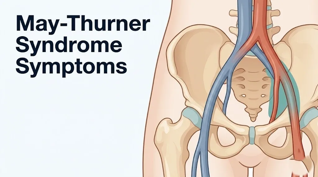
May-Thurner syndrome symptoms typically appear on the left side of the body, producing swelling, heaviness, and pain in the left leg due to a structural compression of the left common iliac vein by the right common iliac artery at the pelvic level. This anatomical arrangement creates a venous outflow problem that most standard medical workups miss because the root cause sits deep in the pelvis, invisible to physical examination alone.
According to a review published in the Journal of Vascular Surgery, up to 22 to 32 percent of adults have some degree of this arteriovenous anatomical overlap, yet most remain asymptomatic. A smaller but clinically important subset develops progressive venous hypertension, thrombosis, or both. Understanding the full symptom spectrum is what separates an early diagnosis from a missed one.
This guide covers every recognized symptom of May-Thurner syndrome with the physiological mechanism behind each one. It also covers how symptoms progress over time, who is most vulnerable, what the emergency thresholds are, and exactly which specialist evaluates and treats this condition.
What Is May-Thurner Syndrome
May-Thurner syndrome is a structural vascular compression disorder in which the right common iliac artery presses on the left common iliac vein, reducing venous drainage from the left leg and pelvis.
The condition was first formally described by Heinrich May and Josef Thurner in 1957, based on a cadaver study that found a fibrotic intraluminal web, called an intraluminal venous spur, in the left common iliac vein of a meaningful proportion of the specimens examined. This spur forms in response to chronic pulsatile mechanical stress from the overlying artery pressing against the vein. The posterior resistance comes from the body of the L4-L5 vertebral segment, which creates a fixed compression point that the vein cannot escape.
The condition is also called Cockett syndrome, named after surgeon Frank Cockett, who described its clinical consequences in the 1960s. The term May-Thurner syndrome is now standard in vascular medicine.
Think of the anatomy like a garden hose being pinched between a pulsating water pipe and a concrete slab. The hose does not burst immediately, but over months to years, the chronic pressure deforms its walls and reduces flow. That reduced flow is what produces every symptom described in this guide.
Key anatomical structures involved:
| Structure | Role in May-Thurner Syndrome | Why It Matters Clinically |
|---|---|---|
| Left common iliac vein (LCIV) | The compressed vessel | Site of reduced venous outflow, spur formation, and DVT risk |
| Right common iliac artery (RCIA) | The compressing structure | Crosses anterior to the LCIV at the iliocaval junction |
| L4-L5 vertebral body | The posterior bony resistance point | Creates the fixed anvil against which the vein is compressed |
| Intraluminal venous spur | Fibrotic endoluminal web from endothelial injury | Reduces luminal area, promotes venous stasis and clot formation |
| Iliocaval junction | Site of arteriovenous crossing | The precise anatomical location where compression occurs |
The anatomical variant is present to some degree in a large portion of the general population. What makes it a clinical syndrome rather than just a normal variant is when it produces measurable venous obstruction, symptoms, or thrombotic complications.
Younger women with a low body mass index are disproportionately represented among symptomatic patients, partly because reduced retroperitoneal fat provides less cushioning between the artery and vein.
May-Thurner Syndrome Symptoms: The Complete Clinical Picture
The recognized symptoms of May-Thurner syndrome include left leg swelling, left leg pain or heaviness, visible left-sided varicose veins, pelvic discomfort, skin changes on the left lower leg, and in acute presentations, the full symptom pattern of deep vein thrombosis.
Not every person with the anatomical compression develops every symptom. The clinical picture varies enormously based on the degree of venous obstruction, whether a clot has formed, the duration of the condition, and the individual’s baseline venous tone. Some people are completely symptom-free. Others present acutely with a massive left-sided deep vein thrombosis as their first recognized sign.
The symptom pattern is almost always left-sided. That anatomical specificity is one of the most diagnostically useful features of May-Thurner syndrome. Right-sided leg swelling, bilateral equal swelling, or symptoms in the right iliac territory point toward a different diagnosis.
Full symptom inventory with mechanism and clinical significance:
| Symptom | Physiological Mechanism | Clinical Significance |
|---|---|---|
| Left leg swelling (edema) | Venous hypertension raises capillary hydrostatic pressure, driving fluid into interstitial space | Most common presenting symptom; often worse after prolonged standing |
| Left leg pain or aching | Venous congestion distends vein walls, activating stretch-sensitive nociceptors in the vessel wall | May be constant, positional, or exertional |
| Left leg heaviness or fatigue | Impaired venous return increases blood volume load in the limb | Frequently misattributed to muscular fatigue or orthopedic causes |
| Left-sided varicose veins | Chronic venous hypertension causes reflux and superficial venous dilation | Visible on physical exam; may be the first sign noticed by patients |
| Pelvic discomfort | Venous congestion in the left iliac and pelvic venous plexus | Can overlap with pelvic congestion syndrome |
| Skin changes (hyperpigmentation, lipodermatosclerosis) | Hemosiderin deposition from chronic venous stasis | Indicates long-standing venous insufficiency |
| Acute DVT symptoms (sudden severe swelling, warmth, redness) | Thrombosis of the iliofemoral venous segment | Medical emergency requiring same-day evaluation |
People with inherited thrombophilia, including Factor V Leiden mutation or antiphospholipid syndrome, face compounded risk because the prothrombotic venous stasis environment produced by May-Thurner compression interacts with their baseline coagulation abnormality.
Left Iliac Vein Compression Symptoms Explained
Left iliac vein compression symptoms arise because reduced venous outflow from the left lower extremity raises pressure throughout the left-sided venous drainage pathway, from the left femoral vein up through the pelvic venous plexus and into the iliac system.
The left common iliac vein drains blood from the entire left leg and a significant portion of the left pelvic organs. When this vessel is mechanically compressed at the iliocaval junction, blood cannot exit the left limb at its normal rate. The system compensates initially by recruiting collateral venous channels, small alternative drainage routes that bypass the point of obstruction. But collaterals have limited capacity.
When collateral compensation is insufficient, pressure builds retrograde into the femoral system and beyond. This sustained high pressure is what produces the characteristic chronic aching, swelling, and heaviness that most patients describe long before any acute thrombotic event.
The degree of compression matters significantly. Research published in Radiology has shown that left iliac vein cross-sectional area reduction of 50 percent or more correlates with symptomatic venous outflow obstruction in most patients, while lesser degrees of compression may remain hemodynamically insignificant.
Symptoms attributable specifically to iliac vein compression (not downstream thrombosis):
- Persistent or intermittent left leg swelling, typically worse at the end of the day or after prolonged sitting or standing
- A sensation of fullness or pressure in the left groin or lower left abdomen
- Visible engorgement of superficial veins on the left inner thigh or left lower abdomen
- Left-sided pelvic aching that is positional and worsened by prolonged sitting
- Mild to moderate left leg fatigue with physical activity that exceeds what the level of exertion would typically produce
Patients who are pregnant or in the postpartum period often experience amplified versions of these symptoms because the pregnant uterus adds additional extrinsic compression to the left iliac vein from above, compounding the existing arterial compression from the right iliac artery.
Left Leg Swelling in May-Thurner Syndrome
Left leg swelling in May-Thurner syndrome is produced by chronic venous hypertension in the left lower extremity, which elevates capillary hydrostatic pressure and drives fluid from the intravascular space into the surrounding interstitial tissue.
This is called pitting edema when mild to moderate. Pressing a finger into the swollen tissue leaves an indentation because the interstitial space is fluid-saturated. In advanced cases or after a large DVT, the swelling can be tense, non-pitting, and extend from the ankle to the groin, a pattern called phlegmasia.
The swelling pattern in May-Thurner syndrome is diagnostically informative. It is almost exclusively unilateral and left-sided. Bilateral equal swelling suggests heart failure, kidney disease, or a systemic protein deficiency rather than a structural venous compression. Right-leg-dominant swelling is unlikely to be May-Thurner syndrome.
According to the American Venous Forum, unilateral left lower extremity swelling in a young woman without a clear cardiac, renal, or hepatic cause warrants imaging evaluation for iliocaval venous obstruction, including May-Thurner syndrome, before a diagnosis of idiopathic edema is accepted.
How the swelling typically progresses through the day:
- Minimal swelling present in the morning after overnight recumbency, when venous drainage is facilitated by horizontal positioning
- Gradual onset of ankle swelling during prolonged sitting, typically beginning by midmorning in sedentary occupations
- Swelling extends to the mid-calf level by early afternoon in more advanced cases
- Maximum swelling and sense of heaviness by late afternoon or evening after a full day of upright activity
- Partial resolution with leg elevation overnight, but baseline swelling increases over weeks to months as venous insufficiency progresses without treatment
In older adults with May-Thurner syndrome, which is less common but documented, the swelling may be incorrectly attributed to age-related venous insufficiency, delaying the structural diagnosis by years.
May-Thurner Syndrome Leg Pain: What It Feels Like
May-Thurner syndrome leg pain is produced by venous distension and increased intraluminal venous pressure activating stretch-sensitive pain receptors in the walls of the left femoral vein and its tributaries, generating a dull, aching, or pressure-type pain localized to the left lower extremity.
This is not the sharp, shooting pain of nerve compression or arterial disease. It is a deep, aching pressure that most patients describe as feeling like the leg is “full” or “congested” rather than injured. The pain often worsens with prolonged standing or sitting and improves with leg elevation or walking, because movement activates the calf muscle pump and partially reduces venous pooling.
The distribution of pain offers a clinical clue. In May-Thurner syndrome, leg pain tends to be diffuse throughout the left thigh and calf rather than focal. Pain concentrated in a specific calf muscle belly and worsening with dorsiflexion of the foot (a positive Homan’s sign) suggests acute deep vein thrombosis in the calf veins rather than pure hemodynamic compression.
A 2021 analysis in the Journal of Vascular and Interventional Radiology reviewing outcomes in patients treated for iliac vein compression noted that leg pain was the second most common presenting complaint after swelling, reported in approximately 60 to 70 percent of symptomatic patients before treatment.
Pain characteristics that help distinguish May-Thurner syndrome from other diagnoses:
- Diffuse left leg aching rather than focal muscle tenderness (distinguishes from muscle strain or compartment syndrome)
- Worsens with prolonged dependency of the leg and improves with elevation (distinguishes from arterial insufficiency, which worsens with elevation)
- No associated lumbar or sciatic radiation pattern (distinguishes from lumbar radiculopathy at L4-L5 or L5-S1)
- May be accompanied by ipsilateral pelvic heaviness or groin fullness (suggests iliac-level venous congestion rather than isolated femoral disease)
- Pain onset or worsening correlated with starting oral contraceptive therapy (suggests estrogen-mediated venous tone reduction compounding mechanical compression)
Men with May-Thurner syndrome, who represent a minority of cases (estimated 30 to 35 percent of symptomatic presentations), tend to present at older ages with more severe venous obstruction before seeking care, likely because the condition is less frequently suspected in male patients by clinicians.
Key Takeaway: Left-sided leg swelling and a deep aching pressure that worsens with prolonged sitting or standing and improves with leg elevation is a symptom pattern that warrants imaging evaluation for venous outflow obstruction, particularly in women of reproductive age.
Iliac Vein Blockage Symptoms You Should Know
Iliac vein blockage symptoms range from subtle chronic heaviness to sudden massive swelling depending on whether the obstruction is gradual hemodynamic restriction or acute thrombotic occlusion of the iliofemoral venous segment.
Gradual iliac vein blockage from the mechanical compression of May-Thurner syndrome produces a slow-onset venous insufficiency picture. The body compensates by developing venous collaterals, which partially reroute blood around the obstruction. This compensation can mask the severity of the structural problem for months to years. During this phase, symptoms remain chronic and progressive but not catastrophic.
Acute iliac vein blockage, meaning the development of a thrombus that abruptly occludes a significant portion of the iliac vein lumen, produces a dramatically different presentation. The left leg swells rapidly, over hours to days, with warmth, redness, skin tightening, and severe pain. This is acute iliofemoral deep vein thrombosis and it is a medical emergency, not a condition to monitor at home.
The distinction between these two presentations matters because they require different urgency levels and different treatments.
Comparing gradual vs. acute iliac vein obstruction in May-Thurner syndrome:
| Feature | Gradual Compression (No Clot) | Acute Thrombosis (Iliofemoral DVT) |
|---|---|---|
| Swelling onset | Weeks to months, worse at end of day | Hours to days, rapid and severe |
| Pain character | Dull aching, positional, relieved by elevation | Severe, constant, not relieved by elevation |
| Skin appearance | Normal or mild varicosities | Warm, red or cyanotic, shiny |
| Leg temperature | Normal or mildly warm | Warm early; cool if blood flow severely compromised |
| Systemic symptoms | None | Fever possible; severe cases may show cardiovascular signs |
| Urgency | Outpatient specialist evaluation within weeks | Emergency evaluation same day |
The National Heart Lung and Blood Institute notes that iliofemoral DVT carries a higher risk of pulmonary embolism than calf-vein DVT because the clot burden is larger and the proximal venous segments connect directly to the inferior vena cava and pulmonary circulation.
Women taking combined oral contraceptive pills containing estrogen face approximately three to four times the baseline risk of DVT compared to non-users, and May-Thurner syndrome substantially amplifies that risk further by adding a structural venous stasis factor.
Pelvic Pain and May-Thurner Syndrome
Pelvic pain is an underrecognized symptom of May-Thurner syndrome, produced by venous congestion in the left pelvic venous plexus when outflow obstruction at the iliac level backs up pressure into the draining pelvic veins.
The left iliac vein does not just drain the left leg. It also drains the left pelvic floor, left uterine and ovarian venous plexus in women, and portions of the left pelvic visceral venous network. When the LCIV is chronically obstructed, pressure propagates retrograde into these pelvic tributaries, producing a pattern of venous congestion in the pelvis that can mimic gynecological conditions, including endometriosis and ovarian cyst-related pain.
This pelvic venous congestion, when severe enough, meets the diagnostic criteria for pelvic congestion syndrome, a condition where chronically dilated and refluxing pelvic veins produce chronic pelvic pain. May-Thurner syndrome is now recognized as a structural driver of secondary pelvic congestion syndrome in a subset of patients.
A review published in the Journal of Vascular Surgery identified May-Thurner anatomy as an underlying structural finding in a portion of women diagnosed with pelvic congestion syndrome who had been treated symptomatically for years without resolution. Addressing the iliac compression in those patients reduced both the pelvic and leg symptoms.
Characteristics of pelvic pain in May-Thurner syndrome:
- Dull, aching pelvic pressure rather than sharp or cramping pain
- Worsens with prolonged standing or sitting and improves with lying down
- Often left-sided or left-predominant, though can be felt centrally as pelvic heaviness
- May worsen premenstrually when pelvic blood flow increases cyclically
- Can be associated with dyspareunia (pain with intercourse), particularly with deep penetration on the left side
- Not reliably explained by gynecological examination or pelvic ultrasound findings
Diagnosing the pelvic component requires cross-sectional imaging, specifically computed tomography venography or magnetic resonance venography, rather than standard pelvic ultrasound, which does not adequately visualize the iliac venous system.
May-Thurner Syndrome Skin Changes
Skin changes in May-Thurner syndrome are a sign of long-standing, inadequately treated venous hypertension in the left lower extremity, representing the downstream tissue-level consequences of chronic elevated venous pressure.
The skin changes follow a progression. Early in the course, the skin of the left lower leg may appear subtly erythematous (faintly red) or feel slightly indurated (firmer than normal). Over months to years of ongoing venous hypertension, the extravasation of red blood cells from capillaries under high pressure leads to the breakdown of hemoglobin in the tissue. The iron-containing breakdown product, hemosiderin, deposits in the skin, producing a brown or rusty discoloration that does not fade with elevation. This is called hemosiderin deposition or hyperpigmentation and it is a reliable marker of chronic venous disease.
In more advanced cases, the skin undergoes structural changes affecting the dermis and subcutaneous fat. Chronic inflammation produces fibrosis of the subcutaneous tissue, a process called lipodermatosclerosis, which causes the lower leg skin to feel hard, woody, and contracted. The leg can take on an inverted bottle-neck appearance where the ankle is constricted by fibrotic tissue while the calf remains swollen above it.
The most severe skin manifestation is a venous stasis ulcer: a wound that develops on the gaiter area (the lower leg above the ankle), typically on the medial surface. These ulcers are notoriously difficult to heal without treating the underlying venous hypertension.
Progression of skin changes in May-Thurner syndrome:
| Stage | Skin Finding | Mechanism | Reversibility |
|---|---|---|---|
| Early | Mild erythema, ankle swelling | Elevated capillary hydrostatic pressure | High with treatment |
| Intermediate | Hemosiderin pigmentation, varicose veins | Capillary red blood cell extravasation, hemoglobin breakdown | Partial with treatment |
| Advanced | Lipodermatosclerosis, skin thickening | Chronic subcutaneous fibrosis from venous inflammation | Limited; structural change persists |
| Severe | Venous stasis ulceration | Skin breakdown from ischemic hypoxia in chronically congested tissue | Requires specialist wound care |
Men over age 50 with unrecognized May-Thurner syndrome are disproportionately represented among patients presenting with venous stasis ulceration because the diagnosis is often not considered in male patients and the venous disease progresses further before evaluation.
Key Takeaway: Brown skin discoloration on the inner lower left leg that does not fade, accompanied by hardening of the skin, is a sign of advanced chronic venous disease and warrants evaluation by a vascular specialist for underlying venous outflow obstruction, not just topical wound care.
Asymptomatic May-Thurner Syndrome: When There Are No Symptoms
Asymptomatic May-Thurner syndrome refers to the anatomical state in which the right common iliac artery overlies and compresses the left common iliac vein but produces no clinically detectable symptoms, no measurable venous outflow obstruction, and no thrombotic events.
This is genuinely common. A post-mortem anatomical study by May and Thurner in 1957 identified the left iliac vein compression anatomy in approximately 22 percent of cadaveric specimens. More recent imaging studies using computed tomography venography in living patients have found some degree of iliac vein narrowing at the crossing point in an even higher percentage of individuals, depending on the threshold of compression defined.
The critical question is not whether the anatomy exists but whether it is hemodynamically significant. Most people with the anatomical variant never develop symptoms. The transition from an anatomical variant to a clinical syndrome requires additional factors: sufficient degree of venous lumen reduction, inadequate collateral compensation, a prothrombotic trigger, or hormonal factors that reduce venous wall tone and increase compressibility.
Asymptomatic May-Thurner anatomy found incidentally during imaging for another reason generally does not require treatment. The Society for Vascular Surgery clinical guidance indicates that treatment decisions should be based on clinical symptoms and hemodynamic assessment, not anatomical finding alone.
Factors that can convert asymptomatic May-Thurner anatomy into a symptomatic clinical syndrome:
- Starting combined oral contraceptive therapy containing ethinyl estradiol (reduces venous wall tone)
- Pregnancy (adds uterine compression and increases coagulability)
- Prolonged immobility (long-haul travel, bed rest, surgery recovery)
- Diagnosis with an inherited thrombophilia (Factor V Leiden, prothrombin G20210A mutation)
- Significant weight loss reducing retroperitoneal fat cushioning
- Development of scoliosis or lumbar spinal deformity altering the compression angle
Patients who are told they have May-Thurner anatomy incidentally should have an informed conversation with their primary care physician or a vascular specialist about these risk factors and whether prophylactic measures, such as avoiding certain contraceptive formulations or using compression hosiery during high-risk periods, are appropriate for their situation.
May-Thurner Syndrome Complications
The primary complications of May-Thurner syndrome are iliofemoral deep vein thrombosis, post-thrombotic syndrome, pulmonary embolism, and the development of chronic venous insufficiency with progressive skin and soft tissue changes.
Complications arise when the venous outflow obstruction is severe enough or prolonged enough to overwhelm the body’s compensatory mechanisms. The most feared acute complication is pulmonary embolism arising from a large iliofemoral clot. Because iliofemoral DVTs involve a large volume of clot in a proximal venous segment directly connected to the inferior vena cava, the risk of embolization to the pulmonary arteries is substantially higher than with isolated calf vein thrombosis.
A 2020 systematic review published in the Journal of Vascular Surgery found that iliofemoral DVT associated with May-Thurner syndrome was associated with a higher rate of post-thrombotic syndrome than DVT from other causes, even with standard anticoagulation treatment, because the underlying structural obstruction persists and continues to produce venous hypertension even after the clot is resolved.
The rarest and most severe acute complication is phlegmasia cerulea dolens, a condition in which massive iliofemoral thrombosis completely obstructs venous outflow from the entire left lower extremity. Blood cannot exit the limb. The leg becomes massively swollen, deeply cyanotic (blue-purple), and intensely painful. Without emergency intervention, arterial inflow to the limb is compromised, leading to venous gangrene and potential limb loss. This is a vascular emergency.
Summary of complications by severity:
| Complication | Mechanism | Urgency Level |
|---|---|---|
| Chronic venous insufficiency | Progressive venous hypertension and valve damage | Outpatient specialist management |
| Post-thrombotic syndrome | Scarring and venous valve damage from prior DVT | Ongoing specialist management |
| Iliofemoral DVT | Thrombosis of the compressed iliac-femoral segment | Same-day emergency evaluation |
| Pulmonary embolism | Embolization of iliofemoral clot to pulmonary arteries | 911 emergency |
| Phlegmasia cerulea dolens | Complete venous occlusion with circulatory compromise | 911 emergency, vascular surgery |
Iliofemoral DVT and May-Thurner Syndrome
Iliofemoral deep vein thrombosis in the context of May-Thurner syndrome occurs when venous stasis from chronic iliac vein compression reaches the threshold at which the coagulation cascade is activated in the slow-moving blood, producing a clot that extends from the iliac vein down through the common femoral and often into the superficial femoral vein.
This is not a peripheral calf DVT. Iliofemoral DVT involves the largest venous segments of the lower extremity. The clot burden is high. The left common iliac vein, external iliac vein, and common femoral vein may all be involved simultaneously, producing a dramatic clinical presentation.
The National Heart Lung and Blood Institute reports that DVT affects approximately 600,000 Americans annually, with iliofemoral DVT representing the most severe form. Among young women presenting with a first left-sided iliofemoral DVT without an obvious provoking factor, May-Thurner syndrome is now considered a major underlying structural cause that should be investigated systematically, particularly when thrombophilia testing is negative.
Clinically, the acute presentation includes sudden onset of severe left leg swelling, warmth, erythema (redness) of the skin, left groin pain, and in some cases a palpable cord along the inner thigh representing the thrombosed femoral vein.
What to tell the emergency provider if you suspect iliofemoral DVT:
- State the exact onset time of symptom worsening and describe the rate of swelling progression
- Describe any recent provoking factors: long travel, surgery, bed rest, new medication, pregnancy
- Disclose all medications, including oral contraceptives and hormone therapy, with start dates
- Report any prior DVT or pulmonary embolism in yourself or immediate family members
- Ask specifically whether imaging will evaluate the iliac vein level, not just the femoral and popliteal veins, since standard compression ultrasound is less reliable above the inguinal ligament
Young women under age 45 presenting to an emergency department with a first left-sided DVT should have a discussion with their treating physician about May-Thurner syndrome evaluation once the acute event is stabilized, because treating the DVT without addressing the underlying structural compression significantly increases the risk of recurrence.
Key Takeaway: A left-sided deep vein thrombosis in a young woman without a clear provoking factor should trigger evaluation for May-Thurner syndrome, because treating the DVT without addressing the underlying iliac vein compression substantially raises the likelihood of recurrence.
Post-Thrombotic Syndrome After May-Thurner
Post-thrombotic syndrome is a chronic complication that develops in a significant proportion of patients after iliofemoral DVT, characterized by persistent venous hypertension from damaged venous valves and residual luminal narrowing even after the acute clot resolves.
When a large DVT forms in the iliofemoral segment, the clot organizes over weeks to months. The inflammatory process that resolves the clot also scars the venous valve leaflets, preventing them from closing properly. Incompetent valves allow blood to flow backward (reflux) down the vein rather than forward toward the heart. This reflux compounds the existing outflow obstruction from May-Thurner compression, creating a doubly compromised venous drainage situation.
The result is chronic, often severe, venous insufficiency that persists years after the original DVT. Patients experience ongoing left leg swelling, pain, heaviness, skin changes, and in severe cases venous stasis ulceration, even after anticoagulation treatment for the DVT is complete.
Research published in the Journal of Vascular Surgery found that up to 50 percent of patients with iliofemoral DVT develop clinically significant post-thrombotic syndrome within five years, and that the risk is higher in patients with May-Thurner anatomy because the structural obstruction continues to elevate venous pressure even after the acute clot burden is reduced.
Symptoms of post-thrombotic syndrome that distinguish it from active DVT:
- Chronic left leg swelling that varies in severity but never fully resolves, unlike acute DVT swelling that typically improves somewhat with anticoagulation
- Left leg aching and heaviness that follows the same end-of-day pattern as the original pre-DVT symptoms
- Skin changes including hyperpigmentation and lipodermatosclerosis on the lower left leg
- Recurrent superficial venous thrombophlebitis in the varicose tributaries of the left leg
- In severe cases, venous ulceration on the medial left ankle region that recurs despite wound care
Compression stockings worn daily, at the gradient level recommended by a vascular specialist (typically 30 to 40 mmHg for post-thrombotic syndrome after iliofemoral DVT), reduce the severity of post-thrombotic syndrome symptoms. They do not treat the structural cause but significantly reduce the daily symptomatic burden.
May-Thurner Syndrome in Women: Why Female Patients Dominate
May-Thurner syndrome disproportionately affects women, with published case series and registry data consistently showing female-to-male ratios ranging from 2:1 to 3:1 among symptomatic patients, and peak incidence concentrated in the reproductive age range of 20 to 45 years.
This sex-specific pattern has a direct physiological explanation. Estrogen, the primary female reproductive hormone produced by the ovaries, acts on venous smooth muscle through estrogen receptors in the vessel wall. Estrogen-receptor activation reduces vascular smooth muscle tone, making veins more distensible and less resistant to external compression. A vein with lower intrinsic wall tone collapses more easily under the pulsatile pressure of the overlying right common iliac artery.
Progestins, whether endogenous from the ovarian progesterone cycle or exogenous from progestin-containing contraceptive formulations, further reduce venous tone through a separate progesterone-receptor mechanism. The combined effect of estrogen and progesterone on venous compliance explains why women taking combined oral contraceptive pills are at substantially elevated risk for symptomatic May-Thurner syndrome compared to non-users.
The pelvic anatomy also contributes. Women have a wider pelvis than men on average. A wider pelvis alters the angle at which the right common iliac artery crosses the left common iliac vein, potentially increasing the contact pressure at the crossing point. The uterus, when enlarged by pregnancy, fibroids, or adenomyosis, adds an additional anterior compressive force on the left iliac vein from below.
Summary of why May-Thurner syndrome presents more in women:
- Estrogen reduces venous smooth muscle tone, increasing vein compressibility
- Progestins compound venous dilation effects
- Oral contraceptive pills containing ethinyl estradiol amplify both venous and thrombotic risk
- Female pelvic anatomy may increase the crossing angle of the artery over the vein
- Uterine mass (pregnancy, fibroids) adds additional pelvic venous compression
- Hormonal cycling during the menstrual phase increases pelvic blood volume and potentially worsens symptoms premenstrually
Women with May-Thurner syndrome who wish to use hormonal contraception should have a detailed conversation with both their gynecologist and their vascular specialist before starting any estrogen-containing formulation, because the combined structural and hormonal thrombotic risk may be prohibitive in symptomatic or previously thrombotic patients.
May-Thurner Syndrome After Pregnancy
May-Thurner syndrome symptoms often become clinically apparent for the first time during pregnancy or in the immediate postpartum period, because pregnancy compounds iliac vein compression through multiple simultaneous mechanisms.
The pregnant uterus grows progressively throughout the second and third trimester, exerting increasing external pressure on the left iliac venous system from below. This uterine compression adds to the existing arterial compression from the right iliac artery, narrowing the effective venous lumen further than at the patient’s baseline. At the same time, blood volume increases by approximately 40 to 50 percent during pregnancy, raising venous return demand precisely when venous outflow is most restricted.
Pregnancy also produces a physiological hypercoagulable state. Clotting factor levels increase (particularly factors VII, VIII, X, and fibrinogen), while natural anticoagulant proteins S and C levels decrease relatively. This prepares the body to handle postpartum hemorrhage but simultaneously raises the risk of venous thrombosis in any area of venous stasis, including the already-compressed left iliac segment.
The postpartum period carries its own distinct risk. After delivery, the uterus involutes rapidly but dehydration, immobility during recovery, and the lingering hypercoagulable state persist for weeks. Women with May-Thurner anatomy who develop a postpartum left-sided DVT should be evaluated for the syndrome specifically, because the structural compression will remain and produce ongoing risk for future pregnancies and for non-pregnant life.
According to the American College of Obstetricians and Gynecologists, venous thromboembolism is one of the leading preventable causes of maternal mortality in the United States. Recognizing May-Thurner syndrome in pregnant patients before a thrombotic event occurs is a meaningful opportunity for risk reduction.
Warning signs during or after pregnancy that may indicate May-Thurner compression has become clinically significant:
- Left leg swelling that is substantially greater than typical pregnancy-related bilateral ankle edema
- Left leg swelling appearing early in the first trimester before uterine size explains it
- Left groin pain or pelvic pressure disproportionate to uterine size and position
- Sudden severe left leg swelling in the postpartum period within the first six weeks after delivery
- Any signs of pulmonary embolism: breathlessness, chest pain, rapid heart rate, in pregnant or postpartum women
Key Takeaway: Pregnant women with left-sided leg swelling and pain that exceeds typical pregnancy edema, or any postpartum woman with sudden left leg swelling and pain, need same-day vascular evaluation, not watchful waiting.
How May-Thurner Syndrome Is Diagnosed
May-Thurner syndrome is diagnosed through a combination of clinical suspicion based on symptom pattern and imaging studies that directly visualize the iliac venous anatomy and measure the degree of compression and venous obstruction.
The diagnostic process typically begins with a venous duplex ultrasound of the left lower extremity, which can identify DVT in the femoral and popliteal veins but is limited in its ability to visualize the iliac segment above the inguinal ligament because bowel gas and depth obstruct ultrasound penetration at that level. A normal duplex ultrasound of the leg does not rule out May-Thurner syndrome.
The gold standard for anatomical diagnosis is intravascular ultrasound (IVUS), which is performed by threading a small ultrasound catheter into the venous system through a femoral or jugular access point. IVUS directly visualizes the iliac vein lumen from the inside, measuring cross-sectional area and identifying the intraluminal spur. Research in Radiology has shown that IVUS identifies hemodynamically significant iliac vein compression more accurately than any external imaging method.
Non-invasive cross-sectional imaging options include computed tomography venography (CTV) and magnetic resonance venography (MRV), both of which provide excellent anatomical visualization of the iliocaval junction and surrounding structures. CTV uses ionizing radiation and iodinated contrast; MRV avoids radiation and uses gadolinium contrast.
Standard diagnostic pathway for May-Thurner syndrome evaluation:
- Clinical assessment: unilateral left leg swelling, pain, DVT history, or unexplained pelvic congestion triggers diagnostic consideration
- Venous duplex ultrasound of the left leg: identifies or excludes femoral and popliteal DVT; limited for iliac visualization
- Cross-sectional venous imaging (CTV or MRV): confirms iliac vein anatomy, measures compression degree, identifies collateral venous channels
- Intravascular ultrasound: performed at the time of planned intervention to confirm hemodynamic significance and guide stent placement
- Thrombophilia evaluation: blood testing for Factor V Leiden, prothrombin G20210A, antiphospholipid antibodies, protein C and S levels, and antithrombin III; performed by a hematologist to identify coexisting coagulation disorders that amplify DVT risk
Patients who have already been treated for a left-sided DVT with anticoagulation and are now being evaluated for the underlying cause should disclose their full anticoagulation history to the imaging team, as some venous mapping studies are affected by current anticoagulant therapy.
May-Thurner Syndrome Treatment and Vascular Specialist Referral
May-Thurner syndrome treatment addresses both the acute thrombotic complication (if present) and the underlying structural venous compression, with the goal of restoring adequate venous outflow from the left lower extremity and preventing recurrent DVT.
For patients presenting with acute iliofemoral DVT, the first priority is anticoagulation to stop clot propagation and reduce pulmonary embolism risk. Anticoagulation options include low-molecular-weight heparin, unfractionated heparin, and direct oral anticoagulants (DOACs). The choice depends on clinical factors including pregnancy status, renal function, and severity of presentation. Anticoagulation alone treats the clot but leaves the underlying structural compression unaddressed.
Catheter-directed thrombolysis (CDT) or pharmacomechanical thrombolysis is used in patients with large iliofemoral DVTs who present within a short enough time window. This approach involves threading a catheter directly into the clot and delivering clot-dissolving drugs locally, reducing the risk of post-thrombotic syndrome compared to anticoagulation alone. A 2023 analysis in the Journal of Vascular and Interventional Radiology found that combined thrombolysis plus iliac vein stenting in May-Thurner-related iliofemoral DVT produced significantly better long-term venous patency than thrombolysis alone.
Following clot removal or dissolution, iliac vein angioplasty and stent placement is the definitive structural treatment for May-Thurner syndrome. A metallic stent is deployed within the left common iliac vein at the point of compression, holding the vein open against arterial pressure. This restores venous outflow and substantially reduces the risk of DVT recurrence.
When and which specialist to see:
- Board-certified vascular surgeon: appropriate for patients with newly diagnosed iliofemoral DVT suspected to be May-Thurner related, or for patients with chronic venous insufficiency and suspected iliac obstruction; request referral from primary care physician
- Interventional radiologist: appropriate for iliac vein stenting evaluation, particularly if DVT occurred and thrombolysis is being considered; often works in collaboration with vascular surgery
- Hematologist: appropriate when thrombophilia testing is needed or when anticoagulation management is complex due to inherited clotting disorders
- Obstetrician-gynecologist or maternal-fetal medicine specialist: appropriate for pregnant patients or those in the postpartum period with suspected May-Thurner syndrome related DVT
Any patient who has experienced a left-sided iliofemoral DVT and has not yet been evaluated for May-Thurner syndrome should ask their primary care physician for a referral to a vascular specialist. The question to ask: “Given that my DVT was left-sided and iliofemoral, should I be evaluated for May-Thurner syndrome anatomy with venous imaging?”
Emergency Symptoms: When to Call 911 or Go to the ER
Certain symptoms associated with May-Thurner syndrome and its complications require immediate emergency evaluation. Do not wait to see if these resolve on their own.
Call 911 or go to the nearest emergency room immediately if you experience:
- Sudden shortness of breath that cannot be explained by exertion: this may indicate pulmonary embolism from an iliofemoral clot traveling to the lungs
- Sharp chest pain, particularly on one side, that worsens with breathing: a recognized presentation of pulmonary infarction from embolism
- Rapid or irregular heartbeat with accompanying breathlessness or light-headedness: suggestive of hemodynamic compromise from large pulmonary embolism
- Coughing up blood or pink-tinged sputum: indicates pulmonary hemorrhage from embolism
- Sudden, severe left leg swelling developing over hours with dark blue or purple skin discoloration: this is the presentation of phlegmasia cerulea dolens, a vascular emergency involving complete venous occlusion
- Loss of sensation or movement in the left leg with severe swelling: indicates circulatory compromise requiring emergency vascular intervention
- Leg pain that becomes unbearable and is not relieved by any position change, accompanied by swelling, warmth, and redness: acute massive iliofemoral DVT requiring emergency anticoagulation and possible thrombolysis
These presentations can indicate pulmonary embolism, massive iliofemoral deep vein thrombosis with circulatory compromise, or phlegmasia cerulea dolens, all of which require emergency medical assessment and vascular surgery consultation, not an urgent care appointment or a phone call to schedule an outpatient visit.
Frequently Asked Questions About May-Thurner Syndrome Symptoms
What are the first signs of May-Thurner syndrome?
The first signs of May-Thurner syndrome are typically left leg swelling and a sense of heaviness or aching in the left lower extremity that worsens after prolonged sitting or standing.
These early symptoms reflect the beginning of venous outflow obstruction from left iliac vein compression, producing mild venous hypertension before a clot has formed.
Some patients first notice visible varicose veins on the left inner thigh or left lower leg before swelling becomes consistent.
Can May-Thurner syndrome cause pelvic pain?
May-Thurner syndrome can cause pelvic pain when venous outflow obstruction at the iliac level backs up pressure into the left pelvic venous plexus, producing pelvic venous congestion.
This pelvic congestion produces a dull, aching left-sided or central pelvic pressure that worsens with prolonged standing and can mimic gynecological conditions including endometriosis.
A review in the Journal of Vascular Surgery identified May-Thurner anatomy as a structural driver of secondary pelvic congestion syndrome in a subset of women with unexplained chronic pelvic pain.
Is May-Thurner syndrome dangerous?
May-Thurner syndrome can be dangerous when it leads to iliofemoral deep vein thrombosis, which carries a high risk of pulmonary embolism due to the proximal location and large clot burden.
The anatomical compression itself, without thrombosis, produces chronic venous insufficiency that worsens over time but is not immediately life-threatening.
The condition becomes a true emergency when acute DVT forms or when clot embolizes to the pulmonary arteries, both of which require immediate medical evaluation.
How do doctors diagnose May-Thurner syndrome?
Doctors diagnose May-Thurner syndrome through clinical assessment of the left-sided symptom pattern combined with imaging studies including computed tomography venography, magnetic resonance venography, or intravascular ultrasound.
Standard leg compression ultrasound is insufficient because it cannot reliably visualize the iliac vein above the inguinal ligament.
Intravascular ultrasound, performed catheter-based at the time of planned treatment, is considered the most accurate method for confirming hemodynamically significant iliac vein compression.
Can you have May-Thurner syndrome without a blood clot?
You can have May-Thurner syndrome without a blood clot; many patients present with chronic left leg swelling, pain, and heaviness from hemodynamic venous obstruction before any thrombosis occurs.
This non-thrombotic presentation is sometimes called non-thrombotic iliac vein lesion (NIVL) and produces symptoms through venous hypertension alone.
Iliac vein stenting can be performed to relieve the structural obstruction in symptomatic non-thrombotic patients as well as in those who have experienced DVT.
What kind of doctor treats May-Thurner syndrome?
May-Thurner syndrome is treated by a board-certified vascular surgeon or an interventional radiologist, depending on whether the primary intervention is open vascular surgery or catheter-based iliac vein stenting.
A hematologist is often involved when coexisting thrombophilia is identified through blood testing, to guide anticoagulation management.
The initial referral typically comes from a primary care physician or emergency medicine physician after venous imaging confirms the diagnosis.
The Bottom Line on May-Thurner Syndrome Symptoms
Unexplained left leg swelling, aching, or heaviness, particularly in a woman of reproductive age, is not a symptom pattern to dismiss as vague or lifestyle-related without vascular imaging. The anatomical compression at the core of May-Thurner syndrome sits too deep in the pelvis to be identified on physical examination. A normal leg ultrasound does not exclude it.
The most important step after recognizing this symptom pattern is asking the right question of the right provider. A board-certified vascular surgeon or interventional radiologist can order computed tomography venography or magnetic resonance venography to evaluate the iliac vein anatomy directly. If the diagnosis is confirmed, structural treatment with iliac vein stenting changes the long-term trajectory of the condition substantially.
You now have the clinical picture, the physiological mechanisms, the emergency thresholds, and the specific specialist type to contact. Use that information to advocate clearly and precisely for the evaluation your symptom pattern warrants.