Early Varicose Veins Symptoms: Signs and Stages in 2026
Early stage varicose veins symptoms typically begin not with the visible twisted veins most people picture, but with subtler sensations: aching legs after standing, a persistent feeling of heaviness or fatigue in the calves, and faint bluish-green veins beginning to show through the skin. Recognizing these early signs matters because venous disease progresses along a defined clinical spectrum, and the window for the simplest interventions is widest before skin changes and complications develop.
According to the Society for Vascular Surgery, chronic venous disease affects an estimated 23 percent of adults in the United States, with varicose veins representing the most visible manifestation of the underlying condition. Research published in the Journal of Vascular Surgery has found that a substantial proportion of people with venous symptoms have measurable venous reflux on duplex ultrasound before their veins become visibly enlarged, which means the symptomatic phase begins earlier than most people and many primary care providers recognize.
This article covers the complete early-stage symptom picture, explains why each symptom happens inside the venous system, walks through the clinical staging framework vascular specialists use, identifies the specific warning signs that require same-day or emergency evaluation, and gives you the information you need to decide when and how to seek care.
Early Stage Varicose Veins Symptoms: What Your Legs Are Telling You
Early stage varicose veins symptoms are primarily sensory before they are visible, presenting as leg aching, heaviness, fatigue, and mild swelling that worsen predictably with prolonged standing and improve with leg elevation.
Most people seeking information about varicose veins focus on what they can see. The clinical reality is that the venous hypertension driving visible varicosity is already generating symptoms in nerves, muscles, and skin tissue before veins become dramatically enlarged or tortuous. The leg that aches after a four-hour shift on your feet but looks nearly normal is already experiencing the upstream consequences of venous valve dysfunction.
The key early symptoms, in order of clinical frequency reported by patients at initial vascular specialist evaluation:
- Leg aching or dull pain, worse after prolonged standing or sitting with legs dependent, relieved by elevation
- Heaviness or fatigue in the calves and thighs, often described as feeling like the legs are weighted or waterlogged
- Mild ankle swelling that appears by late afternoon or evening and resolves overnight
- Itching or burning sensation along visible or pre-visible veins, particularly on the inner calf and ankle
- Visible dilated veins, initially appearing as faint reticular veins or telangiectasias before progressing to classic varicosities
- Restless or cramping legs at night, particularly in the calf muscles
- Skin sensitivity or a crawling sensation along the course of the great saphenous vein on the inner leg
Women experience early-stage varicose vein symptoms at approximately twice the rate of men, based on multiple population studies. This is not simply because women seek care more readily. The underlying biology, specifically the effects of progesterone on venous wall smooth muscle tone and relaxin on valve tissue collagen integrity, directly accelerates the process of valve incompetence in women of reproductive age.
What Are Varicose Veins and How Do They Develop
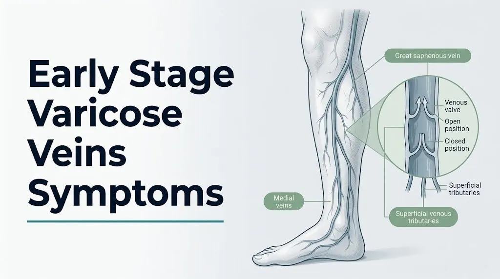
Varicose veins are abnormally dilated, tortuous superficial veins that develop when the bicuspid venous valve leaflets within the great saphenous vein or small saphenous vein fail to close completely, allowing blood to flow backward against gravity.
Think of healthy venous valves like a pair of hinged trapdoors inside a pipe. When blood flows upward toward the heart, the trapdoors open. When the heart relaxes and gravity pulls the blood downward, the trapdoors snap shut, preventing backflow. In varicose vein disease, those trapdoors no longer seal properly. Blood pools above each incompetent valve, pressing against the next valve down, which eventually fails too, in a cascading progression down the leg.
The structural failure begins at the level of the valve leaflet itself. Research published in Phlebology has identified that dysfunctional venous valves show reduced collagen content, altered matrix metalloproteinase activity (specifically elevated MMP-1 and MMP-9), and smooth muscle cell loss in the valve sinus. These changes reduce leaflet stiffness and prevent complete coaption during the relaxation phase of the cardiac cycle.
The resulting retrograde blood flow, called venous reflux, is measurable by duplex ultrasound as a reversed flow lasting more than 0.5 seconds during the Valsalva maneuver or calf compression release. That 0.5-second threshold is the internationally accepted clinical definition of pathological reflux, established by the American Venous Forum.
Once reflux is established, the column of blood that accumulates in the superficial venous system exerts hydrostatic pressure that increases with the height of the column from the reflux point to the ankle. At the ankle, where the hydrostatic column is longest, pressure is highest. This explains precisely why early varicose vein symptoms are worst at the ankle and lower calf, and why they worsen progressively through the day as the refluxing blood column grows.
People with a first-degree relative (parent or sibling) with varicose veins face a substantially elevated risk due to inherited patterns of venous wall collagen composition and valve leaflet structure. Clinical observation suggests heritability of venous disease in the range of 80 percent, though large-scale genetic studies confirming specific loci are still ongoing.
CEAP Classification: How Varicose Vein Disease Is Staged
The CEAP classification (Clinical, Etiological, Anatomical, Pathophysiological) is the internationally accepted staging framework for chronic venous disease, used by vascular surgeons, phlebologists, and vascular medicine specialists to describe disease severity, guide treatment decisions, and communicate consistently in clinical research.
Understanding CEAP tells you what your symptoms mean in clinical terms and where you fall in the disease spectrum.
| CEAP Stage | Clinical Description | Typical Symptoms | Action Threshold |
|---|---|---|---|
| C0 | No visible or palpable venous disease | Symptoms may be present; no visible findings | Evaluation if symptoms affect daily function |
| C1 | Telangiectasias (spider veins) or reticular veins | Mild cosmetic concern; possible mild itching | Discuss with primary care physician |
| C2 | Varicose veins present | Aching, heaviness, itching, mild swelling | Referral to phlebologist or vascular surgeon |
| C3 | Edema without skin changes | Persistent ankle and leg swelling | Prompt evaluation by vascular specialist |
| C4a | Skin pigmentation or eczema | Brownish skin discoloration, dermatitis | Evaluation within weeks; not emergency |
| C4b | Lipodermatosclerosis or white atrophy | Painful, thickened, discolored skin | Prompt specialist evaluation |
| C5 | Healed venous ulcer | Scar from previous ulceration | Ongoing specialist management |
| C6 | Active venous ulcer | Open wound on lower leg or ankle | Urgent specialist evaluation |
According to the American Venous Forum, most people presenting for their first varicose vein consultation are at C2 or C3. C4 through C6 disease represents advanced chronic venous insufficiency requiring active clinical management and carries risk of venous ulceration if left unaddressed.
The critical practical point: early stage varicose veins symptoms correspond to C1 through C2 on the CEAP scale. These are not just cosmetic findings. Untreated C2 disease progresses to C3 and beyond in a proportion of patients, particularly those who continue prolonged occupational standing without compression support, those who gain weight, and women who have additional pregnancies.
Men are more frequently diagnosed at C3 or above, because they tend to present to vascular specialists later in the disease course than women, often after skin changes have already developed.
Key Takeaway: The CEAP classification stages chronic venous disease from C0 (no visible changes) through C6 (active ulcer); early stage varicose veins are C1 to C2, and progressing beyond C3 is preventable with appropriate evaluation and management.
Early Visual Signs of Varicose Veins
The earliest visible signs of varicose vein development are not the large, ropy, twisted veins most people associate with the condition. They are subtle vascular changes that precede classic varicosities by months to years.
Telangiectasias, commonly called spider veins, are the earliest visible manifestation of superficial venous disease. They appear as clusters of fine red, blue, or purple vessels less than 1 millimeter in diameter, most often on the thigh, behind the knee, or on the inner ankle. They represent dilated dermal capillaries and post-capillary venules responding to increased local venous pressure, not reflux in a major trunk vein. Telangiectasias alone are classified as CEAP C1 disease.
Reticular veins are slightly larger, appearing as flat or mildly raised blue-green vessels 1 to 3 millimeters in diameter visible just beneath the skin surface. They often feed clusters of telangiectasias. Reticular veins represent dilation of the subdermal venous plexus in response to elevated venous pressure from deeper sources.
The transition to classic varicose veins (CEAP C2) involves dilation of the superficial truncal veins themselves, primarily the great or small saphenous vein and their named tributaries. These appear as raised, tortuous, blue-green cordlike structures greater than 3 millimeters in diameter. They are most visible on the inner thigh, medial calf, and posterior calf.
To assess whether a visible vein warrants clinical evaluation, observe the following:
- Stand for 5 to 10 minutes to allow maximum venous filling before examining your legs
- Examine the inner thigh from the groin to the knee in good lighting for raised or tortuous veins
- Check the medial (inner) calf from the knee to the ankle for both flat reticular veins and raised varicosities
- Check the back of the calf for the small saphenous vein territory
- Note any vein that is raised above the skin surface, tender to touch, or feels warm, as these features require clinical evaluation to exclude thrombosis
Inner ankle clusters of spider veins, called corona phlebectatica, are a specific finding that vascular specialists recognize as a clinical indicator of underlying saphenous reflux and elevated venous pressure, even when no larger varicosities are visible. Their presence at the ankle should prompt evaluation by a primary care physician with referral to a board-certified vascular surgeon or phlebologist if symptoms are present.
Varicose Veins Aching, Heaviness, and Leg Fatigue
Leg aching and heaviness are the most consistent early sensory symptoms of varicose vein disease, reported by the majority of patients with CEAP C2 disease who present for specialist evaluation.
The mechanism is not mysterious. When venous valve incompetence allows blood to pool in the superficial venous system, the elevated hydrostatic pressure stretches the venous wall and compresses surrounding tissue. Distension of the venous adventitia (the outer layer of the vein wall) activates stretch-sensitive nociceptors, the pain-sensing nerve endings in connective tissue surrounding the veins. The result is a dull, diffuse aching that tracks along the course of the affected vein rather than localizing to a single point.
The aching follows a predictable daily pattern that distinguishes it from musculoskeletal leg pain. Venous aching is worst after prolonged standing or sitting with legs dependent, builds progressively through the day, and reliably improves with leg elevation above the level of the heart or with walking (which activates the calf muscle pump and reduces venous pooling). Musculoskeletal pain from conditions like plantar fasciitis or calf muscle strain does not follow this pattern of positional relief.
According to the Society for Vascular Surgery Clinical Practice Guidelines, leg symptoms that improve with leg elevation and worsen with dependency are clinically consistent with venous origin and should be evaluated with duplex ultrasonography to assess for underlying reflux, even when visible varicose veins are not yet apparent.
The heaviness sensation reflects a different but related mechanism: the weight of the pooled blood volume in the incompetent superficial venous system pressing against the leg tissue. A normal saphenous venous system holds a relatively small blood volume. When reflux allows venous blood to accumulate in the dilated tributaries, the added mass is genuinely measurable in some patients as increased leg circumference by end of day.
Older adults over 65 may attribute this aching and heaviness to arthritis or general aging rather than venous disease, which delays appropriate diagnosis and treatment. A careful positional pattern history (worse when standing, better when lying down with legs up) is the key distinguishing question a primary care physician should ask.
Varicose Veins Swelling and Ankle Edema
Ankle and lower leg swelling (edema) associated with varicose veins develops when elevated venous pressure in the capillary bed forces fluid out of the intravascular space and into the surrounding interstitial tissue.
The physiology follows Starling forces. In a healthy capillary bed, the balance between hydrostatic pressure (pushing fluid out) and oncotic pressure (pulling fluid in) keeps fluid within the vessel. When venous hypertension from reflux elevates capillary hydrostatic pressure, this balance shifts toward net fluid filtration into the tissue. The result is pitting edema, most visible at the ankle by late afternoon, that temporarily resolves overnight when the legs are horizontal and gravity no longer contributes to the hydrostatic load.
Varicose vein-associated edema is characteristically pitting (meaning finger pressure leaves a temporary indentation), bilateral or asymmetric (typically worse on the side with greater venous disease), and positional (almost fully resolved after a night of recumbent sleep). These features distinguish it from cardiac edema (typically bilateral and accompanied by shortness of breath), from lymphedema (non-pitting, does not fully resolve overnight), and from acute DVT swelling (sudden onset, often dramatically unilateral, painful).
The National Heart, Lung, and Blood Institute notes that bilateral leg edema has multiple potential causes, including cardiac, renal, hepatic, and venous etiologies, and that distinguishing venous edema from other forms requires clinical examination and, in many cases, further testing.
To track whether your swelling pattern is consistent with venous disease:
- Measure your ankle circumference with a fabric tape measure at the same point each morning before getting out of bed (resting overnight measurement)
- Measure again at the end of the day after the longest standing period
- Record the difference between morning and evening measurements over two weeks
- Note whether the swelling is equal in both legs or consistently worse on one side
- Report any difference greater than 1 centimeter between morning and evening measurements, or any sudden increase in swelling without a positional explanation, to your primary care physician
Pregnant women develop ankle edema from multiple overlapping mechanisms including compression of the inferior vena cava by the gravid uterus, increased plasma volume, and progesterone-driven venous dilation. Distinguishing normal pregnancy edema from symptomatic varicose vein edema can be clinically difficult, and a board-certified obstetrician-gynecologist should evaluate any ankle swelling in pregnancy that is rapidly progressive, unilateral, or accompanied by calf tenderness.
Key Takeaway: Varicose vein ankle swelling is positional and pitting, predictably worsening through the day and resolving overnight; sudden, unilateral, or severe swelling that does not follow this pattern requires same-day evaluation to exclude deep vein thrombosis.
Varicose Veins Itching, Burning, and Skin Irritation
Itching and burning along the course of varicose veins or over the skin overlying dilated superficial veins is a recognized symptom of venous disease that most people misattribute to dry skin, an allergic reaction, or a rash.
The mechanism involves local inflammatory mediators released by venous hypertension. When elevated pressure in the dilated superficial venous system causes red blood cells and plasma proteins to leak into the surrounding dermal tissue, the breakdown products of extravasated cells (including hemosiderin, the iron-containing remnant of degraded red blood cells) activate mast cells and trigger histamine release. Histamine is the primary biochemical driver of itch in dermal tissue. In early-stage disease, this reaction is mild and intermittent, producing a localized itch that patients often scratch without realizing it is vascular in origin.
The burning sensation has a different mechanism. Distension of the venous adventitia and the surrounding connective tissue activates not just pressure-sensitive nociceptors but also thermoreceptive nerve fibers, which interpret the local inflammatory environment as thermal stimulus. This produces the classic burning sensation patients describe along the course of a dilated vein, typically on the inner calf.
Skin changes in early varicose vein disease stop at the level of localized itching and minor skin sensitivity. When itching progresses to dermatitis with weeping, scaling skin, this represents stasis dermatitis, a CEAP C4a finding that requires evaluation by a primary care physician and likely referral to a board-certified vascular surgeon.
According to the European Journal of Vascular and Endovascular Surgery, itching associated with chronic venous disease is present in an estimated 35 to 50 percent of patients with varicose veins and is often undertreated because its venous origin is not recognized by patients or primary care providers.
People with a personal or family history of eczema or contact dermatitis may find that varicose vein-associated skin inflammation resembles and overlaps with their pre-existing skin condition, making it harder to attribute the itching to venous disease specifically. A dermatologist and a vascular specialist working collaboratively may be needed to sort this out in complex cases.
Varicose Veins Symptoms at Night and Restless Legs
Nocturnal leg symptoms associated with varicose veins, including cramping, restlessness, and a crawling or prickling sensation, are among the most frequently reported but least recognized early manifestations of venous disease.
The underlying physiology relates to what happens in venous circulation during sleep. During waking hours, walking and muscle contractions activate the calf muscle pump (venous sinuses within the soleus and gastrocnemius muscles that propel blood upward with each contraction). At night, lying horizontally removes gravity’s contribution to pooling, but it also eliminates the calf muscle pump’s contribution to venous return. In a patient with venous reflux, this can allow venous pressure redistribution in ways that activate stretch-sensitive and mechanoreceptive nerve fibers in the venous adventitia and surrounding musculature.
The cramping is produced by a related but distinct mechanism. Elevated local venous pressure affects the microcirculation to calf muscle fibers, reducing oxygen delivery at the tissue level. Hypoxic calf muscle tissue generates lactic acid and pro-inflammatory metabolites that lower the threshold for motor neuron excitability, predisposing the gastrocnemius and soleus muscles to spontaneous cramping contractions.
Restless legs syndrome (RLS) and venous disease share significant symptom overlap, and research published in Phlebology has found that a proportion of patients diagnosed with RLS have underlying venous insufficiency as a contributing or primary cause. The key distinguishing feature is that RLS symptoms are typically bilateral, involve a compulsion to move the legs, and are not primarily localized along the course of a specific vein. Venous-origin nocturnal symptoms are more often unilateral or asymmetric, correlating with the side of greater venous disease, and produce cramping or localized aching rather than the urge-to-move characteristic of true RLS.
A person who has been told they have restless legs syndrome but has visible varicose veins or daytime venous symptoms should ask their primary care physician about duplex ultrasonography to assess for underlying reflux before assuming the diagnosis is purely neurological.
Varicose Veins Skin Changes and Advanced Progression
Skin changes in varicose vein disease represent the transition from CEAP C2 to C4 disease, signaling that venous hypertension has been sustained long enough to cause structural changes in the dermis and subcutaneous tissue.
The progression begins with hemosiderin deposition, the brownish-yellow discoloration of skin above the ankle and medial calf that results from red blood cells leaking through capillary walls and being broken down in the tissue. The iron in hemoglobin is converted to hemosiderin, which is then engulfed by tissue macrophages and remains in the dermis as a permanent pigment. This produces the characteristic brownish staining of the medial gaiter area (the area above the inner ankle) seen in C4a disease.
Lipodermatosclerosis develops at C4b. The venous hypertension-driven inflammation activates fibroblasts in the subcutaneous fat layer, producing progressive fibrosis that replaces normal elastic adipose tissue with dense, contracted scar tissue. The skin becomes indurated, thickened, and painful, often with an inverted champagne bottle appearance of the lower leg where the fibrotic contraction narrows the calf above the ankle. Lipodermatosclerosis significantly increases the risk of venous ulceration with minor trauma and represents a vascular emergency threshold that requires prompt evaluation by a board-certified vascular surgeon.
An active venous ulcer (C6 disease) is an open wound, typically on the medial gaiter area, that fails to heal because the underlying venous hypertension continuously impairs tissue oxygenation and repair capacity. The National Heart, Lung, and Blood Institute estimates that venous leg ulcers affect approximately 1 percent of the general population and account for the majority of all chronic leg ulcers, representing a substantial clinical and quality-of-life burden.
Reaching C4-C6 disease is not inevitable. Research published in the Journal of Vascular Surgery indicates that treatment of underlying saphenous reflux at the C2 stage substantially reduces the risk of progression to advanced skin disease and ulceration.
Varicose Veins During Pregnancy and Hormonal Risk Factors
Pregnancy is the most concentrated convergence of varicose vein risk factors that a woman can experience: elevated progesterone, elevated relaxin, expanded blood volume, increased venous pressure from the growing uterus, and reduced venous return from inferior vena cava compression.
Progesterone, produced initially by the corpus luteum and then by the placenta, acts on smooth muscle cells throughout the body, including those in venous walls. It reduces vascular smooth muscle tone, allowing venous walls to distend more easily under pressure. This is physiologically necessary during pregnancy to accommodate the 40 to 50 percent expansion in blood volume that occurs, but it also allows existing venous valves to stretch and prevents new valve leaflets from coapting effectively.
Relaxin, a peptide hormone produced by the corpus luteum and later the placenta, remodels collagen and connective tissue throughout the body to allow pelvic ligament and cervical tissue relaxation for delivery. The same collagen-remodeling effects occur in venous valve leaflets, reducing their structural integrity and increasing reflux risk.
The gravid uterus compresses the inferior vena cava and iliac veins, the major venous trunks returning blood from the legs to the heart. This compression raises venous pressure throughout the lower extremity venous system, adding an obstructive component to the hormonal component of varicose vein development in pregnancy.
According to clinical observation published in Obstetrics and Gynecology, varicose veins are present in an estimated 30 to 40 percent of pregnant women, with prevalence increasing with each successive pregnancy. Many pregnancy-related varicosities improve within three to six months postpartum as hormonal levels normalize and blood volume decreases, but some persist and worsen with subsequent pregnancies.
Women planning future pregnancies who have symptomatic varicose veins should be evaluated by a board-certified vascular surgeon before conception, to discuss whether treatment before pregnancy would reduce progression risk, and to establish baseline imaging for comparison.
Key Takeaway: Pregnancy amplifies varicose vein development through three simultaneous mechanisms: progesterone-driven venous wall relaxation, relaxin-driven valve leaflet collagen remodeling, and inferior vena cava compression from the growing uterus.
Varicose Veins Risk Factors: Who Develops Them and Why
The risk factors for varicose vein development operate through three broad pathways: structural factors that affect venous wall and valve integrity, hemodynamic factors that increase venous pressure, and hormonal factors that weaken vein wall resistance.
Structural and genetic risk factors:
- Family history: clinical observation consistently shows an 80 to 90 percent rate of first-degree relative with varicose veins among affected patients, suggesting a heritable component to venous wall collagen composition and valve leaflet structure
- Female sex: women develop varicose veins at approximately twice the rate of men across all age groups, attributable to hormonal effects on venous wall tone described in the pregnancy section
- Age: prevalence increases with age as venous wall elastin content declines with normal connective tissue aging; adults over 50 show markedly higher prevalence than those under 30
Hemodynamic and lifestyle risk factors:
- Prolonged occupational standing: hairdressers, surgeons, teachers, nurses, and retail workers who stand 6 or more hours daily face elevated venous pressure exposure across each working day
- Obesity: elevated intraabdominal pressure from adipose tissue compresses the iliac and femoral veins, increasing venous pressure throughout the lower extremity venous system
- Sedentary lifestyle: reduced calf muscle pump activity allows venous blood to pool in the lower extremity superficial venous system during prolonged sitting
Hormonal risk factors:
- Multiple pregnancies: cumulative hormonal and mechanical stress with each pregnancy
- Oral contraceptive use: combined oral contraceptives containing estrogen increase venous distensibility and have been associated with elevated venous disease risk in some epidemiological studies
- Postmenopausal hormone therapy: exogenous estrogen and progesterone in menopausal hormone therapy maintains hormonal influence on venous wall tone beyond the reproductive years
Men with varicose veins are less likely to have identifiable hormonal contributors and more likely to have occupational, hemodynamic, or family history-driven disease. They also present with more advanced disease at first evaluation due to delayed care-seeking, making risk factor recognition important for earlier male presentation.
Varicose Veins vs Deep Vein Thrombosis: Knowing the Difference
Varicose veins and deep vein thrombosis (DVT) can produce overlapping symptoms including leg aching, swelling, and a palpable, tender cord in the leg, but they are distinct conditions with very different urgency levels, and confusing them can be dangerous.
DVT is a blood clot forming in the deep venous system of the leg (femoral vein, popliteal vein, or calf deep veins) and carries risk of pulmonary embolism if the clot breaks off and travels to the pulmonary arteries. Varicose veins involve only the superficial venous system and do not directly cause pulmonary embolism through the same mechanism, though they are associated with a modestly elevated DVT risk in epidemiological data.
| Feature | Varicose Veins | Deep Vein Thrombosis |
|---|---|---|
| Vein system involved | Superficial (great/small saphenous tributaries) | Deep (femoral, popliteal, calf deep veins) |
| Onset of swelling | Gradual, builds through the day | Sudden, often within hours to a day |
| Swelling pattern | Bilateral or positional; resolves overnight | Typically unilateral and persistent |
| Pain character | Dull aching, worse with standing | Calf or thigh tenderness to pressure |
| Skin changes | Visible tortuous veins, possible discoloration | Redness, warmth over affected area |
| Associated risk | Superficial thrombophlebitis | Pulmonary embolism |
| Positional relief | Improves substantially with elevation | Does not reliably improve with elevation |
| Urgency | Routine to urgent depending on severity | Emergency evaluation required |
Superficial thrombophlebitis represents a middle clinical scenario: a blood clot forming within a varicose vein rather than the deep system. It produces a painful, warm, firm cord along the course of a varicose vein with surrounding redness and tenderness. While superficial thrombophlebitis alone does not carry the immediate pulmonary embolism risk of DVT, research published in the Journal of Vascular Surgery found that concurrent DVT is present in 6 to 44 percent of patients presenting with superficial thrombophlebitis, depending on the extent and location of the superficial clot. This is why superficial thrombophlebitis requires same-day evaluation by a physician and duplex ultrasonography to exclude concurrent DVT.
Any person with varicose veins who develops sudden new unilateral leg swelling, calf tenderness, or redness with warmth should be evaluated in an emergency room the same day, not scheduled for a routine appointment.
How Varicose Veins Are Diagnosed
Varicose veins are diagnosed through a combination of clinical history, physical examination, and venous duplex ultrasonography, the non-invasive imaging test that directly visualizes venous anatomy and measures venous reflux.
The clinical examination involves inspection of the legs in a standing position to assess vein distribution, size, and pattern. The physician palpates visible varicosities for tenderness, warmth, or cord-like firmness suggesting thrombosis. Historical assessment includes symptom pattern (positional worsening and relief), duration, family history, occupational standing, and prior treatments or procedures.
The Trendelenburg test is a bedside clinical examination that assesses whether the saphenofemoral junction or perforating veins are the primary source of reflux. It involves elevating the leg to empty the superficial veins, applying manual occlusion at the groin, having the patient stand, and observing the pattern of venous refilling. While informative, the Trendelenburg test has lower accuracy than duplex ultrasonography and is used as a screening complement, not a standalone diagnostic tool.
Duplex ultrasonography combines B-mode (structural) ultrasound imaging with Doppler flow measurement. The examining sonographer or vascular surgeon can directly visualize the great saphenous vein, small saphenous vein, and perforating veins while measuring reflux duration during standardized compression maneuvers. Reflux lasting more than 0.5 seconds (for truncal veins) is the diagnostic threshold for pathological venous incompetence, per American Venous Forum guidelines.
The diagnostic workup for a first presentation of varicose veins should include:
- Complete lower extremity venous duplex mapping from the groin to the ankle on both sides
- Assessment of the saphenofemoral junction, saphenopopliteal junction, and major perforating veins
- Deep venous system evaluation to exclude concurrent DVT or post-thrombotic syndrome
- Documentation of reflux duration at each incompetent valve segment
- Correlation of imaging findings with symptom pattern and CEAP clinical stage
A primary care physician can initiate the evaluation and order duplex ultrasonography. Interpretation and treatment planning require a board-certified vascular surgeon or phlebologist with experience in venous disease management.
Varicose Veins Treatment Options Overview
Treatment for varicose veins is directed at eliminating venous reflux by closing or removing the incompetent vein segment, redirecting blood flow through competent deep and perforating veins, and reducing the venous hypertension that drives symptom progression.
Non-interventional management includes graduated compression stockings (medical grade, typically 20 to 30 mmHg for early symptomatic disease or 30 to 40 mmHg for more advanced presentations), leg elevation, regular walking to activate the calf muscle pump, and weight management. Compression therapy reduces venous pooling and the hydrostatic pressure at the ankle. A systematic review published in the Cochrane Database of Systematic Reviews found that compression therapy reduces varicose vein symptoms and edema but does not eliminate reflux or prevent disease progression when used alone without interventional treatment.
| Treatment Approach | Mechanism | Best Suited For | Provider |
|---|---|---|---|
| Compression stockings | Reduces venous pooling, lowers hydrostatic pressure | C2-C3 symptom management, post-procedure care | Primary care physician |
| Sclerotherapy | Chemical agent injures vein wall, causes fibrosis and closure | Telangiectasias, reticular veins, small varicosities (C1-C2) | Phlebologist or vascular surgeon |
| Endovenous laser ablation (EVLA) | Laser energy delivered inside vein causes thermal closure | Great or small saphenous vein reflux (C2-C6) | Board-certified vascular surgeon or phlebologist |
| Radiofrequency ablation (RFA) | Radiofrequency energy heats vein wall causing closure | Great or small saphenous vein reflux (C2-C6) | Board-certified vascular surgeon or phlebologist |
| Ambulatory phlebectomy | Surgical removal of tributary varicosities through micro-incisions | Visible tributary varicosities after axial reflux treatment | Board-certified vascular surgeon |
| Mechanochemical ablation (MOCA) | Combined mechanical disruption and sclerosant delivery | Saphenous vein reflux, alternative to thermal ablation | Board-certified vascular surgeon or phlebologist |
The Society for Vascular Surgery Clinical Practice Guidelines recommend endovenous thermal ablation (EVLA or RFA) as the preferred treatment for great saphenous vein reflux over open surgical ligation, based on randomized controlled trial evidence showing comparable efficacy with lower complication rates and faster recovery.
Pregnant women with varicose veins should use graduated compression stockings throughout pregnancy and postpartum. Interventional treatment is deferred until at least three months after delivery to allow natural postpartum vein regression and to avoid exposing the developing fetus to procedural risks.
When to See a Doctor for Varicose Vein Symptoms
Knowing the right clinical threshold for a medical appointment versus watchful waiting is the most practically useful guidance this article can provide for someone in the early stages of varicose vein disease.
Schedule an appointment with your primary care physician within the next two to four weeks if:
- You have visible varicose veins and any of the sensory symptoms described in this article: aching, heaviness, itching, burning, or swelling
- Your ankle or calf swelling is new and has persisted for more than two weeks despite elevation and compression
- You have nocturnal cramping or restlessness in the legs that is affecting your sleep quality
- You have a first-degree relative with significant varicose vein disease and you are beginning to notice similar vein patterns on your own legs
- You are pregnant and have developed visible varicosities or new leg swelling beyond what your obstetrician considers expected
Request referral to a board-certified vascular surgeon or phlebologist if:
- Your primary care physician confirms varicose veins on examination and your symptoms affect your daily activities
- Compression stockings have not adequately controlled your symptoms after three months of consistent use
- You have developed skin discoloration, dermatitis, or thickening of the skin above the ankle
- You have had an episode of superficial thrombophlebitis in a varicose vein
Bring the following to your appointment to make the evaluation as efficient as possible:
- A written symptom diary noting when symptoms are worst, which positions improve or worsen them, and how long they have been present
- A list of all medications including any hormonal contraceptives or hormone therapy
- Your occupational standing history (hours per day, how many years)
- Any prior imaging of your legs if available
Emergency Symptoms: When to Call 911 or Go to the ER
Certain symptoms associated with varicose vein disease and venous insufficiency require immediate emergency evaluation. Do not wait to see if these resolve on their own.
Call 911 immediately if you experience:
- Active bleeding from a varicose vein that does not stop with direct pressure within five minutes: a ruptured variceal vein on the skin surface can bleed rapidly, particularly in older adults on anticoagulant medications; apply firm continuous direct pressure with a clean cloth and call 911 without releasing pressure to search for help
- Sudden onset of shortness of breath, chest pain, or coughing up blood, particularly in combination with leg swelling or calf tenderness: this combination suggests pulmonary embolism, a potentially fatal complication of DVT that requires immediate emergency medical treatment
Go to the nearest emergency room immediately (do not wait for a scheduled appointment) if you experience:
- Sudden, severe unilateral leg swelling with calf or thigh tenderness and warmth: this presentation is consistent with acute deep vein thrombosis until proven otherwise by duplex ultrasonography
- A varicose vein that has become acutely red, hot, swollen, and exquisitely tender along its full course: this suggests acute superficial thrombophlebitis, which requires same-day duplex ultrasonography to exclude concurrent DVT
- Sudden severe new leg pain out of proportion to your usual varicose vein symptoms, particularly after a period of prolonged immobility such as a long flight or bed rest
- A leg wound or sore above the ankle that has not healed within two weeks: this may represent a venous ulcer (C6 disease) requiring active wound management and vascular evaluation
These presentations can indicate variceal hemorrhage, deep vein thrombosis with pulmonary embolism risk, or acute venous occlusion and require emergency medical assessment, not an urgent care appointment or a phone call.
Frequently Asked Questions About Early Stage Varicose Veins Symptoms
What do early stage varicose veins feel like before you can see them?
Early stage varicose veins feel like a deep, diffuse leg ache and a persistent sense of heaviness or fatigue in the calves that builds through the day with standing and relieves with leg elevation.
Some people notice a burning or crawling sensation along the inner calf or thigh before any veins become visibly enlarged, which reflects early venous hypertension irritating the nerve endings in the venous adventitia.
These sensory symptoms are often present at CEAP C0 to C1 disease, before the vein has dilated enough to be visible or palpable on examination.
Can varicose veins go away on their own without treatment?
Varicose veins caused by underlying venous valve incompetence do not resolve spontaneously because the structural failure of the valve leaflets that drives reflux does not self-repair in adults.
Pregnancy-related varicose veins may improve substantially within three to six months postpartum as progesterone levels normalize and blood volume decreases, though they may recur with subsequent pregnancies.
Without interventional treatment targeting the refluxing vein segment, varicose veins typically remain stable or progressively worsen over years to decades, with a proportion of untreated patients developing advanced skin changes.
What is the difference between varicose veins and spider veins?
Spider veins (telangiectasias) are tiny dilated dermal capillaries and post-capillary venules less than 1 millimeter in diameter, appearing as fine red or purple surface vessels, classified as CEAP C1 disease.
Varicose veins are larger, raised, tortuous superficial veins greater than 3 millimeters in diameter representing dilation of the great or small saphenous vein and their major tributaries, classified as CEAP C2 disease.
Both can occur together and both may reflect underlying venous reflux, though spider veins alone are more often cosmetic while varicose veins are more consistently associated with symptomatic venous disease requiring clinical evaluation.
Are varicose veins dangerous or just a cosmetic problem?
Varicose veins are a clinical condition with a spectrum of severity ranging from cosmetic concern at CEAP C1 to a genuine health risk at CEAP C4 through C6, where skin breakdown, chronic wounds, and substantially elevated DVT risk are documented complications.
The Society for Vascular Surgery classifies symptomatic varicose veins (CEAP C2 with symptoms) as a condition warranting evaluation and treatment, not a purely cosmetic issue.
The specific danger of untreated advanced varicose vein disease includes venous ulceration, variceal hemorrhage from traumatic rupture, and the modestly elevated DVT risk associated with venous insufficiency confirmed in epidemiological cohort data.
How do I know if my leg pain is from varicose veins or a blood clot?
Varicose vein pain is typically a dull, diffuse ache that builds through the day with standing and reliably improves with leg elevation and walking; it usually affects both legs to some degree.
Deep vein thrombosis presents with sudden onset of pain, swelling, and tenderness typically in one leg, does not reliably improve with elevation, and may be accompanied by redness and warmth over the affected area.
Any sudden new unilateral leg swelling with calf tenderness, particularly after prolonged immobility, requires emergency room evaluation the same day to exclude DVT, and should not be assumed to be varicose vein-related without duplex ultrasonography confirmation.
At what stage of varicose veins should I see a doctor?
You should see a primary care physician when you have any visible varicose veins combined with leg symptoms including aching, heaviness, swelling, or itching, which corresponds to CEAP C2 disease.
Referral to a board-certified vascular surgeon or phlebologist is appropriate when symptoms affect your quality of life, when compression therapy has not adequately controlled symptoms after three months, or when any skin changes appear on the lower leg or ankle.
Waiting until you develop skin discoloration, lipodermatosclerosis, or an open wound before seeking evaluation allows CEAP C4 through C6 disease to develop, which is harder to treat and carries more complication risk than C2 disease addressed early.
Closing
Early stage varicose veins symptoms are the body’s clearest early signal that venous valve function is failing, and that signal deserves a clinical response rather than dismissal as a cosmetic nuisance. The aching, heaviness, and mild swelling that begin at CEAP C1 and C2 are telling you that venous hypertension is already building in your lower extremity veins, and that the window for the simplest interventions is open right now.
The single most actionable step is a primary care physician appointment with a request for lower extremity venous duplex ultrasonography. That test will tell you whether reflux is present, which vein segment is affected, and how much venous hypertension your legs are working against every day you stand.
Bring your symptom pattern to that appointment: when the aching starts, which positions relieve it, how long the swelling takes to resolve overnight, and whether your symptoms have worsened over the past year. That clinical picture, paired with duplex imaging, gives a vascular specialist everything needed to stage your disease accurately and discuss whether compression alone is appropriate or whether interventional treatment would stop the progression before it reaches your skin.