Pinched Vagus Nerve in Neck Symptoms: 2026 Guide
Pinched vagus nerve in neck symptoms include heart palpitations, nausea, dizziness, episodes of near-fainting, hoarseness, difficulty swallowing, and neck discomfort, but the clinical reality is more precise and more important than most online sources acknowledge. What most people calling this a “pinched” vagus nerve are experiencing is vagal nerve irritation or dysfunction rather than the kind of mechanical nerve compression that happens with a herniated disc pressing on a spinal nerve root.
The distinction matters clinically. The National Institute of Neurological Disorders and Stroke describes the vagus nerve (cranial nerve X) as the longest cranial nerve in the body, carrying both sensory and parasympathetic motor signals to and from the heart, lungs, gastrointestinal tract, and multiple other organ systems. Because this nerve regulates heart rhythm, digestion, blood pressure, and voice production simultaneously, symptoms from its dysfunction can appear anywhere in the body and are frequently mistaken for unrelated cardiac, gastrointestinal, or psychiatric conditions.
This article covers the complete symptom profile of vagal nerve irritation and compression at the neck level, explains the exact physiological reason each symptom occurs, identifies the conditions most likely to mimic or overlap with vagal dysfunction, flags which symptoms require emergency evaluation, and tells you exactly which type of specialist to see and why.
Pinched Vagus Nerve in Neck Symptoms: What the Evidence Actually Shows
The symptoms attributed to a pinched vagus nerve in the neck reflect dysfunction of a nerve that simultaneously controls heart rhythm, gut motility, laryngeal movement, and autonomic blood pressure regulation, producing a symptom profile that spans multiple body systems at once.
This multi-system involvement is what makes vagal dysfunction both distinctive and frequently misdiagnosed. A person experiencing heart palpitations, nausea, and hoarseness simultaneously is not having three unrelated problems. They may be experiencing dysfunction of a single nerve that controls all three functions through separate anatomical branches.
The complete symptom profile associated with vagal nerve dysfunction at the cervical level includes:
- Heart palpitations, slow heartbeat (bradycardia), or episodes of rapid heartbeat (tachycardia)
- Nausea, bloating, and early satiety from slowed gastric motility
- Dizziness or lightheadedness, particularly when standing quickly
- Episodes of near-fainting or fainting (vasovagal syncope)
- Drop in blood pressure when moving from lying to standing (orthostatic hypotension)
- Hoarseness or a changed, weakened voice (dysphonia)
- Difficulty or discomfort when swallowing (dysphagia)
- A sensation of fullness, pressure, or discomfort in the neck
- Ear pain or discomfort (from the auricular branch of the vagus, also called Arnold’s nerve)
- Unexplained cough triggered by touch near the ear canal (Arnold’s nerve cough reflex)
- Fatigue that tracks with autonomic dysfunction patterns
- Anxiety or panic-like symptoms driven by autonomic nervous system dysregulation
The American Academy of Neurology emphasizes that these symptoms require objective evaluation before attributing them to vagal dysfunction, because each symptom on this list can also indicate a cardiac, thyroid, neurological, or gastrointestinal condition that requires different treatment entirely.
Vagus Nerve Anatomy in the Neck: Why Location Matters
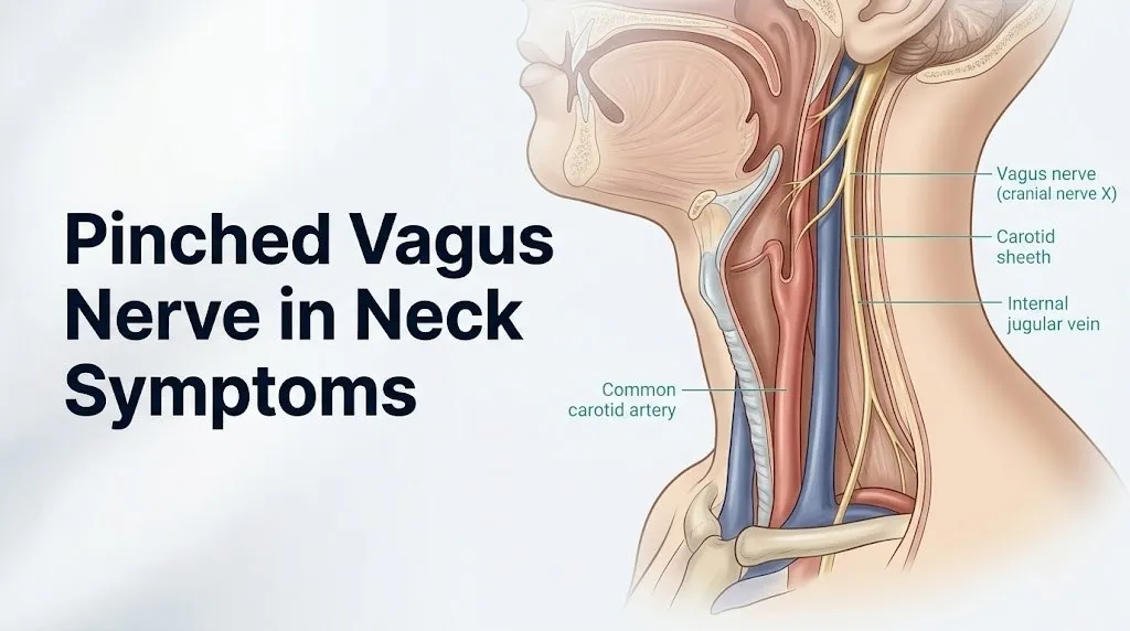
The vagus nerve exits the skull at the jugular foramen, a narrow opening between the occipital bone and the petrous portion of the temporal bone, descending through the neck enclosed within the carotid sheath.
The carotid sheath is a cylindrical fascial compartment that runs the length of the neck. It contains three structures traveling together: the common carotid artery (and the internal carotid artery in the upper portion), the internal jugular vein, and the vagus nerve itself. The vagus nerve travels between the artery (medially) and the vein (laterally), running vertically downward toward the thorax.
This anatomy explains why the vagus nerve can be affected by pathology in the neck. Any process that compresses or distorts the carotid sheath and its contents, including enlarged lymph nodes, carotid artery aneurysm, penetrating neck trauma, or a mass arising from adjacent structures, can mechanically affect the vagus nerve. However, the vagus nerve in the neck is not anatomically adjacent to the cervical intervertebral discs or the cervical nerve root foramina in a way that would allow standard cervical disc herniation to compress it directly. This is the most clinically important distinction that most online sources fail to make.
Think of the carotid sheath like a cable conduit running down the side of the neck, separate from the bony channel that carries the spinal cord and nerve roots. A herniated disc at C5-C6 can compress the C6 nerve root in its foramen without touching the vagus nerve, which runs in a completely separate anatomical compartment several centimeters away. The symptoms of C6 radiculopathy (arm pain, finger tingling) and vagal dysfunction (palpitations, nausea) are therefore physiologically distinct, even though the neck is the shared anatomical region.
The Cleveland Clinic describes the vagus nerve’s cervical descent as beginning at the jugular foramen and continuing within the carotid sheath to the level of the clavicle, where it enters the thoracic cavity to continue its course to the heart, lungs, and abdominal organs.
Can the Vagus Nerve Actually Be Pinched in the Neck?
True mechanical compression (pinching) of the vagus nerve in the neck can occur, but it is rare and typically caused by specific pathological processes rather than by common cervical disc disease.
Confirmed causes of genuine vagal nerve compression in the neck, documented in peer-reviewed case series and case reports, include: enlarged cervical lymph nodes from lymphoma or metastatic cancer pressing on the carotid sheath contents; carotid artery aneurysm or pseudoaneurysm expanding and distorting the carotid sheath; paraganglioma (carotid body tumor) arising at the carotid bifurcation; penetrating trauma to the neck; severe cervical osteophyte formation in advanced spondylosis that indirectly displaces adjacent soft tissue structures; and post-surgical scarring from prior neck surgery including thyroidectomy, carotid endarterectomy, or lymph node dissection.
What most people searching for “pinched vagus nerve” are actually describing is vagal nerve irritation or functional dysregulation, which can be triggered by neck muscle tension patterns (particularly chronic tension of the sternocleidomastoid muscle and scalene muscles compressing adjacent vascular structures), inflammatory processes near the carotid sheath, or autonomic dysregulation from other causes that produces identical symptoms through central rather than peripheral mechanisms.
The clinical distinction has real diagnostic consequences. If your vagal symptoms are caused by true structural compression from a neck mass, the appropriate evaluation is urgent neck imaging and specialist evaluation. If your symptoms reflect functional autonomic dysregulation (the far more common scenario), the appropriate pathway is autonomic function testing, tilt-table evaluation, and cardiology or neurology assessment.
According to a reference framework from the National Institute of Neurological Disorders and Stroke, disorders of cranial nerve X (ICD-10 code G52.2) encompass both structural and functional causes, and the distinction between them requires clinical examination and targeted imaging rather than symptom pattern alone.
Key Takeaway: A true “pinched” vagus nerve from a neck mass or vascular pathology is a specific, often urgently evaluated finding, while functional vagal dysregulation producing similar symptoms is far more common and follows a different diagnostic pathway entirely.
Core Symptoms of Vagus Nerve Irritation in the Neck
The core symptoms of vagus nerve irritation in the neck reflect the nerve’s dual role as both a sensory reporter from the body’s organs and a parasympathetic controller of those organs’ function.
Approximately 80% of vagal nerve fibers are afferent (sensory), carrying information upward from the heart, lungs, gastrointestinal tract, and neck structures to the brainstem’s nucleus tractus solitarius. The remaining 20% are efferent (motor/parasympathetic), carrying commands downward from the dorsal motor nucleus of the vagus to the heart, airway, and gut. Irritation or compression of the vagus nerve at the cervical level can disrupt both directions of signal flow simultaneously.
The most consistently reported core symptoms include:
| Symptom | Physiological Mechanism | Which Vagal Branch or Function Involved |
|---|---|---|
| Heart palpitations | Disrupted parasympathetic modulation of the sinoatrial node | Efferent fibers to cardiac SA node via right vagus |
| Nausea or bloating | Slowed gastric motility from reduced efferent parasympathetic drive | Efferent fibers to gastric smooth muscle via dorsal motor nucleus |
| Dizziness or lightheadedness | Disrupted baroreceptor reflex integration in nucleus tractus solitarius | Afferent fibers from carotid sinus baroreceptors |
| Near-fainting (presyncope) | Reflex vasovagal arc: bradycardia plus vasodilation | Afferent trigger plus efferent cardiac and vascular response |
| Hoarseness | Reduced motor input to intrinsic laryngeal muscles | Recurrent laryngeal nerve (branch of vagus) |
| Difficulty swallowing | Reduced pharyngeal muscle coordination | Pharyngeal branch of vagus |
| Neck pressure or pain | Direct periosteal or fascial irritation near carotid sheath | Local sensory afferent fibers |
| Ear pain or cough | Arnold’s nerve (auricular branch) irritation | Auricular branch of vagus supplying external ear canal |
Women are disproportionately affected by functional vagal dysregulation syndromes compared to men, a pattern that clinical epidemiological data from Dysautonomia International attributes to sex-based differences in autonomic nervous system regulation, estrogen effects on cardiac chronotropy, and the higher prevalence of connective tissue disorders (such as hypermobile Ehlers-Danlos syndrome) among females.
Vagus Nerve and Heart Palpitations or Irregular Heartbeat
Heart palpitations from vagal dysfunction occur because the vagus nerve is the primary parasympathetic regulator of the sinoatrial (SA) node, the heart’s natural pacemaker located in the right atrium.
Under normal conditions, the right vagus nerve delivers a continuous stream of acetylcholine to muscarinic M2 receptors on the SA node, which slows the intrinsic firing rate of the node. This is the physiological basis of what cardiologists call resting vagal tone: a healthy heart rate at rest (60 to 80 beats per minute) is slower than the SA node’s intrinsic rate (approximately 100 beats per minute) precisely because vagal tone continuously brakes it. When vagal input is disrupted, irregular, or abruptly changed, the SA node receives inconsistent braking signals, producing the sensation of palpitations, skipped beats, or a heart rate that feels unpredictably fast or slow.
Acute vagal surges (such as those triggered by pressing on the neck, bearing down, or a sudden emotional response) can produce brief but pronounced bradycardia that the person experiences as a forceful thud or a momentary sensation that their heart “stopped.” This is the basis of the vasovagal response and explains why certain neck movements or positions can precipitate palpitation episodes in people with vagal hypersensitivity.
The American Heart Association identifies vagal hypertonia (excessive vagal activity) as a recognized contributor to reflex bradycardia and vasovagal syncope, and distinguishes it from pathological cardiac arrhythmias such as atrial fibrillation or heart block, both of which require a different diagnostic and treatment approach entirely.
People taking beta-adrenergic blocking medications (beta-blockers such as metoprolol or atenolol) may have masked or altered palpitation patterns because these drugs blunt the sympathetic counterbalance to vagal input, making vagal dysregulation symptoms potentially more pronounced or differently timed.
Quick Tip:
- Record palpitation episodes by timing: when they occur, how long they last, whether they resolve with position change, and whether they are accompanied by dizziness or near-fainting.
- Bring this written log to your cardiologist appointment; palpitation timing and triggers are the primary initial clues in determining whether the cause is vagal, arrhythmic, or anxiety-driven.
- People with a prior diagnosis of Wolff-Parkinson-White syndrome or other accessory pathway conditions should not assume palpitations are vagal in origin without cardiological confirmation.
Vagus Nerve Symptoms Involving Nausea and Digestive Problems
Nausea and digestive disruption from vagal dysfunction are produced by the nerve’s direct parasympathetic control over gastric motility, esophageal coordination, and gastrointestinal secretion.
The vagus nerve provides approximately 75% of all parasympathetic input to the abdominal organs. Efferent vagal fibers descend from the dorsal motor nucleus of the vagus in the brainstem through the thorax and into the abdomen, where they synapse on the enteric nervous system’s postganglionic neurons within the walls of the esophagus, stomach, small intestine, and ascending and transverse colon. These fibers stimulate smooth muscle peristalsis, promote gastric acid secretion, and regulate the lower esophageal sphincter tone.
When vagal efferent output is reduced or disrupted, gastric motility slows. Food that should move from the stomach into the small intestine within 1 to 4 hours (depending on composition) remains in the stomach longer. This produces early satiety (feeling full after only small amounts of food), bloating, upper abdominal discomfort, and nausea. In severe or chronic cases, this pattern meets the clinical definition of gastroparesis (delayed gastric emptying), which the Mayo Clinic defines as a condition where the stomach cannot empty itself of food normally, producing chronic nausea, vomiting, and poor nutritional intake.
The esophageal component of vagal dysfunction produces difficulty swallowing (dysphagia) and the sensation that food is catching or sticking in the lower chest or throat, which patients frequently describe as something “stuck” below the breastbone.
People with longstanding diabetes mellitus are at particular risk for vagal neuropathy affecting the gastrointestinal tract because chronically elevated blood glucose produces oxidative stress that damages the myelin sheath of vagal efferent fibers over time, producing diabetic gastroparesis as a recognized complication.
Key Takeaway: Nausea and digestive symptoms from vagal dysfunction reflect the nerve’s direct control over gastric motility; when vagal output is disrupted, the stomach simply empties more slowly, producing the classic pattern of early fullness, bloating, and persistent nausea after eating.
Dizziness, Fainting, and Blood Pressure Drops from Vagal Dysfunction
Dizziness and fainting from vagal dysfunction are produced by disruption of the baroreceptor reflex arc, a physiological feedback loop that the vagus nerve helps regulate to maintain blood pressure during position changes and physical stress.
Baroreceptors are stretch-sensitive sensory receptors located in the walls of the carotid sinus (at the bifurcation of the common carotid artery in the neck) and in the aortic arch. These receptors detect changes in blood vessel wall stretch, which reflects blood pressure changes. Afferent vagal fibers (the carotid sinus nerve, a branch of cranial nerve IX/glossopharyngeal, which communicates functionally with vagal afferents at the nucleus tractus solitarius) carry this pressure information to the brainstem continuously.
When blood pressure drops (for example, when standing from sitting), the nucleus tractus solitarius integrates the baroreceptor signal and triggers a sympathetic compensatory response: heart rate increases, blood vessels constrict, and blood pressure is restored within seconds. When vagal afferent input is disrupted, this integration fails. The compensatory response is delayed or insufficient, blood pressure drops transiently, cerebral perfusion decreases, and the person experiences dizziness, visual graying, and, in more severe cases, loss of consciousness (vasovagal syncope).
The vasovagal syncope mechanism is essentially the opposite: paradoxical vagal activation produces simultaneous bradycardia (from efferent vagal output to the SA node) and peripheral vasodilation, collapsing both cardiac output and vascular resistance at once. The person becomes pale, diaphoretic (diaphoresis, or sudden cold sweating), nauseated, and loses consciousness. Recovery is typically rapid when lying flat, which restores cerebral perfusion.
The American Heart Association classifies vasovagal syncope as the most common cause of fainting in otherwise healthy individuals, responsible for approximately 1 in 3 cases of syncope evaluated in emergency departments, according to data reviewed in the Annals of Internal Medicine.
Older adults with vagal dysfunction face higher injury risk from syncope episodes because falls from fainting can produce hip fractures, head trauma, and subdural hematomas.
Voice Changes and Swallowing Difficulty from Vagal Nerve Involvement
Hoarseness and difficulty swallowing are among the most anatomically specific symptoms of vagal nerve involvement in the neck, because they are produced by named branches of the vagus nerve that supply the larynx and pharynx.
The recurrent laryngeal nerve is a branch of the vagus that supplies motor innervation to all intrinsic muscles of the larynx except the cricothyroid muscle. On the left side, the recurrent laryngeal nerve loops under the aortic arch before ascending back to the larynx. On the right side, it loops under the right subclavian artery. Both ascending portions travel in a groove between the trachea and esophagus in the neck before entering the larynx. This anatomical course means that any process affecting the vagus nerve in the neck or the upper chest (not just the neck) can interrupt recurrent laryngeal nerve function.
When the recurrent laryngeal nerve is compressed or irritated, the vocal folds (vocal cords) on the affected side cannot fully abduct or adduct during speech and breathing. The result is a weak, breathy, or hoarse voice quality (dysphonia) that does not resolve with rest or hydration. In bilateral recurrent laryngeal nerve involvement, stridor (a high-pitched breathing noise from inadequate vocal fold abduction during inspiration) can develop.
The pharyngeal branches of the vagus supply the constrictor muscles of the pharynx and contribute to the swallowing reflex coordination. Disruption of pharyngeal branch function produces dysphagia: food or liquid may not be propelled efficiently into the esophagus, or the timing of the swallowing reflex may be altered, creating a risk of aspiration (food or liquid entering the airway rather than the esophagus).
Any new onset of hoarseness lasting more than 2 to 3 weeks, particularly in a person who smokes, has a history of neck surgery, or has a palpable neck mass, warrants evaluation by a board-certified otolaryngologist (ENT specialist) with laryngoscopy to directly visualize vocal fold movement. This is not a symptom to attribute to vagal dysfunction without excluding laryngeal pathology, thyroid disease, and lung apex mass (Pancoast tumor) first.
Vagus Nerve and Anxiety or Panic-Like Symptoms
Anxiety and panic-like symptoms from vagal dysfunction are physiologically real and mechanistically distinct from primary anxiety disorders, even though the two can coexist and are frequently confused with each other.
The vagus nerve carries dense afferent (sensory) projections from the heart, lungs, and gastrointestinal tract upward to the brainstem’s nucleus tractus solitarius, which then relays signals to the insular cortex, the amygdala, and the anterior cingulate cortex, all of which are core structures in the brain’s interoceptive and emotional processing systems. When vagal afferent signals are distorted or dysregulated (for example, during a period of heightened vagal tone or baroreceptor instability), the brain’s emotional processing centers receive unusual interoceptive input and interpret it as a threat signal.
The result feels like anxiety or panic: a sense of apprehension, rapid or pounding heartbeat, shortness of breath, sweating, and a feeling that something is wrong without a clear external cause. This presentation can be nearly identical to generalized anxiety disorder or panic disorder, but its physiological origin is autonomic rather than primarily psychological.
Clinically, the distinction is important because treating autonomic-origin anxiety-like symptoms with benzodiazepines or SSRI therapy alone will not address the underlying vagal dysregulation. Research published in JAMA Neurology has noted that patients with conditions such as POTS and dysautonomia frequently receive primary anxiety disorder diagnoses before autonomic dysfunction is identified, delaying appropriate treatment by an average of several years.
Pregnant women with autonomic dysregulation or a history of vasovagal syncope may notice a worsening of anxiety-like vagal symptoms during the first trimester, when progesterone-driven vasodilation reduces systemic vascular resistance and compounds orthostatic stress on the autonomic system.
Key Takeaway: Anxiety or panic-like symptoms that accompany heart palpitations, nausea, or dizziness in the absence of a clear psychological trigger should be evaluated for autonomic nervous system dysfunction before assuming a primary psychiatric cause.
What Causes Vagus Nerve Irritation or Compression in the Neck
Vagus nerve irritation or compression in the neck is caused by any process that physically impinges on the carotid sheath contents, chronically activates vagal afferents via neck muscle tension or cervical inflammation, or damages the nerve directly through surgical, traumatic, or vascular mechanisms.
Causes ranked by frequency from clinical observational data:
| Cause | Mechanism of Vagal Involvement | Population Most Affected |
|---|---|---|
| Chronic neck muscle tension (sternocleidomastoid, scalene) | Indirect compression of carotid sheath contents; afferent vagal activation | Adults with desk-based work, poor cervical posture |
| Cervical lymphadenopathy (enlarged lymph nodes) | Direct carotid sheath compression from adjacent lymph node expansion | Any age; infection, inflammatory disease, or malignancy |
| Post-surgical scarring (thyroidectomy, carotid surgery, neck dissection) | Scar tissue encases or tethers recurrent laryngeal or vagal nerve | Adults post-head/neck surgery |
| Carotid artery aneurysm or pseudoaneurysm | Expanding vascular wall directly displaces vagus nerve within carotid sheath | Adults over 50; hypertension, atherosclerosis |
| Carotid body tumor (paraganglioma) | Mass at carotid bifurcation compresses vagus and adjacent structures | Adults 40 to 70 |
| Penetrating neck trauma | Direct nerve transection or contusion | Any age following injury |
| Advanced cervical spondylosis with severe osteophyte formation | Osteophytes compress esophagus or displace soft tissue near carotid sheath | Adults over 60 |
| Inflammatory/autoimmune neuropathy | Inflammatory demyelination of vagal nerve fibers | Adults with viral illness prodrome, autoimmune disease |
| Diabetic autonomic neuropathy | Oxidative damage to vagal efferent myelin over time | Adults with longstanding type 2 diabetes |
Functional vagal dysregulation without structural nerve compression is the most common mechanism overall, produced by dysautonomia, POTS, post-viral autonomic dysfunction (including post-COVID-19 autonomic syndrome), and chronic stress-induced sympathovagal imbalance.
Cervical Spondylosis, Disc Disease, and Vagal Nerve Involvement
Cervical spondylosis and herniated cervical discs do not typically compress the vagus nerve directly, but they can contribute to vagal symptom patterns through indirect mechanisms involving adjacent vascular and soft tissue structures.
Cervical spondylosis refers to age-related degenerative changes of the cervical intervertebral discs, vertebral bodies, and facet joints, including disc space narrowing, endplate sclerosis, and osteophyte (bone spur) formation. When osteophytes form anteriorly (on the front surface of the cervical vertebral bodies), they can compress the esophagus or distort adjacent soft tissue compartments, potentially impinging on carotid sheath contents. This is most reported at the C5-C6 and C6-C7 levels, which are the segments with the highest degenerative change frequency according to the American Academy of Orthopaedic Surgeons.
A herniated nucleus pulposus at cervical levels compresses ipsilateral nerve roots in the neural foramina, producing cervical radiculopathy with arm pain, numbness, and weakness in a dermatomal and myotomal distribution. This is not vagal compression. However, the same inflammatory environment that surrounds a herniated disc, including elevated prostaglandins and interleukin-6 in the epidural and paraspinal space, can produce systemic autonomic sensitization that worsens pre-existing vagal dysregulation.
The practical implication: a person with known cervical disc disease who also experiences palpitations, nausea, and dizziness should not assume their vagal symptoms are caused by the disc disease. Both conditions can coexist, but they require separate evaluation pathways. The disc disease evaluation falls to a board-certified orthopedic spine surgeon or neurological spine surgeon; the autonomic symptom evaluation falls to a board-certified neurologist or cardiologist.
Older adults with advanced cervical spondylosis involving anterior osteophytes large enough to compress the esophagus should be evaluated specifically for dysphagia aortica or dysphagia spondylotica as a contributing mechanism for swallowing symptoms, rather than attributing dysphagia exclusively to vagal dysfunction.
Vagal Dysfunction vs Cervical Radiculopathy: Telling Them Apart
Vagal dysfunction and cervical radiculopathy are two distinct conditions that can co-occur in the neck but produce entirely different symptom profiles through completely separate anatomical pathways.
Cervical radiculopathy occurs when a cervical nerve root is compressed within its neural foramen by a herniated disc, osteophyte, or inflamed ligamentum flavum. The compressed nerve root carries sensory and motor fibers to a specific dermatomal and myotomal territory. The result is unilateral pain radiating from the neck into the shoulder, arm, forearm, or fingers, following a predictable pattern: C6 radiculopathy produces thumb and index finger numbness; C7 radiculopathy produces middle finger numbness and triceps weakness; C8 radiculopathy produces ring and pinky finger numbness and grip weakness.
Vagal dysfunction produces none of these limb symptoms. Its symptom territory is the autonomic and visceral organ systems, not the arms or hands.
| Feature | Vagal Dysfunction | Cervical Radiculopathy |
|---|---|---|
| Primary symptom territory | Heart, gut, throat, blood pressure | Neck, shoulder, arm, hand |
| Numbness or tingling | Absent (or non-dermatomal) | Present; dermatomal distribution |
| Arm or hand weakness | Absent | May be present (grip, biceps, triceps) |
| Heart palpitations | Characteristic feature | Not a feature |
| Nausea or bloating | Characteristic feature | Not a feature |
| Voice changes or hoarseness | May be present (vagal branch) | Absent (spinal nerve does not supply larynx) |
| Reproduction with neck movement | Possible (positional vagal activation) | Yes; often worsens with specific neck positions |
| Diagnostic confirmation | Autonomic function tests, Holter, tilt-table | MRI cervical spine, EMG, nerve conduction velocity |
| Primary specialist | Neurologist, cardiologist, autonomic specialist | Orthopedic or neurological spine surgeon, physiatrist |
The overlap that confuses patients is neck pain. Both conditions can produce neck pain. But the downstream symptoms are physiologically distinct. A reader experiencing both arm numbness and heart palpitations may have both conditions simultaneously, which is not uncommon in adults with degenerative cervical disease who also have autonomic nervous system vulnerabilities.
Key Takeaway: If your predominant symptoms are arm or hand numbness and weakness, the cause is far more likely cervical radiculopathy than vagal dysfunction; if your predominant symptoms are palpitations, nausea, and fainting, the evaluation should focus on autonomic function rather than spinal imaging alone.
POTS, Dysautonomia, and the Vagus Nerve Connection
Postural orthostatic tachycardia syndrome (POTS) and broader dysautonomia are the most clinically common conditions producing the full symptom profile that people attribute to a “pinched vagus nerve,” and understanding the connection clarifies why these symptoms span so many body systems simultaneously.
POTS is a form of dysautonomia characterized by an excessive heart rate increase of 30 or more beats per minute (or a rate exceeding 120 beats per minute) within 10 minutes of moving from lying to standing, without a corresponding significant drop in blood pressure, combined with symptoms of orthostatic intolerance. The condition reflects impaired autonomic compensation for postural change, involving abnormal sympathetic activation, reduced baroreflex sensitivity, and altered vagal tone in a dysregulated interplay that the autonomic nervous system cannot normalize.
Dysautonomia International reports that POTS affects an estimated 1 to 3 million Americans, with females comprising approximately 80% of diagnosed cases. The average delay from symptom onset to diagnosis is approximately 5.5 years, according to data reviewed in the Autonomic Neuroscience journal, primarily because the multisystem symptom profile is repeatedly attributed to anxiety, deconditioning, or other diagnoses before autonomic function testing is performed.
The vagal connection in POTS involves impaired vagal withdrawal response upon standing. In a person without POTS, standing triggers appropriate sympathetic activation and vagal withdrawal, maintaining blood pressure and moderating heart rate. In POTS, this withdrawal is excessive or dysregulated, producing the compensatory tachycardia that defines the condition alongside the full range of nausea, dizziness, cognitive fogging (frequently called “brain fog”), and fatigue that POTS patients experience.
Post-COVID-19 autonomic syndrome, documented in multiple case series published in JAMA Neurology and The Lancet, produces a POTS-like presentation with vagal dysregulation in a subset of individuals following SARS-CoV-2 infection, suggesting that viral damage to autonomic nerve fibers (including vagal afferents) is a mechanism of post-acute COVID symptoms in this population.
Emergency Symptoms: When to Call 911 or Go to the ER
Certain symptoms associated with vagal nerve dysfunction or cervical nerve compression require immediate emergency evaluation. Do not wait to see if these resolve on their own.
Call 911 or go to the nearest emergency room immediately if you experience:
- Sudden onset of a heart rate below 40 beats per minute or above 150 beats per minute, confirmed by pulse or heart rate monitoring, especially with dizziness or chest discomfort (indicating potentially dangerous cardiac arrhythmia requiring immediate evaluation and possible intervention)
- Loss of consciousness with injury, prolonged unconsciousness, or failure to recover orientation within 1 to 2 minutes after fainting (indicating a syncopal episode with complication risk)
- New onset of complete voice loss (aphonia) combined with difficulty breathing (stridor), suggesting bilateral recurrent laryngeal nerve compromise with airway risk
- Sudden difficulty breathing combined with neck swelling or a rapidly enlarging pulsatile neck mass (suggesting carotid aneurysm, hematoma, or vascular emergency with vagal compression)
- Severe difficulty swallowing with drooling or inability to manage saliva, indicating complete pharyngeal dysfunction with aspiration risk
- Chest pain combined with palpitations and dizziness, which cannot be distinguished from a cardiac emergency without electrocardiographic evaluation
- Sudden onset of facial drooping, arm weakness, speech difficulty, or vision changes alongside neck pain (indicating possible stroke, which requires emergency evaluation and not a “wait and see” approach)
- Fainting episode in a person with known cardiac disease, diabetes, or on anticoagulant or antiarrhythmic medication (higher risk of dangerous underlying cause)
These presentations can indicate cardiac arrhythmia, vascular emergency, airway compromise, or acute neurological event and require emergency medical assessment, not an urgent care appointment or a phone call.
How Vagus Nerve Dysfunction Is Diagnosed
Diagnosing vagus nerve dysfunction requires a layered clinical approach, because no single test confirms “vagal pinching” as a diagnosis, and each potential contributing cause requires a different diagnostic tool.
The evaluation typically proceeds in this sequence:
- Complete medical history and neurological examination: the physician assesses symptom pattern, onset, triggers, and associated symptoms; a neurological examination evaluates cranial nerve function (including gag reflex, palate elevation, vocal quality, and swallowing), cardiovascular exam including lying and standing blood pressure and heart rate
- 12-lead electrocardiogram (ECG): performed first to exclude primary cardiac arrhythmia as a cause of palpitations; identifies bradycardia, heart block, accessory pathways, or ST-T changes
- Holter monitor (24 to 48 hour or extended ECG recording): captures intermittent rhythm changes that a brief ECG misses; documents heart rate variability (HRV), which is a quantitative measure of vagal tone
- Tilt-table test: the patient is secured to a motorized table and moved from flat to 70-degree upright position while heart rate and blood pressure are continuously monitored; the most sensitive diagnostic test for POTS and vasovagal syncope
- Autonomic function testing battery: includes quantitative sudomotor axon reflex test (QSART), deep breathing heart rate variability, and Valsalva maneuver with beat-to-beat blood pressure monitoring; evaluates both sympathetic and parasympathetic autonomic function
- MRI of the neck with contrast: evaluates the carotid sheath, adjacent lymph nodes, vertebral bodies, and soft tissue masses for structural compression of vagal or adjacent structures; indicated when a neck mass, vascular abnormality, or structural compression is clinically suspected
- Laryngoscopy: direct visualization of vocal fold movement by a board-certified otolaryngologist; indicated for any hoarseness persisting more than 2 to 3 weeks to exclude vocal fold paralysis from recurrent laryngeal nerve compromise
- Gastric emptying study (nuclear medicine scintigraphy): the gold standard for diagnosing gastroparesis when vagal-related gastrointestinal dysfunction is suspected; the patient eats a standardized radiolabeled meal and gastric emptying rate is measured at 1, 2, and 4 hours
According to the Mayo Clinic, tilt-table testing and autonomic function battery testing together provide the most complete functional assessment of the vagus nerve’s cardiovascular regulatory role, and are the recommended first-line tests when autonomic dysfunction is the primary clinical hypothesis.
Treatment Approaches and When to See a Specialist
Treatment of vagal nerve dysfunction at the neck level is directed by the underlying cause, and no single intervention applies across the full spectrum of conditions that produce this symptom profile.
The appropriate specialist depends entirely on which symptoms and which suspected mechanism dominate the clinical picture:
| Symptom Focus | Suspected Mechanism | Specialist to See | First Test to Request |
|---|---|---|---|
| Heart palpitations, fainting, irregular rate | Cardiac vagal dysregulation, arrhythmia | Board-certified cardiologist | Holter monitor, ECG |
| Dizziness, fainting, blood pressure drops | POTS, vasovagal syncope, dysautonomia | Board-certified neurologist or autonomic specialist | Tilt-table test, autonomic battery |
| Hoarseness, voice changes, 2 or more weeks | Recurrent laryngeal nerve involvement | Board-certified otolaryngologist (ENT) | Laryngoscopy |
| Nausea, bloating, early satiety | Gastroparesis, vagal GI dysfunction | Board-certified gastroenterologist | Gastric emptying study |
| Neck mass, vascular pulsation, rapidly progressive symptoms | Structural compression; urgent evaluation needed | Emergency medicine or primary care physician immediately | CT neck with contrast, neck ultrasound |
| Anxiety-like symptoms with autonomic features | Autonomic dysregulation, POTS | Board-certified neurologist; autonomic specialist | Tilt-table test, autonomic function testing |
| All of the above combined | Systemic dysautonomia | Autonomic disorder specialist; multidisciplinary team | Full autonomic battery, tilt-table, cardiac workup |
For functional vagal dysregulation without structural compression, evidence-reviewed management approaches discussed in clinical literature (not recommendations) include: increased fluid and sodium intake to expand blood volume in POTS; compression garments for orthostatic intolerance; physical reconditioning programs for deconditioning-related dysautonomia; pharmacological approaches (fludrocortisone, midodrine, beta-blockers) prescribed and managed by a board-certified cardiologist or autonomic specialist; and dietary modification for gastroparesis symptoms under gastroenterologist guidance.
For structural vagal compression from a confirmed neck mass, lymphadenopathy, or vascular anomaly, management depends on the underlying cause and is directed by the relevant specialist (oncologist, vascular surgeon, or head and neck surgeon).
Frequently Asked Questions About Pinched Vagus Nerve in Neck Symptoms
What does a pinched vagus nerve in the neck feel like?
Vagus nerve irritation in the neck typically produces a combination of heart palpitations, nausea, dizziness, near-fainting, hoarseness, and difficulty swallowing, along with a sensation of pressure or discomfort in the neck.
Because the vagus nerve controls multiple organ systems simultaneously, the symptom pattern tends to span the heart, gut, throat, and blood pressure regulation rather than producing isolated pain in one area.
Some people also experience ear pain or an unexplained cough triggered by pressure near the external ear canal, produced by the auricular branch of the vagus nerve (Arnold’s nerve).
Can the vagus nerve actually be pinched in the neck?
True mechanical compression of the vagus nerve in the neck can occur, but it requires a specific structural cause such as an enlarged lymph node, carotid artery aneurysm, carotid body tumor, or post-surgical scarring.
Routine cervical disc herniation does not compress the vagus nerve, because the vagus runs within the carotid sheath several centimeters away from the spinal nerve root foramina where disc herniations occur.
Most people using the phrase “pinched vagus nerve” are experiencing functional vagal dysregulation rather than true structural compression, and the distinction requires clinical examination and often neck imaging to clarify.
What causes vagus nerve irritation in the neck?
The most common causes of vagal irritation in the neck are chronic neck muscle tension affecting adjacent vascular structures, enlarged cervical lymph nodes from infection or inflammatory disease, post-surgical scarring from prior neck surgery, and functional dysautonomia including POTS and post-viral autonomic syndrome.
Less common but more urgent causes include carotid artery aneurysm, carotid body tumor (paraganglioma), and penetrating neck trauma.
Diabetic autonomic neuropathy can produce vagal dysfunction throughout the course of the nerve without any structural neck pathology.
Is a pinched vagus nerve in the neck dangerous?
Vagal dysfunction can be dangerous when it produces significant bradycardia (heart rate below 40 beats per minute), recurrent fainting with injury risk, complete vocal fold paralysis affecting breathing, or severe gastroparesis causing malnutrition.
The underlying cause of vagal compression matters more than the symptom severity alone: a carotid aneurysm or cervical lymphoma causing vagal compression is a medical urgency requiring prompt diagnosis and intervention.
Functional vagal dysregulation without a structural cause is rarely immediately dangerous but significantly impairs quality of life and warrants thorough evaluation by a board-certified neurologist or cardiologist.
How is vagus nerve dysfunction in the neck diagnosed?
Vagal nerve dysfunction is diagnosed through a combination of 12-lead ECG, Holter monitoring, tilt-table testing, autonomic function testing, and MRI of the neck when structural compression is suspected.
Laryngoscopy is indicated when hoarseness is present to directly assess vocal fold movement and exclude recurrent laryngeal nerve paralysis.
A gastric emptying study (nuclear scintigraphy) is the standard diagnostic test when gastroparesis from vagal gastrointestinal dysfunction is clinically suspected.
When should I go to the ER for vagus nerve symptoms?
Go to the emergency room immediately if you experience a heart rate below 40 beats per minute, loss of consciousness with injury, sudden complete voice loss with difficulty breathing, severe difficulty swallowing your own saliva, chest pain with palpitations, or a rapidly enlarging pulsatile neck mass with any new neurological symptom.
Fainting in a person with known cardiac disease or on antiarrhythmic medication also warrants emergency evaluation rather than a routine appointment.
Sudden onset of facial drooping, arm weakness, or speech difficulty alongside neck symptoms requires calling 911 immediately, because these signs suggest a stroke, which is a time-critical emergency.
Closing
The symptom pattern people describe as a “pinched vagus nerve in the neck” is real and physiologically grounded, but the label oversimplifies a clinical picture that spans the heart, gut, throat, and autonomic nervous system. Getting an accurate diagnosis requires working with the right specialists in the right sequence.
Start with a primary care physician who can order a baseline ECG and refer you to cardiology for Holter monitoring if palpitations are your dominant symptom, to neurology for tilt-table testing if fainting or dizziness dominates, or to otolaryngology for laryngoscopy if hoarseness has lasted more than two to three weeks. If you have a palpable neck mass, neck swelling, or rapidly progressive symptoms alongside any of the above, that moves to the front of the line.
The reader who leaves this article understanding that vagal dysfunction produces symptoms across multiple organ systems simultaneously, that true cervical vagal compression requires specific structural pathology, and that palpitations with fainting is an emergency threshold rather than something to monitor for a few days, has the clinical information they need to make confident decisions about when and where to seek care.