Gallbladder Symptoms: What They Feel Like in 2026
Gallbladder symptoms range from episodic cramping pain in the upper right abdomen to severe, unrelenting pain with fever and jaundice that requires emergency surgery. Understanding the difference between a manageable episode and a medical emergency can be the most practically important thing you take from this article.
According to the National Institute of Diabetes and Digestive and Kidney Diseases (NIDDK), approximately 10 to 15 percent of adults in the United States have gallstones, and about 1 to 3 percent develop symptoms each year. That translates to roughly 20 million Americans living with this condition at any given time, making gallbladder disease one of the most common reasons for emergency abdominal surgery in the developed world.
This article covers every major gallbladder symptom with the physiological explanation behind each one, how early symptoms differ from advanced ones, which populations are at higher risk and why, and exactly when specific symptoms mean you need to go to the emergency room rather than wait for a scheduled appointment.
Gallbladder Symptoms: What They Are and Why They Happen
Gallbladder symptoms occur when bile flow through the biliary system is obstructed, restricted, or associated with inflammation of the gallbladder wall, triggering a cascade of visceral pain, nausea, and systemic responses depending on severity.
The gallbladder is a small, pear-shaped organ sitting on the undersurface of the right lobe of the liver. Its job is to store and concentrate bile, a digestive fluid the liver produces continuously to emulsify dietary fats. When you eat a meal containing fat or protein, cells in the lining of the duodenum (the first segment of the small intestine) release a hormone called cholecystokinin (CCK). CCK signals the gallbladder to contract, squeezing bile through the cystic duct into the common bile duct and then into the small intestine.
This works smoothly until a gallstone, a hardened crystalline deposit made primarily of cholesterol or bile pigments, forms inside the gallbladder and blocks the cystic duct. When CCK still signals the gallbladder to contract but bile has nowhere to go, pressure builds inside the gallbladder rapidly. That pressure-driven distension is what produces the pain known as biliary colic, and it is the engine behind most gallbladder symptoms people experience.
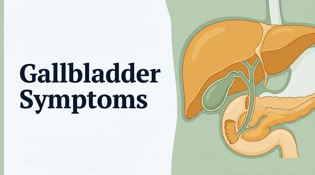
Think of the gallbladder like a water balloon connected to a pipe with a closed valve. The water (bile) keeps building pressure with nowhere to drain. The balloon stretches. That stretching generates the cramping, wave-like pain in the upper right abdomen that most people experience during a gallbladder episode.
Not all gallbladder symptoms are caused by stones. Acalculous cholecystitis (gallbladder inflammation without stones), biliary dyskinesia (impaired gallbladder motility), and sphincter of Oddi dysfunction can all produce similar symptom patterns. The NIDDK notes that acalculous cholecystitis accounts for approximately 5 to 10 percent of acute cholecystitis cases and tends to occur in critically ill patients or those on total parenteral nutrition.
Individual variation note: People with diabetes mellitus and underlying autonomic neuropathy may experience blunted visceral pain signals. Their gallbladder symptoms can present at a lower apparent severity than the actual degree of gallbladder inflammation, making the clinical picture misleading and potentially dangerous.
What Does Gallbladder Pain Feel Like?
Gallbladder pain typically feels like a deep, cramping, or squeezing pressure in the upper right or upper middle abdomen, often building steadily over 15 to 30 minutes before reaching peak intensity, and sometimes radiating to the right shoulder or right shoulder blade.
The quality of the pain is distinct. Unlike the constant, pressure-like discomfort of a stomach ache, gallbladder pain tends to build in intensity rather than arrive suddenly. Most people describe it as a tight gripping sensation, sometimes with a burning quality, located just below the right rib cage or in the upper middle abdomen (the epigastric region). The pain is visceral in origin, arising from distension of the gallbladder wall’s smooth muscle and from stretch of the hepatic capsule overlying the liver as inflammation develops.
The radiating quality of the pain has a specific anatomical explanation. When inflammation or distension of the gallbladder irritates the inferior surface of the diaphragm, the phrenic nerve (arising from cervical spinal levels C3 through C5) transmits pain signals that the brain misinterprets as coming from the right shoulder. This is called referred pain, and it is one of the most reliable indicators that the biliary system is involved rather than the stomach, intestine, or kidneys.
| Pain Quality | Clinical Description | Anatomical Reason |
|---|---|---|
| Location | Right upper quadrant, epigastric | Gallbladder sits under right liver lobe |
| Character | Cramping, squeezing, pressure | Visceral pain from gallbladder wall distension |
| Radiation | Right shoulder, right scapula | Phrenic nerve irritation from diaphragm |
| Onset | Gradual over 15 to 30 minutes | Progressive cystic duct obstruction |
| Peak intensity | 1 to 2 hours after onset | Maximum intraluminal pressure |
| Duration | 30 minutes to 6 hours (biliary colic) | Stone may shift, relieving obstruction |
Individual variation note: Older adults above age 65 may report the pain as more of a dull ache rather than a sharp or cramping sensation. A 2021 review published in the Journal of the American Geriatrics Society found that classic right upper quadrant tenderness is absent in up to 60 percent of older adults with confirmed acute cholecystitis, making age-related atypical presentation a genuine diagnostic challenge.
Gallbladder Attack Symptoms
A gallbladder attack, clinically called biliary colic or acute cholecystitis depending on duration and severity, produces a recognizable cluster of symptoms including sudden right upper quadrant pain, nausea, vomiting, and sometimes sweating and chills.
The full symptom picture during a gallbladder attack typically includes:
- Severe right upper quadrant or epigastric pain building over 15 to 60 minutes, often reaching a level described as 7 to 10 out of 10 in intensity
- Nausea, caused by activation of the vagus nerve (cranial nerve X) by biliary distension, which also triggers the vomiting center in the brainstem
- Vomiting, which may be forceful but does not relieve the pain (distinguishing it from vomiting caused by gastrointestinal obstruction)
- Diaphoresis (profuse sweating) from the autonomic nervous system’s response to severe visceral pain
- Pain radiating to the right shoulder or right shoulder blade, via phrenic nerve irritation of the diaphragm
- Inability to find a comfortable position, as the pain is visceral and positional changes offer little relief
- Bloating or abdominal distension, from associated gastrointestinal slowing
Vomiting that does not relieve the pain is one of the most clinically useful distinctions between a gallbladder attack and a gastrointestinal source of discomfort. In gastrointestinal conditions like gastroparesis or food poisoning, vomiting often provides partial relief. In biliary colic, the pain source is the gallbladder wall and duct, not the stomach. Emptying the stomach changes nothing about the obstruction.
According to the American College of Gastroenterology, the majority of biliary colic episodes peak in intensity within 1 to 2 hours and resolve within 6 hours as the gallstone shifts and relieves the cystic duct obstruction. Pain lasting more than 6 hours suggests the obstruction is persistent and acute cholecystitis has likely developed.
Individual variation note: People taking opioid medications for other conditions may have altered gallbladder motility due to the effect of opioids on the sphincter of Oddi, potentially changing the character and timing of their symptoms in ways that make self-assessment less reliable.
Key Takeaway: Gallbladder pain builds gradually to a severe peak, does not improve with vomiting, and radiates to the right shoulder because the phrenic nerve transmits diaphragmatic irritation as shoulder pain; if the pain lasts more than 6 hours, it is no longer just biliary colic.
Symptoms of a Gallbladder Attack vs. Other Abdominal Pain
The symptoms of a gallbladder attack are distinct from other sources of abdominal pain in their location, timing pattern, relationship to fatty meals, and the specific combination of right-sided radiation and persistent pain that does not respond to antacids or passing gas.
Several conditions share surface similarities with gallbladder attacks, and the clinical picture often requires imaging to differentiate. The most common sources of confusion include:
| Condition | Pain Location | Key Distinguishing Feature | Diagnostic Test |
|---|---|---|---|
| Gallbladder attack (biliary colic) | Right upper quadrant, epigastric | Triggered by fatty food, lasts 1 to 6 hours | Abdominal ultrasound |
| Acute cholecystitis | Right upper quadrant | Persistent more than 6 hours, fever, Murphy’s sign | Ultrasound + CBC + CRP |
| GERD or peptic ulcer | Epigastric, behind sternum | Burning quality, improves with antacids | Endoscopy |
| Acute pancreatitis | Epigastric, radiating to back | Band-like pain, worse leaning forward, elevated lipase | CT scan + lipase |
| Right-sided kidney stone | Flank, radiating to groin | Colicky, hematuria | CT scan + urinalysis |
| Liver abscess or hepatitis | Right upper quadrant | Dull aching, gradual onset, liver enzyme elevation | Liver function tests + ultrasound |
| Myocardial infarction | Chest, radiating to jaw or left arm | Associated shortness of breath, diaphoresis | ECG + troponin |
The antacid test is a practical self-check many gastroenterologists reference. If your upper abdominal pain improves meaningfully within 15 minutes of taking an antacid, a gastrointestinal acid source is more likely than the gallbladder. Gallbladder pain does not respond to antacids because the mechanism has nothing to do with stomach acid.
Individual variation note: In individuals with a prior history of gastroesophageal reflux disease (GERD), distinguishing gallbladder pain from acid-related pain by symptom quality alone is genuinely difficult. Both conditions can worsen after meals and produce epigastric discomfort. A person with known GERD who develops new or worsening upper right quadrant pain that no longer responds to their usual antacid regimen should have an abdominal ultrasound ordered by their primary care physician to evaluate the biliary system specifically.
Where Is Gallbladder Pain Located?
Gallbladder pain is located primarily in the right upper quadrant of the abdomen, specifically in the area just below the right rib cage, and may also present in the epigastric region (upper middle abdomen) or radiate to the right shoulder, right shoulder blade, or mid-back.
The right upper quadrant is the anatomical neighborhood where the gallbladder sits, suspended just beneath the inferior surface of the right lobe of the liver. Pressing firmly in that area during an active episode produces immediate, significant tenderness. Clinically, a physician assessing this area performs what is called Murphy’s sign: pressing the right subcostal area while asking the patient to breathe in deeply. If the descending diaphragm pushes an inflamed gallbladder against the examining hand and causes the patient to stop inhaling sharply due to pain, Murphy’s sign is positive, which is a reliable clinical indicator of acute cholecystitis.
Pain in the epigastric region (upper middle abdomen, between the two rib margins) is also common because the gallbladder and common bile duct share visceral pain fibers that do not always localize precisely to the right side. Early in an attack, many people initially feel the pain as central upper abdominal discomfort that migrates rightward as the episode progresses.
Boas’ sign refers to right subscapular hyperesthesia, an area of increased skin sensitivity behind the right shoulder blade, which occurs in some people with acute cholecystitis due to phrenic nerve involvement. It is less commonly discussed in general health content but represents a useful clinical pointer that the pain source is biliary.
Individual variation note: People with obesity may have greater difficulty localizing the pain due to thicker abdominal wall tissue and altered visceral sensation. Their Murphy’s sign may also be harder to elicit on physical examination, which can complicate clinical assessment and make imaging even more essential for accurate diagnosis.
What Triggers a Gallbladder Attack?
A gallbladder attack is most commonly triggered by eating a high-fat or high-protein meal, which causes the duodenum to release cholecystokinin and signal the gallbladder to contract forcefully against a partially or fully obstructed cystic duct.
The fat-meal trigger is the most consistent and clinically well-established precipitant. Dietary fat arriving in the duodenum stimulates I-cells in the duodenal mucosa to secrete cholecystokinin (CCK). CCK then binds to receptors on the gallbladder smooth muscle wall, driving a powerful contraction. When a gallstone is sitting loosely in or near the cystic duct opening, that contraction can drive the stone firmly into the duct, creating the obstruction that generates pain.
Common attack triggers in order of frequency include:
- High-fat meals: fried foods, fast food, fatty meats, cream sauces, full-fat dairy
- Large meals of any composition, which release more CCK than small meals
- Rapid weight loss or fasting, which concentrates bile and promotes stone formation, sometimes precipitating the first attack
- Pregnancy (covered in a dedicated section below)
- Hormonal contraceptives containing estrogen, which increase hepatic cholesterol secretion into bile
- Alcohol in some individuals, which affects gallbladder motility
- Lying down immediately after eating, which may alter bile drainage and pressure dynamics
Less commonly, an attack can occur at rest or during sleep, particularly after a large evening meal. The gallbladder continues responding to CCK signals for several hours after a meal.
According to a 2019 study published in BMJ Open Gastroenterology, the single most consistent dietary trigger reported by patients with symptomatic cholelithiasis was high-fat meal consumption within 2 hours of symptom onset, with 78 percent of symptomatic patients reporting a clear dietary association.
Individual variation note: Individuals on very low-calorie diets (below 800 calories per day) or those who have undergone bariatric surgery are at elevated risk for rapid stone formation. The American College of Gastroenterology notes that ursodeoxycholic acid is sometimes prescribed preventively in post-bariatric patients during rapid weight loss phases to reduce lithogenic bile supersaturation.
Gallbladder Symptoms After Eating
Gallbladder symptoms that occur after eating, particularly after fatty meals, are the hallmark postprandial pattern of biliary colic, with pain onset typically beginning 30 to 90 minutes after a meal and peaking within 1 to 2 hours.
The timing matters clinically. Stomach-related pain from gastritis or a peptic ulcer typically begins during or immediately after eating or, in the case of duodenal ulcers, 2 to 4 hours after eating when stomach acid enters an empty duodenum. Gallbladder pain’s onset at 30 to 90 minutes post-meal reflects the time it takes for dietary fat to reach the duodenum, stimulate CCK release, and produce significant gallbladder contraction pressure.
The typical post-meal symptom pattern in biliary disease includes:
- Eating a meal containing fat or protein
- Feeling normal or mildly bloated for the first 30 minutes
- Developing a gradually building right upper quadrant or epigastric ache beginning at 30 to 90 minutes
- Experiencing nausea, sometimes vomiting, as the pain intensifies over the next 30 to 60 minutes
- Reaching peak pain at 1 to 2 hours post-meal
- Either resolving gradually over the next 1 to 4 hours (biliary colic) or persisting and worsening beyond 6 hours (acute cholecystitis)
Beyond the fat-meal pattern, some people notice that symptoms are accompanied by bloating, belching, and a sensation of fullness that does not resolve normally after the meal. These associated dyspeptic symptoms were historically attributed to the gallbladder but research has complicated that picture. A 2020 study published in the Annals of Internal Medicine found that bloating and belching as isolated symptoms (without the characteristic right upper quadrant pain) are not reliably predictive of gallbladder disease and may represent overlapping functional gastrointestinal symptoms.
Individual variation note: People with irritable bowel syndrome (IBS) frequently experience post-meal abdominal symptoms including cramping, bloating, and right-sided discomfort that can mimic biliary colic. An abdominal ultrasound is the only way to definitively determine whether the gallbladder is involved. Assuming IBS is the cause of post-meal right upper quadrant pain without imaging is a clinical mistake that delays gallbladder diagnosis.
Key Takeaway: Gallbladder symptoms after eating follow a predictable 30-to-90-minute delay from meal to pain onset, driven by cholecystokinin release stimulating gallbladder contraction against a stone-obstructed duct; this timing pattern distinguishes gallbladder pain from most stomach-based discomfort.
Biliary Colic Symptoms
Biliary colic refers specifically to the pain episode produced by transient, intermittent obstruction of the cystic duct by a gallstone, causing gallbladder distension that resolves when the stone shifts back into the gallbladder, with the full episode lasting between 30 minutes and 6 hours.
The word “colic” is technically a misnomer in biliary disease. True colic in the intestinal or ureteral sense involves rhythmic peristaltic waves that produce crescendo-decrescendo pain cycles. Biliary “colic” is actually more of a sustained, pressure-type pain that reaches a plateau rather than cycling in waves. It builds, stays at high intensity for 1 to 2 hours, then gradually fades as the stone moves. This distinction is clinically useful because patients who describe their pain as “coming in waves every few minutes” are more likely describing intestinal pain than gallbladder pain.
Key biliary colic features that clinicians use to identify the pattern:
- Onset to peak: 15 to 60 minutes
- Duration: 30 minutes to 6 hours (median approximately 1 to 2 hours)
- Resolution: Gradual, over 30 to 90 minutes, as stone moves
- Frequency: Episodic, weeks to months between episodes (variable)
- Post-episode: Patient feels normal between episodes
- Associated features: Nausea, vomiting, diaphoresis, inability to get comfortable
- Fever: Absent (fever indicates progression to acute cholecystitis)
The between-episode normal feeling is one of the most important clinical features. A person with biliary colic feels completely well between attacks. They eat normally, have no persistent abdominal discomfort, and their physical examination is unremarkable. This “symptom-free interval” pattern is what distinguishes biliary colic from acute cholecystitis, in which the pain is persistent and worsens over time.
The American College of Gastroenterology states that approximately 1 to 2 percent of patients with asymptomatic gallstones become symptomatic each year, and that once symptomatic biliary colic begins, the annual risk of complications (acute cholecystitis, choledocholithiasis, cholangitis) is approximately 1 to 3 percent per year.
Individual variation note: In sickle cell disease, gallstones are formed from excess bilirubin rather than cholesterol (pigment stones), and biliary colic can be difficult to distinguish from a sickle cell vasoocclusive pain crisis. Both produce severe abdominal pain. Evaluation by a hematologist and gastroenterologist jointly is often needed in this population.
Acute Cholecystitis Symptoms
Acute cholecystitis is gallbladder inflammation caused by persistent cystic duct obstruction and develops when the transient pressure event of biliary colic does not resolve, leading to ischemia, bacterial invasion, and progressive inflammation of the gallbladder wall.
The clinical transition from biliary colic to acute cholecystitis is defined by duration. Pain persisting beyond 6 hours that is worsening rather than resolving, accompanied by fever (typically above 38°C / 100.4°F) and right upper quadrant tenderness on physical examination, meets the clinical criteria for acute cholecystitis per the 2018 Tokyo Guidelines for diagnosis and severity grading of acute cholecystitis.
Symptoms that distinguish acute cholecystitis from uncomplicated biliary colic:
- Pain lasting more than 6 hours without significant improvement
- Fever (low-grade initially, spiking with bacterial colonization)
- Chills and rigors (indicating systemic inflammatory response)
- Right upper quadrant tenderness on palpation, often with guarding
- Positive Murphy’s sign (inability to breathe in deeply when the right subcostal area is pressed)
- Anorexia (complete loss of appetite, distinct from the mild nausea of biliary colic)
- Elevated white blood cell count (leukocytosis above 10,000 to 12,000 cells/microliter)
- Elevated C-reactive protein (CRP above 3 mg/dL per Tokyo Guidelines criteria)
A 2022 study published in JAMA Surgery found that the median time from symptom onset to emergency surgery in patients with gangrenous cholecystitis (the most severe form) was 48 to 72 hours, emphasizing that delayed presentation significantly increases complication risk and surgical complexity.
Individual variation note: Older adults above age 70 with acute cholecystitis frequently present without fever and without a positive Murphy’s sign, even with confirmed acute cholecystitis on imaging. The Tokyo Guidelines specifically note that the diagnostic criteria should be applied with greater imaging reliance in elderly patients because clinical findings are unreliable in this population. An older adult with new right upper quadrant discomfort and any systemic signs warrants urgent imaging, not watchful waiting.
Chronic Gallbladder Symptoms
Chronic cholecystitis produces a pattern of repeated mild-to-moderate biliary colic episodes over months or years, leading to progressive gallbladder wall thickening, fibrosis, and impaired motility, with symptoms that are less dramatic than acute cholecystitis but persistent and disruptive.
Unlike the dramatic, severe pain of a full gallbladder attack, chronic gallbladder disease presents more subtly. Many people live with it for years before receiving a diagnosis. The chronic pattern includes:
- Recurring right upper quadrant or epigastric discomfort after meals, typically rated 3 to 6 out of 10 in intensity
- Persistent nausea, particularly after fatty food, that has become a near-daily experience
- Early satiety (feeling full quickly during meals), from impaired gallbladder motility affecting overall digestive function
- Mild, intermittent upper abdominal bloating
- Occasional right shoulder aching without a clear cause
- Intolerance to fried foods, cream-based sauces, or fatty meats that was not previously present
The mechanism of chronic cholecystitis involves repeated low-grade inflammatory insults to the gallbladder wall from intermittent stone movement. Over time, the wall becomes thickened with fibrous tissue, and the gallbladder loses its contractile efficiency. Even partial stones or gallbladder sludge (a semi-solid mix of cholesterol crystals, calcium salts, and bile) can perpetuate this pattern without causing discrete, dramatic attacks.
According to the Mayo Clinic, chronic cholecystitis can also cause biliary dyskinesia, a condition in which the gallbladder contracts abnormally (either too slowly or without coordination) even in the absence of stones. The hallmark finding on a HIDA scan (hepatobiliary iminodiacetic acid scintigraphy) is a reduced gallbladder ejection fraction below 35 percent at 30 minutes post-CCK stimulation.
Individual variation note: Women in the perimenopausal and postmenopausal age range may notice worsening of chronic gallbladder symptoms coinciding with hormonal changes, as the shift in estrogen levels affects bile composition. Those taking hormone replacement therapy (HRT) containing estrogen have a documented higher risk of gallstone formation and symptom progression, as noted in the Women’s Health Initiative observational data.
Key Takeaway: Chronic gallbladder disease is often mistaken for persistent digestive sensitivity or IBS; the distinguishing feature is a specific right upper quadrant or epigastric pattern that worsens predictably after fatty meals and produces recurring nausea that does not resolve with typical antacid or dietary approaches.
Gallbladder Symptoms That Come and Go
Gallbladder symptoms that come and go, with symptom-free intervals between episodes, represent the classic natural history of cholelithiasis with intermittent biliary colic, where a gallstone temporarily blocks the cystic duct and then moves back into the gallbladder, restoring normal bile flow until the next episode.
This intermittent pattern is one of the most confusing aspects of gallbladder disease for patients. They feel entirely normal between attacks and reasonably wonder whether the episode they experienced was really significant. The answer is clinically clear: any episode of right upper quadrant pain lasting 30 minutes or more, particularly if accompanied by nausea, deserves evaluation by a primary care physician with an abdominal ultrasound ordered. Waiting to see if it happens again is not a safe strategy once a first episode has occurred.
The natural history of untreated symptomatic gallstones tends to involve escalating episode frequency and severity over time. What starts as one episode every few months often progresses to more frequent attacks. The American College of Gastroenterology reports that among patients with symptomatic biliary colic who decline surgical treatment, approximately 30 to 50 percent will experience a significant complication (acute cholecystitis, choledocholithiasis, or cholangitis) within 10 years.
Between-episode periods can be completely normal, lasting days, weeks, or months. During these intervals, physical examination findings are typically absent, which is another reason why imaging is the cornerstone of diagnosis rather than physical examination alone. The gallbladder looks and feels normal from the outside when a stone is quietly sitting within the gallbladder rather than lodged in the duct.
Individual variation note: People who have had a first gallbladder episode during fasting for a medical procedure or during an extended period without eating may be at risk for another episode if they return immediately to high-fat eating. A gradual dietary reintroduction with low-fat meals is prudent after any suspected biliary episode, while awaiting ultrasound results from their primary care physician.
Gallbladder Attack Symptoms in Women
Women experience gallbladder attack symptoms at approximately 2 to 3 times the rate of men, driven by estrogen-induced increases in hepatic cholesterol secretion into bile and progesterone-mediated reductions in gallbladder motility that promote bile stasis and stone formation.
The hormone-gallbladder connection is well-documented. Estrogen increases the amount of cholesterol the liver secretes into bile without a proportional increase in bile salts and phospholipids, making the bile supersaturated with cholesterol and prone to crystal formation. Progesterone slows gallbladder contraction rate and reduces the completeness of emptying after each contraction, leaving bile to stagnate and concentrate further. Together, these hormonal effects explain why women of reproductive age, pregnant women, and women using estrogen-containing oral contraceptives all carry a significantly elevated gallstone risk.
According to the NIDDK, the prevalence of gallstones in women aged 20 to 60 is approximately 9 to 15 percent, compared to 4 to 6 percent in age-matched men. By age 60, the gap narrows but women still maintain a higher lifetime risk.
Symptom presentation in women is generally similar to the general population, but several nuances are worth noting:
- Women are more likely to present with associated nausea and vomiting as prominent features
- Younger women with gallbladder disease more often have a strong family history, which is a clinically useful diagnostic pointer
- Women with polycystic ovary syndrome (PCOS) have an elevated gallstone risk due to both insulin resistance and the hormonal profile of the condition
- Women using combined oral contraceptives (containing both estrogen and progestin) have approximately a 1.5-fold increased risk of symptomatic gallstones compared to non-users, per data published in Contraception journal
Individual variation note: Women who have recently completed a pregnancy are at elevated risk for their first symptomatic biliary episode in the postpartum period, as the elevated estrogen and progesterone levels of pregnancy dramatically increase stone formation risk, and the hormonal shift after delivery can precipitate an attack.
Gallbladder Symptoms in Pregnancy and Older Adults
Gallbladder symptoms in pregnancy require special clinical attention because altered anatomy, limited imaging options, and restricted treatment choices make the diagnostic and management picture substantially more complex than in the non-pregnant population.
In pregnancy: The enlarging uterus progressively displaces abdominal organs, including the gallbladder and bile ducts, altering pain location and potentially making the right upper quadrant pain feel higher or differently located than expected. The elevated estrogen and progesterone levels of pregnancy increase both stone formation and impaired gallbladder emptying. According to the American College of Obstetricians and Gynecologists (ACOG), symptomatic cholelithiasis complicates approximately 0.05 to 0.3 percent of pregnancies and is the second most common non-obstetric cause of hospitalization during pregnancy.
Key considerations in pregnant patients include:
- Ultrasound is the safe, radiation-free imaging choice and remains the first-line diagnostic test throughout all trimesters
- Fever and right upper quadrant pain in a pregnant woman require prompt emergency evaluation to differentiate acute cholecystitis from HELLP syndrome (hemolysis, elevated liver enzymes, low platelets), preeclampsia, and acute fatty liver of pregnancy, all of which share overlapping symptoms
- Laparoscopic cholecystectomy in the second trimester is the safest surgical window if operation is needed
- Call 911 or go to the emergency room immediately if a pregnant woman has fever, right upper quadrant pain, and either elevated blood pressure or visual disturbances, as this pattern may indicate HELLP or preeclampsia rather than gallbladder disease
In older adults: Gallbladder disease in adults above age 70 presents differently in several clinically important ways. Fever is often absent or low-grade even with severe acute cholecystitis. Right upper quadrant pain may be mild, vague, or described as “general discomfort.” The classical Murphy’s sign may be negative despite active gallbladder inflammation.
A 2020 retrospective cohort study published in the British Journal of Surgery found that older adults with acute cholecystitis were significantly more likely to present with complications (gangrenous cholecystitis, perforation) at the time of diagnosis compared to younger patients, precisely because their attenuated symptoms led to delayed evaluation.
Gallbladder Infection Symptoms and Fever
Fever, chills, and rigors in the context of gallbladder disease indicate that bacterial infection has entered the biliary system, a development that transforms a surgical problem into a potential septic emergency requiring urgent intervention.
When the cystic duct remains obstructed for more than several hours, the stagnant bile inside the gallbladder becomes a medium for bacterial colonization. The most common organisms are gram-negative enteric bacteria: Escherichia coli, Klebsiella pneumoniae, Enterococcus faecalis, and Bacteroides fragilis. As these bacteria multiply and the gallbladder wall becomes increasingly inflamed and ischemic, the systemic inflammatory response activates, producing the fever and chills that are clinical indicators of transition from uncomplicated cholecystitis to infected cholecystitis.
Fever-associated gallbladder symptoms requiring urgent evaluation:
- Temperature above 38°C (100.4°F) with right upper quadrant pain lasting more than 6 hours
- Shaking chills (rigors) indicating bacteremia (bacteria entering the bloodstream)
- Jaundice (yellowing of the skin or whites of the eyes) indicating that stones have migrated into the common bile duct, blocking bile drainage from the liver
- Dark, tea-colored urine from bilirubin accumulating in the bloodstream and being excreted by the kidneys
- Clay-colored or pale stools from the absence of bile pigments in the intestine
- Right upper quadrant pain combined with fever and jaundice together constitute Charcot’s triad, the classic clinical picture of ascending cholangitis (infection ascending into the bile ducts), which is a medical emergency
Reynolds’ pentad represents the most severe presentation: Charcot’s triad plus hypotension (low blood pressure) and altered mental status (confusion). This indicates biliary sepsis with systemic septic shock, which carries a mortality risk requiring immediate intensive care management.
According to a 2021 guideline update from the American College of Gastroenterology, ascending cholangitis diagnosed by Charcot’s triad carries mortality rates of 13 to 88 percent if untreated, with mortality risk directly related to delay in biliary decompression via endoscopic retrograde cholangiopancreatography (ERCP) or surgical drainage.
Individual variation note: Immunocompromised patients, including those on chronic corticosteroid therapy, those undergoing chemotherapy, or those with HIV/AIDS, may not mount a fever even in the presence of severe biliary infection. The absence of fever in this population does not rule out serious infection. Any immunocompromised person with new right upper quadrant pain and any systemic symptom change warrants same-day emergency evaluation.
Key Takeaway: Fever and chills during a gallbladder episode are not simply “part of the attack”; they signal bacterial infection of the biliary system and mark a clinical transition from biliary colic to acute infectious cholecystitis or cholangitis, both of which require urgent medical evaluation, not watchful waiting at home.
When to Go to the ER for Gallbladder Pain
Go to the emergency room for gallbladder pain when any of the following are present: pain lasting more than 6 hours without improvement, fever above 38°C (100.4°F), jaundice, chills or rigors, hypotension, confusion, or any pain severe enough that normal functioning is impossible.
The threshold question most people ask is whether they should call their doctor in the morning or go to the emergency room tonight. The clinical answer depends on specific features of the episode, not overall pain severity.
Go to the emergency room immediately for:
- Pain that has lasted more than 6 continuous hours without clear resolution
- Pain accompanied by fever (even low-grade) and chills
- Yellowing of the skin or the whites of the eyes (scleral icterus / jaundice)
- Dark urine (tea or cola color) with pale or clay-colored stools
- Pain accompanied by hypotension (feeling faint, dizzy, or having a racing heart)
- Confusion or altered mental status in someone with known gallbladder disease
- Pain that is the “worst you have ever felt” in the upper abdomen
- Chest pain or left arm pain accompanying the upper right abdominal episode (requires cardiac evaluation to rule out myocardial infarction)
- Pregnancy with fever and right upper quadrant pain (rule out HELLP and preeclampsia)
- Hard, rigid abdomen (peritoneal signs suggesting gallbladder perforation)
Call your primary care physician the next business day for:
- A first episode of right upper quadrant pain lasting 30 minutes to 6 hours that resolved completely
- Recurrent mild-to-moderate episodes without fever, jaundice, or persistent pain
- Ongoing post-meal right upper quadrant discomfort without acute symptoms
Individual variation note: People with diabetes should use a lower threshold for emergency evaluation because autonomic neuropathy may blunt their pain perception, meaning their reported pain severity may underestimate the degree of gallbladder inflammation. A diabetic patient with even moderate right upper quadrant discomfort, any low-grade fever, and nausea should be evaluated the same day rather than waiting.
How a Gallbladder Problem Is Diagnosed
A gallbladder problem is diagnosed through a combination of clinical history, physical examination findings (including Murphy’s sign), abdominal ultrasound as the primary imaging test, and blood tests measuring markers of inflammation and bile duct obstruction.
The diagnostic process typically unfolds in this order:
- Clinical history: The physician asks about the character, location, timing, duration, and triggers of the pain, associated symptoms (nausea, vomiting, fever, jaundice), dietary relationship, and prior episodes
- Physical examination: Palpation of the right upper quadrant for tenderness and Murphy’s sign; examination of the skin and eyes for jaundice; assessment of vital signs for fever and tachycardia
- Abdominal ultrasound: The first-line imaging test for gallbladder disease. An abdominal ultrasound detects gallstones with approximately 95 percent sensitivity, identifies gallbladder wall thickening (above 3 mm suggests cholecystitis), pericholecystic fluid, and bile duct dilation. It is radiation-free, widely available, and inexpensive.
- Blood tests: A complete blood count (CBC) looks for leukocytosis. Liver function tests (alkaline phosphatase, ALT, AST, bilirubin) identify bile duct obstruction or hepatic involvement. C-reactive protein (CRP) and erythrocyte sedimentation rate (ESR) confirm systemic inflammation.
- HIDA scan: If ultrasound findings are equivocal or biliary dyskinesia is suspected, a hepatobiliary iminodiacetic acid (HIDA) scan evaluates gallbladder ejection fraction. An ejection fraction below 35 percent after CCK stimulation suggests biliary dyskinesia.
- CT scan: Ordered when complications (gallbladder perforation, abscess, gangrenous cholecystitis, or cholangitis) are suspected or when ultrasound findings are insufficient.
- MRCP (magnetic resonance cholangiopancreatography): A non-invasive magnetic resonance imaging technique used specifically to visualize the bile duct system and identify common bile duct stones without radiation or contrast injection into the duct system.
- ERCP: Used when common bile duct stones need to be removed endoscopically at the time of diagnostic imaging.
The provider who first orders this workup is typically a primary care physician or an emergency medicine physician. A gastroenterologist manages complex biliary conditions, biliary dyskinesia, and sphincter of Oddi dysfunction. A general surgeon or hepatobiliary surgeon makes the decision about and performs laparoscopic cholecystectomy (surgical gallbladder removal), which is the definitive treatment for symptomatic cholelithiasis and acute cholecystitis.
Individual variation note: Pregnant women should not undergo CT scans for gallbladder evaluation unless no alternative exists and the clinical situation is an emergency, due to fetal radiation exposure risk. MRCP is the preferred advanced imaging option in pregnancy when ultrasound findings are insufficient and bile duct involvement is suspected.
Emergency Symptoms: When to Call 911 or Go to the ER
Certain symptoms associated with gallbladder disease require immediate emergency evaluation. Do not wait to see if these resolve on their own.
Call 911 or go to the nearest emergency room immediately if you experience:
- Right upper quadrant or epigastric pain lasting more than 6 continuous hours without improvement, particularly with fever above 38°C (100.4°F) or chills, which indicates acute cholecystitis or biliary infection requiring urgent surgical or procedural management
- Yellowing of the skin or the whites of the eyes (jaundice), which indicates bile duct obstruction and potentially ascending cholangitis
- Dark, tea-colored urine combined with pale or clay-colored stools, both of which signal bilirubin accumulating in the blood due to blocked bile drainage
- Shaking chills or rigors accompanying upper abdominal pain, which indicate bacteria have entered the bloodstream (bacteremia) from an infected biliary system
- Sudden onset of extremely severe upper abdominal pain with a rigid, board-like abdomen, which may indicate gallbladder perforation and peritonitis
- Any of the above in combination with confusion, disorientation, or sudden altered mental status, which constitute Reynolds’ pentad and indicate septic shock
- Upper right abdominal pain accompanied by chest pain, shortness of breath, or left arm pain in a person with cardiac risk factors, until myocardial infarction has been excluded by ECG and troponin testing
- A pregnant woman with right upper quadrant pain, fever, and elevated blood pressure, until HELLP syndrome and preeclampsia have been excluded by obstetric emergency evaluation
These presentations can indicate complicated acute cholecystitis, ascending cholangitis, gallbladder perforation, or biliary sepsis, all of which require emergency medical assessment, intravenous antibiotics, and often emergency surgery or biliary drainage, not an urgent care appointment or a phone call.
Frequently Asked Questions About Gallbladder Symptoms
Can gallbladder symptoms come and go?
Gallbladder symptoms can come and go, particularly in the early stages when bile duct blockages are intermittent rather than persistent.
Gallbladder pain that occurs after fatty meals and resolves within a few hours typically indicates biliary colic, a pattern produced by temporary blockage of the cystic duct by a gallstone that shifts back into the gallbladder.
Pain lasting more than 6 hours, or accompanied by fever, chills, or jaundice, suggests acute cholecystitis and warrants evaluation by a primary care physician or emergency medicine physician the same day.
How long does a gallbladder attack last?
A typical gallbladder attack caused by biliary colic lasts between 30 minutes and 6 hours, with most episodes peaking at 1 to 2 hours and then gradually resolving.
Pain that persists beyond 6 hours without significant improvement indicates the cystic duct obstruction has not resolved and acute cholecystitis has likely developed.
Any pain episode lasting more than 6 hours requires same-day medical evaluation, not home management.
What does a gallbladder attack feel like vs. a heart attack?
A gallbladder attack produces cramping or squeezing pain in the right upper abdomen that may radiate to the right shoulder, typically follows a fatty meal, and is accompanied by nausea and vomiting.
A heart attack typically produces pressure, tightness, or crushing pain in the center or left side of the chest that radiates to the left arm, jaw, or neck, and may be accompanied by shortness of breath, sweating, and a sense of impending doom.
Any upper abdominal or chest pain episode in a person with cardiac risk factors should be evaluated in an emergency room promptly, because distinguishing the two on symptoms alone is not clinically reliable without an ECG and troponin blood test.
Can you have gallbladder symptoms without gallstones?
Yes, gallbladder symptoms can occur without gallstones in conditions including biliary dyskinesia, acalculous cholecystitis, sphincter of Oddi dysfunction, and gallbladder sludge.
Biliary dyskinesia causes right upper quadrant pain identical to biliary colic despite a normal-appearing gallbladder on ultrasound, and is diagnosed by HIDA scan showing a reduced gallbladder ejection fraction below 35 percent.
Acalculous cholecystitis, gallbladder inflammation without stones, accounts for approximately 5 to 10 percent of acute cholecystitis cases per the National Institute of Diabetes and Digestive and Kidney Diseases.
What are the first signs of gallbladder problems?
The first signs of gallbladder problems are usually episodic right upper quadrant or epigastric pain following a fatty meal, accompanied by nausea, that lasts 30 minutes to a few hours and then resolves completely.
Many people notice a new intolerance to fatty foods, persistent mild nausea after eating, or occasional right shoulder aching before they experience a full gallbladder attack.
A first episode of right upper quadrant pain lasting more than 30 minutes warrants a scheduled appointment with a primary care physician and an abdominal ultrasound to evaluate the gallbladder.
When should you go to the ER for gallbladder pain?
Go to the emergency room for gallbladder pain when any of the following are present: pain lasting more than 6 continuous hours, fever above 38°C (100.4°F), chills or rigors, jaundice, dark urine, pale stools, confusion, or a rigid abdomen.
Pain accompanied by chest pain or left arm pain requires immediate emergency evaluation to rule out myocardial infarction, regardless of how certain you are that the cause is the gallbladder.
Pregnant women with right upper quadrant pain and fever or elevated blood pressure should go to the emergency room immediately to rule out HELLP syndrome and preeclampsia.
Closing
Gallbladder symptoms have a clear physiological logic once you understand what the gallbladder does and what happens when a stone blocks its drainage. The right upper quadrant pain, the fat-meal trigger, the nausea that does not resolve with vomiting, the radiating shoulder ache: each one has a specific anatomical mechanism behind it. Knowing the mechanism helps you assess your own symptoms with genuine clinical clarity rather than guesswork.
The most practically important distinction to carry from this article is the 6-hour threshold. Biliary colic that resolves within 6 hours, without fever, without jaundice, without chills: call your primary care physician and get an abdominal ultrasound scheduled. Pain that persists past 6 hours, or arrives with fever, jaundice, rigors, or a rigid abdomen: that is a same-day emergency room situation, not a waiting game.
If what you have read here sounds like your experience, your next step is a conversation with your primary care physician, specifically requesting an abdominal ultrasound. That single test provides more diagnostic clarity about your gallbladder than any symptom checklist can.